Abstract
Background
The question of which type of prosthetic aortic valve leads to the best outcomes in patients in their 60s remains controversial. We examined the hemodynamic and clinical outcomes of aortic valve replacement in sexagenarians according to the type of prosthesis.
Methods
We retrospectively reviewed 270 patients in their 60s who underwent first-time aortic valve replacement from 1995 to 2011. Early and late mortality, major adverse valve-related events, anticoagulation-related events, and hemodynamic outcomes were assessed. The mean follow-up duration was 58.7±44.0 months.
Results
Of the 270 patients, 93 had a mechanical prosthesis (mechanical group), and 177 had a bioprosthesis (tissue group). The tissue group had a higher mean age and prevalence of preoperative stroke than the mechanical group. The groups had no differences in the aortic valve mean pressure gradient (AVMPG) or the left ventricular mass index (LVMI) at 5 years after surgery. In a sub-analysis limited to prostheses in the supra-annular position, the AVMPG was higher in the tissue group, but the LVMI was still not significantly different. There was no early mortality. The 10-year survival rate was 83% in the mechanical group and 90% in the tissue group. The type of aortic prosthesis did not influence overall mortality, cardiac mortality, or major adverse valve-related events. Anticoagulation-related events were more common in the mechanical group than in the tissue group (p=0.034; hazard ratio, 4.100; 95% confidence interval, 1.111–15.132).
Conclusion
The type of aortic prosthesis was not associated with hemodynamic or clinical outcomes, except for anticoagulation-related events.
Keywords: Aortic valve, Surgery, Aortic valve replacement
Introduction
The current guidelines for aortic valve replacement from the American College of Cardiology (ACC), the American Heart Association (AHA), and Society of Thoracic Surgeons suggest age thresholds of 70 and 65 years [1,2]. The implantation of aortic bioprostheses has recently become more common in this relatively young population [3,4]. The 2017 guidelines of the European Society of Cardiology (ESC) suggest a lower age threshold than was presented in the previous version [5,6]. Because the life expectancy of the general population is increasing in most countries, including South Korea, using, such as 60 increases the risk of late structural valvular degeneration and reoperation [7]. It is currently unknown which type of aortic prosthesis is best for patients in their 60s, as previous studies have included populations with a wide range of ages and did not review hemodynamic outcomes according to age [8–13]. The aim of the present study was to investigate hemodynamic outcomes, mortality, valve-related complications, and anticoagulation-related events in patients in their 60s according to the type of aortic prosthesis used.
Methods
1) Selection of patients and prostheses
This study included patients who underwent aortic valve replacement as their first cardiac operation in their 60s, between the dates of January 1995 to December 2011. The exclusion criteria were a Bentall operation, other concomitant cardiac valve replacement, aortic valve replacement for infective endocarditis, and acute coronary syndrome.
The patient’s age at the time of surgery was an important factor for prosthesis selection. We recommended a mechanical aortic prosthesis for patients younger than 65 with no contraindications for chronic anticoagulation. We also considered the patient’s preferences and the likelihood that chronic anticoagulation would be necessary. Our institutional review board of Samsung Medical Center approved the study protocol (IRB approval no. 2012-12-075) and waived the need for consent from patients or their relatives.
2) Follow-up
Standard guidelines were used to define mortality and morbidity [14]. Cardiac-related mortality was defined as a death that was cardiac-related or sudden death without any specific cause. Major valve-related adverse events (MAVREs) included any structural or non-structural prosthesis dysfunction, valve thrombosis, embolism, bleeding, and prosthetic endocarditis. We defined anticoagulation-related events as all thromboembolic and bleeding events.
Follow-up transthoracic echocardiography was performed before discharge and at 1, 3, and 5 years postoperatively. The aortic valve mean pressure gradient (AVMPG, mm Hg) and the left ventricular mass index (LVMI, g/m2) were analyzed to compare early hemodynamic performance and outcomes. Left ventricular end-systolic and diastolic dimensions were obtained in the parasternal view in accordance with the American Society of Echocardiography guidelines [15]. The left ventricular ejection fraction was calculated using the Simpson biplane method. The AVMPG was calculated with the Bernoulli equation. The left ventricular mass was calculated using the formula of Devereux and Reichek [16]. The LVMI was defined as the left ventricular mass divided by the body surface area. Complete echocardiographic data at 1 year after surgery were available for 235 of the 258 patients (91%) and at the 5-year follow-up for 136 (65%) of the 210 patients who survived for longer than 5 years.
To determine the effects of educational status and residence on anticoagulation-related complications, we recorded patients’ place of residence during follow-up and their highest level of education. Place of residence was defined as the place where the patient was living at the time of data collection and included 2 categories: (1) the province in which our institution was located, or (2) any other province. Educational level was also divided into 2 categories: (1) not a high school graduate, or (2) a high school graduate or higher.
Data were acquired by medical record review and direct telephone interviews with patients or their families. Follow-up was completed for all patients. Most patients (84%) received outpatient follow-up at Samsung Medical Center, but some (12%) received outpatient care elsewhere. The National Registry of Births and Deaths was used to verify survival status and cause of death in the remaining 4% of patients.
All patients who received a mechanical valve were placed on lifelong anticoagulation treatment with warfarin. Our target for the international normalized ratio (INR) was between 1.7 and 2.5, depending on the presence of risk factors for thromboembolism, such as atrial fibrillation or a history of cerebral infarction. After achieving a stable INR level, we followed up patients every 2 to 3 months and adjusted the dose of warfarin. Patients who underwent tissue aortic valve replacement were placed on lifelong antiplatelet therapy as tolerated and received strict hypertension control. The mean overall duration of follow-up was 58.7±44.0 months (maximum, 198 months; 1,321 patient-years in total).
3) Surgical technique
Aortic valve replacement was performed under standard cardiopulmonary bypass with bicaval cannulation and moderate hypothermia. Spaghetti or pledgeted 2-0 sutures were used to implant the prosthesis. New-generation mechanical prostheses, such as the St. Jude Medical Regent Valve (St. Jude Medical, St. Paul, MN, USA), the On-X Valve (On-X Life Technologies Inc., Austin, TX, USA), and Sorin Overline (Sorin Biomedica Cardio SpA, Saluggia, Italy), with a supra-annular implantation technique were exclusively used after 2002.
4) Statistical analysis
Descriptive statistics for categorical variables are reported as frequency and percentage and for continuous variables as mean±standard deviation. Categorical variables were compared between groups with the chi-square test or Fisher exact test, and continuous variables were compared with the 2-sample t-test or Wilcoxon rank-sum test, as appropriate. All statistical tests were 2-sided, with an alpha level of 0.05. Event-free survival curves were constructed with Kaplan-Meier estimates and compared with the log-rank test. Cox proportional hazard regression was performed to determine the predictors of overall mortality, cardiac mortality, MAVREs, and anticoagulation-related events. The following variables were entered into the model: age, gender, diabetes, hypertension, history of stroke, coronary artery disease, etiology of aortic valve disease, and New York Heart Association (NYHA) functional class. The stepwise forward method was used for model selection in the multivariate analysis. The results are reported as hazard ratios with 95% confidence intervals. Statistical analysis was carried out using SPSS ver. 17.0 (SPSS Inc., Chicago, IL, USA).
Results
1) Patient characteristics and operative data
Based on our inclusion and exclusion criteria, 270 patients were enrolled in the study. The aortic valve pathologies were mostly degenerative (94%). Most patients had aortic valve stenosis (81%). Ninety-three patients (34%) had a mechanical aortic valve (mechanical group), and 177 (66%) had an aortic tissue prosthesis (tissue group). A bileaflet mechanical valve was used in most (75%) of the patients who received a mechanical aortic prosthesis.
The Medtronic Hall Valve (Medtronic Inc., Minneapolis, MN, USA), which was implanted in 23 patients (25%), was the only tilting disc prosthesis that we used. The St. Jude Medical Regent Valve was implanted in 34 patients (37%), and was the most common implant in the mechanical group. Other types of bileaflet mechanical valves were used in 36 patients (39%). Bovine pericardial valves were used more often than porcine valves (bovine: 92%; porcine: 8%). The Carpentier-Edwards Perimount or Magna Valve (Edwards Lifesciences, Irvine, CA, USA) was implanted in 158 patients (89%) in the tissue group. In both the tissue and mechanical groups, the aortic prosthesis was most often 23 mm in size. The average size of the aortic prosthesis was 22.04±1.63 mm in the mechanical group and 22.74±1.69 mm in the tissue group.
The mean age of the patients in the tissue group was higher than that of the patients in the mechanical group. A history of stroke was more prevalent in the tissue group than in the mechanical group. No other preoperative variables differed between groups. Fifty-four patients (20%) underwent concomitant cardiac procedures. The concomitant cardiac procedures included coronary artery bypass grafting (n=25, 9%), mitral valve repair (n=29, 11%), tricuspid valve repair (n=17, 6%), ascending aorta replacement (n=11, 4%), and a maze procedure (n=20, 7%). A between-group comparison of the baseline characteristics is presented in Table 1.
Table 1.
Patient characteristics
| Variable | Mechanical group (n=93) | Tissue group (n=177) | p-value |
|---|---|---|---|
| Female | 36 (39) | 60 (34) | 0.433 |
| Age (yr) | 62.6±2.3 | 66.2±2.6 | <0.001 |
| Body surface area (m2) | 1.7±0.1 | 1.7±0.2 | 0.121 |
| Diabetes mellitus | 7 (8) | 25 (14) | 0.111 |
| Hypertension | 33 (36) | 73 (41) | 0.357 |
| Atrial fibrillation | 10 (11) | 25 (14) | 0.568 |
| History of stroke | 1 (1) | 12 (7) | 0.039 |
| Coronary artery disease | 11 (12) | 24 (14) | 0.687 |
| Degenerative aortic valve | 88 (95) | 166 (94) | 0.782 |
| New York Heart Association class III or IV | 23 (25) | 36 (20) | 0.407 |
| Ejection fraction (%) | 57.8±1.1 | 58.5±12.1 | 0.647 |
| Bicuspid aortic valve | 48 (52) | 94 (54) | 0.811 |
| Concomitant procedures | |||
| Coronary artery bypass grafting | 5 (5) | 20 (11) | 0.111 |
| Mitral valve repair | 8 (9) | 21 (12) | 0.411 |
| Tricuspid valve repair | 4 (4) | 13 (7) | 0.433 |
| Ascending aorta replacement | 6 (7) | 6 (3) | 0.246 |
| Ascending aorta wrapping | 9 (10) | 35 (20) | 0.033 |
| Modified maze procedure | 7 (8) | 16 (9) | 0.672 |
| Aortic root widening | 1 (1) | 1 (1) | 1.000 |
| Aortic annulus reconstruction | 3 (3) | 4 (2) | 0.635 |
| Left ventricular outflow tract muscle resection | 3 (3) | 10 (6) | 0.377 |
Values are presented as number (%) or mean±standard deviation.
2) Hemodynamic performance and outcomes
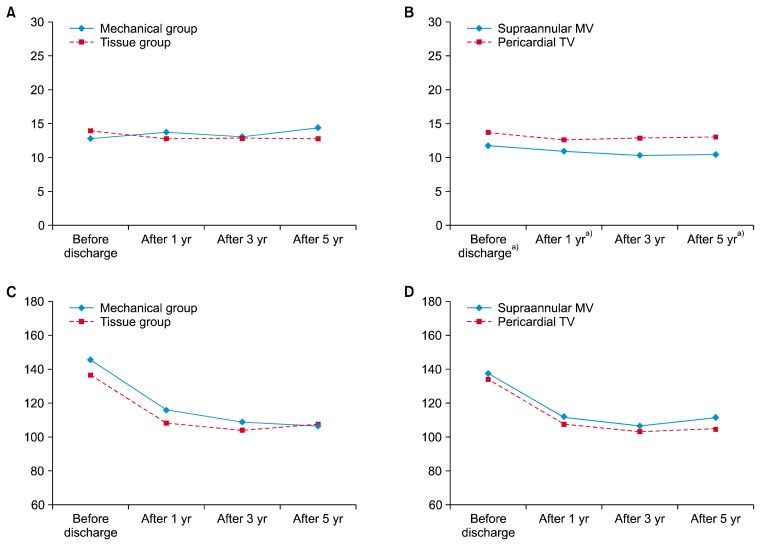
There was 1 patient whose AVMPG found to be over 40 mm Hg. He had received a pericardial aortic prosthesis 14 years ago, and had yet to undergo a reoperation on the closing date of the study. The average AVMPG and LVMI in the mechanical group were not significantly different from the corresponding values in the tissue group. In a sub-analysis limited to prostheses in the supra-annular position, the average AVMPG before discharge and at 1 and 5 years after surgery was lower in the mechanical group than in the tissue group (before discharge: p=0.007; 1 year: p=0.028; and 5 years: p=0.025). The average LVMI did not differ between groups at any time postoperatively. The hemodynamic outcomes are summarized in Table 2 and Fig. 1.
Table 2.
Hemodynamic performance and outcomes
| Variable | Mechanical group | Tissue group | p-value | Mechanical valve at supra-annular position | Tissue valve at supra-annular position | p-value |
|---|---|---|---|---|---|---|
| Aortic valve mean pressure gradient (mm Hg) | ||||||
| Before discharge | 12.9±5.479 | 13.83±4.510 | 0.102 | 11.77±4.220 | 13.73±4.527 | 0.007 |
| Postoperative | ||||||
| At 1 yr | 13.73±7.183 | 12.70±3.874 | 0.270 | 10.92±4.723 | 12.63±3.933 | 0.028 |
| At 3 yr | 13.03±5.331 | 12.84±5.133 | 0.850 | 10.27±3.141 | 12.86±5.270 | 0.066 |
| At 5 yr | 14.32±5.991 | 12.72±4.492 | 0.078 | 10.44±3.034 | 12.95±4.611 | 0.025 |
| Left ventricular mass index (g/m2) | ||||||
| Before discharge | 145.27±46.094 | 136.22±39.974 | 0.106 | 137.26±37.880 | 133.88±38.451 | 0.593 |
| Postoperative | ||||||
| At 1 yr | 115.64±34.009 | 108.11±29.421 | 0.083 | 111.57±24.181 | 107.42±29.309 | 0.412 |
| At 3 yr | 108.53±25.443 | 103.69±30.371 | 0.353 | 106.21±24.006 | 102.83±29.939 | 0.662 |
| At 5 yr | 105.91±26.293 | 106.96±28.098 | 0.841 | 111.27±27.616 | 104.59±25.873 | 0.364 |
Values are presented as mean±standard deviation.
Fig. 1.
(A, B) Average aortic valve mean pressure gradient and (C, D) left ventricular mass index before discharge and at 1, 3, and 5 years after surgery. MV, mechanical valve; TV, tissue valve. a)Indicates a statistically significant difference between groups.
3) Clinical outcomes
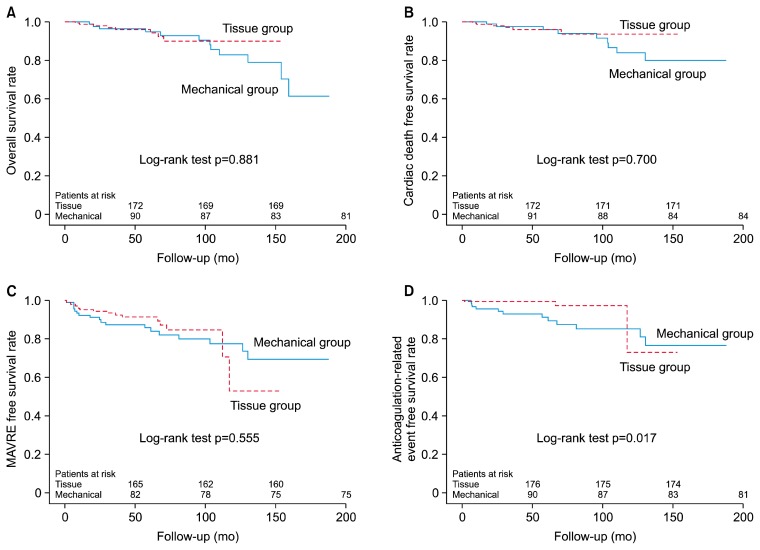
There were no cases of in-hospital mortality or 30-day mortality in either group. Postoperative complications were rare and did not significantly differ between groups. Table 3 summarizes the early clinical outcomes. There were 12 late deaths (13%) in the mechanical group and 8 (4.5%) in the tissue group (p=0.881) (Fig. 2A). The 10-year survival rate was 83% in the mechanical group and 90% in the tissue group. The frequency of cardiac mortality did not significantly differ between the mechanical (n=9, 10%) and tissue groups (n=6, 3%) (p=0.7) (Fig. 2B). The overall incidence of MAVREs was 19% (n=18) in the mechanical group and 10% (n=17) in the tissue group (p=0.555) (Fig. 2C). The incidence of anticoagulation-related events was greater in the mechanical group (p=0.017) (Fig. 2D), but no significant differences were found according to the patient’s place of residence (p=0.861) or level of education (p=0.428). Sixteen patients (6%) had anticoagulation-related events. In the mechanical group, 13 patients (14%) experienced anticoagulation-related events, including 7 bleeding events (44%) and 9 thromboembolic events (56%). In the tissue group, there were 3 anticoagulation-related events (1.7%), including 1 bleeding and 2 thromboembolic events. There were 4 late reoperations, which were performed due to structural tissue prosthesis degeneration (n=1), acute type A aortic dissection (n=1), and prosthetic valve endocarditis (n=2). The case of structural degeneration of the tissue prosthesis occurred in a patient who was on chronic dialysis. Prosthetic endocarditis occurred in 1 patient in each group. All reoperations were successful, with no cases of early mortality.
Table 3.
Early clinical outcomes
| Variable | Mechanical group | Tissue group | p-value |
|---|---|---|---|
| Early mortality | 0 | 0 | |
| Intra-aortic balloon pump | 0 | 2 (1.1) | 0.547 |
| Complete heart block | 1 (1.1) | 0 | 0.344 |
| Reoperation for bleeding | 1 (1.1) | 3 (1.7) | 1.000 |
| Deep sternal wound infection | 1 (1.1) | 0 | 0.344 |
| Paravalvular leakage | 0 | 0 | |
| Myocardial infarction | 0 | 0 | |
| Stroke | 0 | 0 |
Values are presented as number (%).
Fig. 2.
Kaplan-Meier survival curves. (A) Overall survival, (B) cardiac mortality-free survival, (C) MAVRE-free survival, and (D) anticoagulation-related events in the mechanical and tissue groups. MAVRE, major adverse valve related event.
In the univariate analysis, the type of aortic prosthesis was found to be unrelated to overall morality, cardiac mortality, or MAVREs. A mechanical aortic prosthesis and NYHA class III/IV status were risk factors for anticoagulation-related events in the univariate and multivariate analyses. Hypertension and NYHA class III or IV status were independent predictors of cardiac mortality (p=0.042) and MAVREs (p=0.001). The statistical analyses and Kaplan-Meier survival curves are shown in Table 4 and Fig. 2.
Table 4.
Predictors of clinical outcomes
| Variable | Univariate | Multivariate | |
|---|---|---|---|
|
|
|
||
| p-value | p-value | Hazard ratio (95% confidence interval) | |
| Overall mortality | |||
|
| |||
| Coronary artery disease | 0.028 | 0.080 | |
|
| |||
| Mechanical prosthesis | 0.881 | ||
|
| |||
| Cardiac mortality | |||
|
| |||
| Hypertension | 0.022 | 0.042 | 3.061 (1.039–9.013) |
|
| |||
| Mechanical prosthesis | 0.700 | ||
|
| |||
| Major adverse valve-related mortality | |||
|
| |||
| NYHA class III/IV | 0.001 | 0.001 | 2.984 (1.520–5.858) |
|
| |||
| Mechanical prosthesis | 0.556 | ||
|
| |||
| Anticoagulation-related event | |||
|
| |||
| NYHA class III/IV | 0.004 | 0.005 | 4.454 (1.580–12.553) |
|
| |||
| Mechanical prosthesis | 0.028 | 0.034 | 4.100 (1.111–15.132) |
NYHA, New York Heart Association.
Discussion
Current guidelines from North America suggest relatively clear age thresholds of 70 years for bioprostheses and 50 for mechanical prostheses [1]. Moreover, the ESC recommends either mechanical or tissue prostheses between the ages of 60 and 65 [5]. Thus, 60–70 years is the most controversial age range for selecting the type of prosthesis. We performed this study because no previous report has focused on this age group. In contrast to previous studies, our study population did not include patients who underwent a cardiac reoperation or other younger or older patients [8–13]. No previous studies have specifically investigated the age threshold of 65 years that is mentioned in the current guidelines. Herein, we focused on the impact of prosthesis type and age on clinical and hemodynamic outcomes.
Because we only included patients in their 60s who underwent their first cardiac operation, there were lower rates of coronary artery disease, concomitant coronary artery bypass, and poor NYHA status (III/IV) than in the populations described in previous publications [8–11]. In this study, the 10-year survival rate was high: 83% in the tissue group and 90% in the mechanical group. Life expectancy is increasing in many countries in North America, Europe, and Asia [7]. Using tissue prostheses in the young may lead to an increase in structural valve degeneration late in these patients’ lifespan. Cardiac reoperation is becoming safer than before. However, the risk of reoperation is always higher than that of first-time operations. Although transcatheter aortic valve replacement is an option, that technique has several limitations, including the size of the prosthesis and the unavailability of long-term outcomes [17,18]. Thus, some authors have advocated using mechanical prostheses in older patients [10,11,19–22].
We did not find any differences in overall and cardiac mortality or valve-related events between the mechanical and tissue prosthesis groups. However, using a mechanical valve was a predictor of anticoagulation-related events. This is an inevitable drawback of mechanical prostheses, because of the need for postoperative chronic anticoagulation therapy. In the study, educational level and place of residence were not related to anticoagulation-related events. These should not be barriers for choosing to use a mechanical prosthesis. However, a meticulous anticoagulation program is important to improve the outcomes of patients who receive a mechanical aortic valve.
Our recommendation for the choice of an aortic prosthesis is similar to the current ACC/AHA guidelines. In this study, the patient’s age was not related to overall mortality, cardiac mortality, valve-related events, or anticoagulation-related events. This is consistent with the findings of other studies [13,19]. Considering the longer life expectancy in most developed countries, the implantation of tissue prostheses in patients younger than 65 seems to be inappropriate. Tissue prostheses should be recommended after discussion of the bleeding risk due to anticoagulation and possible late reoperations. It should also be noted that quality of life and the costs of follow-up can be affected by the mode of anticoagulation. The inconvenience of frequent follow-up and repeated blood testing, noises from the mechanical valve, and restrictions on food and medicine can decrease patients’ quality of life.
Our target INR was somewhat lower than the levels presented in current recommendations [1,5], because herbal medicines, which usually increase the risk of bleeding [23], are popular in Korea. Although sophisticated INR control has been emphasized recently, point-of-care INR testing and dedicated anticoagulation centers are not yet available in most countries, including Korea. In this study, thromboembolic and bleeding events occurred at about the same frequency (thromboembolic: 56%; bleeding: 44%). These results suggest that our anticoagulation strategy successfully balanced the risks of bleeding and thromboembolism. In the tissue group, the incidence of anticoagulation events was quite low (1.7%).
We experienced 1 case of premature tissue valve failure (0.6%) at 6 years after operation and are following a patient whose AVMPG is over 40 mm Hg. This is consistent with previous studies that have reported low rates of bioprosthesis failure in older patients [24,25]. Most patients with a bioprosthesis were placed on an antiplatelet agent with strict blood pressure control. Statins were also used in some patients due to studies showing their effectiveness [26,27]. In this study, bovine pericardial prostheses were used much more frequently than porcine prostheses. In general, pericardial prostheses are considered to be more durable, with superior hemodynamic performance [25,28,29]. Life expectancy in Korea is rapidly increasing [30], so the choice of a pericardial prosthesis may be reasonable.
In general, mechanical prostheses are considered to have better hemodynamic performance than tissue prostheses [31]. In our study, however, we did not observe hemodynamic superiority of the mechanical prostheses in the overall population. The mechanical group included patients who received an older type of mechanical valve with everting rather than non-everting sutures. Therefore, we compared the early hemodynamic performance between mechanical and tissue prostheses implanted in the supra-annular position with those implanted using the non-everting suture technique. The average AVMPG was lower in patients with a supra-annular mechanical valve than in those with a pericardial tissue valve. However, the LVMI did not differ significantly between groups, suggesting that the difference in AVMPG between groups may not have been clinically significant.
This study has some limitations. The retrospective study design and the small number of patients limit the interpretation of our results. To overcome heterogeneity between the mechanical and tissue groups, we performed multivariate analysis. We did not use a point-of-care INR test, and did we not have a dedicated INR monitoring system. However, monthly follow-up of patients with a stable INR level is a widely accepted strategy at many centers. Finally, the follow-up duration of our study was not long enough to compare structural valve degeneration, which usually occurs 10 to 15 years after surgery.
In conclusion, our study suggests that aortic tissue prostheses are superior to mechanical aortic prostheses regarding anticoagulation-related events, and are not inferior to mechanical aortic prostheses in terms of overall and cardiac mortality, MAVREs, and hemodynamic performance. In this respect, tissue prostheses could be selected for patients younger than 65. However, considering the increase in life expectancy and the likelihood of reoperation, mechanical prostheses are preferable for the elderly. Thus, assessing the risk of bleeding and thromboembolism, as well as choosing the most appropriate anticoagulation strategy, may be key considerations in prosthesis selection.
Acknowledgments
This study was supported by a Grant of the Samsung Vein Clinic Network (Daejeon, Anyang, Cheongju, Cheonan; Fund no. KTCS04-100).
Footnotes
Conflict of interest
No potential conflict of interest relevant to this article was reported.
References
- 1.Nishimura RA, Otto CM, Bonow RO, et al. 2017 AHA/ACC focused update of the 2014 AHA/ACC guideline for the management of patients with valvular heart disease: a report of the American College of Cardiology/American Heart Association Task Force on clinical practice guidelines. J Am Coll Cardiol. 2017;70:252–89. doi: 10.1016/j.jacc.2017.03.011. [DOI] [PubMed] [Google Scholar]
- 2.Svensson LG, Adams DH, Bonow RO, et al. Aortic valve and ascending aorta guidelines for management and quality measures. Ann Thorac Surg. 2013;95(6 Suppl):S1–66. doi: 10.1016/j.athoracsur.2013.01.083. [DOI] [PubMed] [Google Scholar]
- 3.Dunning J, Gao H, Chambers J, et al. Aortic valve surgery: marked increases in volume and significant decreases in mechanical valve use: an analysis of 41,227 patients over 5 years from the Society for Cardiothoracic Surgery in Great Britain and Ireland National database. J Thorac Cardiovasc Surg. 2011;142:776–82. doi: 10.1016/j.jtcvs.2011.04.048. [DOI] [PubMed] [Google Scholar]
- 4.Brown JM, O’Brien SM, Wu C, Sikora JA, Griffith BP, Gammie JS. Isolated aortic valve replacement in North America comprising 108,687 patients in 10 years: changes in risks, valve types, and outcomes in the Society of Thoracic Surgeons National Database. J Thorac Cardiovasc Surg. 2009;137:82–90. doi: 10.1016/j.jtcvs.2008.08.015. [DOI] [PubMed] [Google Scholar]
- 5.Baumgartner H, Falk V, Bax JJ, et al. 2017 ESC/EACTS Guidelines for the management of valvular heart disease. Eur Heart J. 2017;38:2739–91. doi: 10.1093/eurheartj/ehx391. [DOI] [PubMed] [Google Scholar]
- 6.Vahanian A, Baumgartner H, Bax J, et al. Guidelines on the management of valvular heart disease: the Task Force on the Management of Valvular Heart Disease of the European Society of Cardiology. Eur Heart J. 2007;28:230–68. doi: 10.1093/eurheartj/ehl428. [DOI] [PubMed] [Google Scholar]
- 7.Salomon JA, Wang H, Freeman MK, et al. Healthy life expectancy for 187 countries, 1990–2010: a systematic analysis for the Global Burden Disease Study 2010. Lancet. 2012;380:2144–62. doi: 10.1016/S0140-6736(12)61690-0. [DOI] [PubMed] [Google Scholar]
- 8.Ashikhmina EA, Schaff HV, Dearani JA, et al. Aortic valve replacement in the elderly: determinants of late outcome. Circulation. 2011;124:1070–8. doi: 10.1161/CIRCULATIONAHA.110.987560. [DOI] [PubMed] [Google Scholar]
- 9.Brown ML, Schaff HV, Lahr BD, et al. Aortic valve replacement in patients aged 50 to 70 years: improved outcome with mechanical versus biologic prostheses. J Thorac Cardiovasc Surg. 2008;135:878–84. doi: 10.1016/j.jtcvs.2007.10.065. [DOI] [PubMed] [Google Scholar]
- 10.Stassano P, Di Tommaso L, Monaco M, et al. Aortic valve replacement: a prospective randomized evaluation of mechanical versus biological valves in patients ages 55 to 70 years. J Am Coll Cardiol. 2009;54:1862–8. doi: 10.1016/j.jacc.2009.07.032. [DOI] [PubMed] [Google Scholar]
- 11.Vicchio M, Della Corte A, De Santo LS, et al. Tissue versus mechanical prostheses: quality of life in octogenarians. Ann Thorac Surg. 2008;85:1290–5. doi: 10.1016/j.athoracsur.2007.12.039. [DOI] [PubMed] [Google Scholar]
- 12.Weber A, Noureddine H, Englberger L, et al. Ten-year comparison of pericardial tissue valves versus mechanical prostheses for aortic valve replacement in patients younger than 60 years of age. J Thorac Cardiovasc Surg. 2012;144:1075–83. doi: 10.1016/j.jtcvs.2012.01.024. [DOI] [PubMed] [Google Scholar]
- 13.Brennan JM, Edwards FH, Zhao Y, et al. Long-term safety and effectiveness of mechanical versus biologic aortic valve prostheses in older patients: results from the Society of Thoracic Surgeons Adult Cardiac Surgery National Database. Circulation. 2013;127:1647–55. doi: 10.1161/CIRCULATIONAHA.113.002003. [DOI] [PubMed] [Google Scholar]
- 14.Akins CW, Miller DC, Turina MI, et al. Guidelines for reporting mortality and morbidity after cardiac valve interventions. Ann Thorac Surg. 2008;85:1490–5. doi: 10.1016/j.athoracsur.2007.12.082. [DOI] [PubMed] [Google Scholar]
- 15.Lang RM, Bierig M, Devereux RB, et al. Recommendations for chamber quantification: a report from the American Society of Echocardiography’s guidelines and Standards Committee and the Chamber Quantification Writing Group, developed in conjunction with the European Association of Echocardiography, a branch of the European Society of Cardiology. J Am Soc Echocardiogr. 2005;18:1440–63. doi: 10.1016/j.echo.2005.10.005. [DOI] [PubMed] [Google Scholar]
- 16.Devereux RB, Reichek N. Echocardiographic determination of left ventricular mass in man: anatomic validation of the method. Circulation. 1977;55:613–8. doi: 10.1161/01.CIR.55.4.613. [DOI] [PubMed] [Google Scholar]
- 17.Kobayashi J. Changing strategy for aortic stenosis by transcatheter valve treatment in Japan. Circ J. 2013;77:309–10. doi: 10.1253/circj.CJ-12-1546. [DOI] [PubMed] [Google Scholar]
- 18.Maeda K, Kuratani T, Mizote I, et al. Early experiences of transcatheter aortic valve replacement in Japan. Circ J. 2013;77:359–62. doi: 10.1253/circj.CJ-12-0650. [DOI] [PubMed] [Google Scholar]
- 19.Accola KD, Scott ML, Palmer GJ, et al. Surgical management of aortic valve disease in the elderly: a retrospective comparative study of valve choice using propensity score analysis. J Heart Valve Dis. 2008;17:355–64. [PubMed] [Google Scholar]
- 20.De Vincentiis C, Kunkl AB, Trimarchi S, et al. Aortic valve replacement in octogenarians: is biologic valve the unique solution? Ann Thorac Surg. 2008;85:1296–301. doi: 10.1016/j.athoracsur.2007.12.018. [DOI] [PubMed] [Google Scholar]
- 21.Kurlansky PA, Williams DB, Traad EA, et al. The valve of choice in elderly patients and its influence on quality of life: a long-term comparative study. J Heart Valve Dis. 2006;15:180–9. [PubMed] [Google Scholar]
- 22.Lund O, Bland M. Risk-corrected impact of mechanical versus bioprosthetic valves on long-term mortality after aortic valve replacement. J Thorac Cardiovasc Surg. 2006;132:20–6. doi: 10.1016/j.jtcvs.2006.01.043. [DOI] [PubMed] [Google Scholar]
- 23.Ge B, Zhang Z, Zuo Z. Updates on the clinical evidenced herb-warfarin interactions. Evid Based Complement Alternat Med. 2014;2014:957362. doi: 10.1155/2014/957362. [DOI] [PMC free article] [PubMed] [Google Scholar]
- 24.Said SM, Ashikhmina E, Greason KL, et al. Do pericardial bioprostheses improve outcome of elderly patients undergoing aortic valve replacement? Ann Thorac Surg. 2012;93:1868–74. doi: 10.1016/j.athoracsur.2012.01.061. [DOI] [PubMed] [Google Scholar]
- 25.McClure RS, Narayanasamy N, Wiegerinck E, et al. Late outcomes for aortic valve replacement with the Carpentier-Edwards pericardial bioprosthesis: up to 17-year follow-up in 1,000 patients. Ann Thorac Surg. 2010;89:1410–6. doi: 10.1016/j.athoracsur.2010.01.046. [DOI] [PubMed] [Google Scholar]
- 26.Farivar RS, Cohn LH. Hypercholesterolemia is a risk factor for bioprosthetic valve calcification and explantation. J Thorac Cardiovasc Surg. 2003;126:969–75. doi: 10.1016/S0022-5223(03)00708-6. [DOI] [PubMed] [Google Scholar]
- 27.Antonini-Canterin F, Zuppiroli A, Popescu BA, et al. Effect of statins on the progression of bioprosthetic aortic valve degeneration. Am J Cardiol. 2003;92:1479–82. doi: 10.1016/j.amjcard.2003.08.066. [DOI] [PubMed] [Google Scholar]
- 28.Dalmau MJ, Gonzalez-Santos JM, Blazquez JA, et al. Hemodynamic performance of the Medtronic Mosaic and Perimount Magna aortic bioprostheses: five-year results of a prospectively randomized study. Eur J Cardiothorac Surg. 2011;39:844–52. doi: 10.1016/j.ejcts.2010.11.015. [DOI] [PubMed] [Google Scholar]
- 29.Okamura H, Yamaguchi A, Yoshizaki T, et al. Clinical outcomes and hemodynamics of the 19-mm Perimount Magna bioprosthesis in an aortic position: comparison with the 19-mm Medtronic Mosaic Ultra Valve. Circ J. 2012;76:102–8. doi: 10.1253/circj.CJ-11-0728. [DOI] [PubMed] [Google Scholar]
- 30.Yang S, Khang YH, Harper S, Davey Smith G, Leon DA, Lynch J. Understanding the rapid increase in life expectancy in South Korea. Am J Public Health. 2010;100:896–903. doi: 10.2105/AJPH.2009.160341. [DOI] [PMC free article] [PubMed] [Google Scholar]
- 31.Khoo JP, Davies JE, Ang KL, Galinanes M, Chin DT. Differences in performance of five types of aortic valve prostheses: haemodynamic assessment by dobutamine stress echocardiography. Heart. 2013;99:41–7. doi: 10.1136/heartjnl-2012-302256. [DOI] [PubMed] [Google Scholar]


