Abstract
Entomopathogenic nematodes (EPNs) that are symbiotically associated with Xenorhabdus and Photorhabdus bacteria can kill target insects via direct infection and toxin action. There are limited reports identifying such organisms in the National Park of Thailand. Therefore, the objectives of this study were to identify EPNs and symbiotic bacteria from Nam Nao National Park, Phetchabun Province, Thailand and to evaluate the larvicidal activity of bacteria against Aedes aegypti and Ae. albopictus. A total of 12 EPN isolates belonging to Steinernema and Heterorhabditis were obtained form 940 soil samples between February 2014 and July 2016. EPNs were molecularly identified as S. websteri (10 isolates) and H. baujardi (2 isolates). Symbiotic bacteria were isolated from EPNs and molecularly identified as P. luminescens subsp. akhurstii (13 isolates), X. stockiae (11 isolates), X. vietnamensis (2 isolates) and X. japonica (1 isolate). For the bioassay, bacterial suspensions were evaluated for toxicity against third to early fourth instar larvae of Aedes spp. The larvae of both Aedes species were orally susceptible to symbiotic bacteria. The highest larval mortality of Ae. aegypti was 99% after exposure to X. stockiae (bNN112.3_TH) at 96 h, and the highest mortality of Ae. albopictus was 98% after exposure to P. luminescens subsp. akhurstii (bNN121.4_TH) at 96 h. In contrast to the control groups (Escherichia coli and distilled water), the mortality rate of both mosquito larvae ranged between 0 and 7% at 72 h. Here, we report the first observation of X. vietnamensis in Thailand. Additionally, we report the first observation of P. luminescens subsp. akhurstii associated with H. baujardi in Thailand. X. stockiae has potential to be a biocontrol agent for mosquitoes. This investigation provides a survey of the basic diversity of EPNs and symbiotic bacteria in the National Park of Thailand, and it is a bacterial resource for further studies of bioactive compounds.
Introduction
Xenorhabdus and Photorhabdus are Gram-negative bacteria of the family Enterobacteriaceae and are symbiotically associated with the entomopathogenic nematodes (EPNs), Steinernema and Heterorhabditis respectively [1]. The infective juvenile stages (IJs) of EPNs containing symbiotic bacteria in their midguts live in the soil of diverse ecological systems [2]. The nematodes can kill the target insects via direct infection through the promotion of the secondary metabolites and toxins produced by symbiotic bacteria. When the IJs of nematodes enter the mouth, anus or spiracles of insect hosts, symbiotic bacteria are released from their intestines into the hemocoel of target insects [3]. Subsequently, the symbiotic bacteria produce several toxins or secondary metabolites, killing the insects by induction of immunosuppression and pervasion of the hemolymph [4]. Finally, the symbiotic bacteria replicate rapidly and cause septicemia in insects [5]. In addition, several secondary metabolites produced by the symbiotic bacteria have been reported as bioactive compounds with activities including cytotoxic, antimicrobial, antiparasitic and insecticidal ones [6, 7].
Earlier studies on the larvicidal activity of symbiotic bacteria have demonstrated that Photorhabdus insect-related (Pir) protein had a high toxicity against Aedes aegypti and Aedes albopictus, a main vector of Dengue virus [8]. In addition, cell suspensions of P. luminescens and X. nematophila were orally toxic to Ae. aegypti [9]. The mortality rate of Ae. aegypti was further increased when the Cry4Ba protein from Bacillus thuringiensis and suspensions of P. luminescens and X. nematophila were mixed [10]. The mortality of Ae. aegypti larvae was 20% when fed with Cry4Ba (5 ηg/ml) alone. At 48 h after treatment, enhancement of toxicity was observed by combining Cry4Ba toxin with X. nematophila (108 CFU) or P. luminescens (108 CFU), which showed 95% and 85% of larval mortality respectively [10]. In addition, cell suspensions of Xenorhabdus nematophila together with B. thuringiensis subsp. israelensis strongly promote the mortality rate of Ae. aegypti and Culex pipiens pallens [11]. Recently, the fluid cultured forms of X. nematophila and P. luminescens rapidly caused death of Ae. aegypti within a few hours and caused delays in the development of pupa and adults [12]. This suggests that Xenorhabdus and Photorhabdus bacteria are the natural resources for searching for bioactive compounds. However, species identifications of EPNs and their symbiotic bacteria are important fields of research for studies in agricultural and medical fields.
At present, EPNs have been identified in several geographical areas with approximately 100 species of Steinernema and 26 species of Heterorhabditis [13–27]. However, the diversity and application of both EPNs and their symbionts have not been thoroughly studied in several countries, including Thailand. In our country, six species of Steinernema have been found in several provinces: S. siamkayi, S. websteri, S. minutum, S. khoisanae, S. scarabaei and S. kushidai. Additionally, 6 species of Heterorhabditis, including H. indica, H. baujardi, H. bacteiophora, H. somsookae, H. gerrardi and H. zealandica, were recorded in Thailand [15, 28–33]. Symbiotic bacteria have been discovered worldwide, with approximately 24 species of Xenorhabdus and 5 species of Photorhabdus. In Thailand, three species of Xenorhabdus were reported, including X. stockiae, X. miraniensis and X. japonica. Additionally, 3 species with 6 subspecies of Photorhabdus, including P. luminescens subsp. akhurstii, P. luminescens subsp. hainanensis, P. luminescens subsp. laumondii, P. asymbiotica subsp. australis, P. luminescens subsp. namnaonensis and P. temperata subsp. temperata, were also recorded across the country [15, 33–36].
Most surveyed locations for EPNs and their associated microbes in Thailand were roadside, with minor amounts surveyed from fruit crops, rice fields and river banks [15, 31, 32]. The first novel species of EPN in Thailand, S. siamkayi, was isolated from soil samples in Phetchabun province [28]. Subsequently, a novel subspecies of symbiotic bacteria, P. luminescens subsp. namnaonensis, was isolated from H. baujardi from the Nam Nao district in Phetchabun province [36]. This suggests that EPNs and their symbionts may be diverse in Phetchabun province, a mountainous region of central Thailand. Therefore, other areas in Phetchabun province should be explored for EPNs and their symbiotic bacteria. Nam Nao National Park includes the mountainous forests of Phetchabun and Chaiyaphum provinces in Thailand. The flora consists mainly of dry dipterocarps, mixed deciduous, hill evergreens, vast bamboo groves, pine forests and some grassland areas. Despite the interesting location for the surveying of EPNs and their symbiotic bacteria, there is only one report of these organisms in the National Park (Mae Wong) of Thailand. This area is abundant with insect hosts and might lead to the recovery of several species of EPNs and symbiotic bacteria. In addition, four organisms were newly recorded in Mae Wong National Park, namely H. zealandica, S. kushidai, X. japonica and P. temparata subsp. temperata. Therefore, the objectives of this study were to isolate and identify EPNs and their associated Xenorhabdus and Photorhabdus from Nam Nao National Park of Thailand and to determine the larvicidal activity of these bacteria against Ae. aegypti and Ae. albopictus.
Materials and methods
Collection of soil samples
The methods for soil collection in Nam Nao National Park of Phetchabun province were approved and permitted by the Department of National Park, Wildlife and Plant Conservation, Thailand (Permission number 0907.4/8245). Overall, 940 soil samples were collected from 188 soil sites in Nam Nao National Park between February 2014 and July 2016. At each site, five soil samples (300–500 g) were collected using a hand shovel. The soil collection process was performed according to Thanwisai et al. [15]. The latitude and altitude of each soil site were determined using a GPS navigator (Garmin nüvi 1250, Taiwan). The moisture and pH of each soil sample were recorded using the soil pH and moisture meter Takemura DM15 (Takemura Electronic Works, Chiba, Japan). The soil temperature of each sample was also measured using a soil survey instrument (Yancheng Kecheng Optoelectronic Technology, Jiangsu, China). Soil texture was also noted. All soil samples were transported in ambient temperature to the Department of Microbiology and Parasitology, Faculty of Medical Science, Naresuan University, Phitsanulok province, Thailand for the isolation of EPNs. Soil parameters (soil temperature, pH and moisture) of samples with and without EPNs were compared statistically using the Mann-Whitney test in the SPSS Program, Version 17 (P < 0.05).
Identification of entomopathogenic nematodes
A baiting technique [37] using Galleria mellonella larvae followed by a White trap [38] was performed to isolate EPNs from soil samples. The infective juvenile stages (IJs) of EPNs emerged from the G. mellonella cadaver and moved to water. The IJs were collected in a culture flask and cleaned by a sedimentation technique, along with several changes of sterile distilled water. The clean IJs were kept in aerated water and stored at 13 °C in a refrigerator. To increase the number and confirm the entomopathogenicity of EPNs, a new G. mellonella larva was infected with IJs.
The genomic DNA of EPNs was extracted from the IJs as described previously [15]. The genomic DNA was dissolved in 20 μl of sterile distilled water and stored at –20 °C for future use.
A PCR-based analysis and DNA sequencing were conducted to identify Steinernama and Heterorhabditis species. The primers used for amplifying Steinernama 28S rDNA were 539_F (5’GGATTTCCTTAGTAACTGCGAGTG-3’) and 535_R (5’–TAGTCTTTCGCCCCTATACCCTT-3’) [39]. The PCR conditions consisted of pre-denaturation at 95 °C for 1 min, followed by 35 cycles of denaturation at 94 °C for 1 min, annealing at 55 °C for 30 s, extension at 72 °C for 45 s, and a final extension at 72 °C for 7 min. The size of amplification fragment was 870 bp.
The primers used for amplifying Heterorhabditis internal transcribe spacer (ITS) were18S_F (5’-TGATTACGTCCCTGCCCTTT-3’) and 26S_R (5’-TTTCACTCGCCGTTACTAAGG-3’) or TW81_F (5’-GTTTCCGTAGGTGAACCTGC-3’) and AB28_R (5’-ATATGCTTAAGTTCAGCGGGT-3’) [40]. The PCR conditions for Heterorhabditis consisted of predenaturation at 95 °C for 1 min, followed by 35 cycles of denaturation at 94 °C for 30 s, annealing at 50 °C for 1 min, extension at 72 °C for 1 min and a final extension at 72 °C for 7 min. The PCR product was varied between 800–1,100 bp.
A total of 30 μl of the PCR mixture of each Steinernama and Heterorhabditis consisted of 3 μl of 10x buffer (1x), 4.2 μl of 25 mM MgCl2 (3.5 mM), 0.6 μl of 10 mM dNTPs (200 μM), 1.2 μl of 5 μM forward primer (0.8 μM), 1.2 μl of 5 μM reverse primer (0.8 μM), 0.3 μl of 2.5 unit Taq DNA polymerase (0.1 U/ml), 7.5 μl of DNA solution (20–200 ηg) and 12 μl of sterile distilled water. Thermocycling for Steinernama and Heterorhabditis was performed in the thermal cycler (Applied Biosystems, Pittsburgh, PA, USA). PCR products were verified by electrophoresis on 1.2% TBE-buffered agarose gel. The PCR products were purified using a Gel/PCR DNA Fragment Extraction Kit (Geneaid Biotech Ltd., New Taipei, Taiwan) as recommended by the manufacturer. The DNA sequencing for both directions was analyzed by the Macrogen Inc. service in Korea.
Isolation and identification of symbiotic bacteria
Symbiotic bacteria were isolated from the hemolymph of dead G. mellonella larvae, which had been infected with the IJs of EPNs. The dead larvae of G. mellonella were surface-sterilized by dipping into absolute ethanol for 1 min and placed in a sterile petri dish to dry. Sterile forceps were used to open the 3rd segment from the head of G. mellonella. A sterile loop was used to touch hemolymph and streaked on nutrient agar supplemented with bromothymol blue and triphenyl-2,3,5-tetrazolium chloride (NBTA). Colonies of Xenorhabdus and Photorhabdus were blue and green on NBTA after four days of incubation in the dark at room temperature. A single colony of each isolate of symbiotic bacteria was inoculated in 3 ml of Luria-Bertani (LB) broth and incubated with shaking at 180 rpm overnight (approximately 18–24 h). The genomic DNA of symbiotic bacteria was extracted from bacterial pellets using the Blood/Cell DNA Mini Kit (Geneaid Biotech Ltd., New Taipei, Taiwan). The genomic DNA of symbiotic bacteria was stored at –20 °C for further use in PCR.
PCR-based analysis and recA gene sequencing was performed to identify Xenorhabdus and Photorhabdus species. Primers used were recA_F (5’-GCTATTGATGAAAATAAACA-3’) and recA_R (5’–RATTTTRTCWCCRTTRTAGCT-3’) to obtain an 890 pb amplicon [41]. The PCR conditions for Xenorhabdus and Photorhabdus were based on a previous report by Thanwisai el at. (2012) [15]. A total of 30 μl of the PCR reaction contained 3 μl of 10x buffer (1x), 4.2 μl of 25 mM MgCl2 (3.5 mM), 0.6 μl of 10 mM dNTPs (200 μM), 1.2 μl of 5 μM forward primer (0.8 μM), 1.2 μl of 5 μM reverse primer (0.8 μM), 0.3 μl of 2.5 unit Taq DNA polymerase (0.1 U/ml), 3 μl of genomic DNA solution (20–200 ηg) and 16.5 μl of sterile distilled water. PCR was conducted in a thermal cycler (Applied Biosystems, Pittsburgh, PA, USA). The thermal profile was as previously described [15]. PCR products were visualized on a ethidium bromide-stained 1.2% agarose gel and purified using a PCR clean-up Gel Extraction Kit (Macherey-nagel, Düren, Germany). The DNA sequencing for both directions was analyzed by Macrogen Inc. service in Korea.
Sequencing and phylogenetic analysis
To identify species of EPNs (Steinernema and Heterorhabditis) and symbiotic bacteria (Xenorhabdus and Photorhabdus), comparison of the partially edited nucleotide sequences (recA, ITS and 28S rDNA) was performed using the BLASTN program from NCBI. A cut-off of ≥ 97% identity was considered for the same species. The nucleotide sequences were aligned using ClustalW, and a phylogenetic tree was constructed using the maximum likelihood method with the MEGA Version 7.0 program [42].
Bioassay for larvicidal activity
In this study, five isolates of symbiotic bacteria were selected for using in the bioassay. The symbiotic bacteria, including P. luminescens subsp. akhurstii (bNN121.4_TH), X. stockiae (bNN112.3_TH), X. japonica (bNN165.4_TH), X. vietnamensis (bNN167.2_TH) and X. vietnamensis (bNN167.3_TH), were used for determining the oral toxicity against Ae. aegypti and Ae. albopictus larvae.
The eggs of Ae. aegypti and Ae. albopictus (laboratory strain) on a filter paper were purchased from the Taxonomy and Reference Museum of the Department of Medical Sciences at the National Institute of Health of Thailand, Ministry of Public Health, Nonthaburi Province, Thailand. Aedes eggs were placed into distilled water to allow hatching of first instar larvae, which were fed with minced food pets. The late third and early fourth instar larvae were used in the bioassays.
Escherichia coli ATCC® 25922 and distilled water were used as controls for the bioassays. Escherichia coli ATCC® 25922 was inoculated in the LB broth and incubated at 37°C for 24 h, while each isolate of Xenorhabdus and Photorhabdus was inoculated on 5YS medium (5% (w/v) yeast extract, 0.5% (w/v) NaCl, 0.05% (w/v) NH4H2PO4, 0.05% (w/v) K2HPO4 and 0.02% (w/v) MgSO4.7H2O) and incubated at room temperature for 48 h. All broth cultures of bacteria were centrifuged at 10,000 x g for 10 min. The supernatant was discarded and the pellets were suspended in sterile distilled water to an OD600 of 0.1 with a spectrophotometer (Metertech SP-880, Taiwan).
Suspensions containing Xenorhabdus and Photorhabdus was evaluated for toxicity against the larvae (third late to early fourth instars) of Ae. aegypti and Ae. albopictus. Distilled water and a suspension of E. coli ATCC® 25922 were used as controls. In each bioassay, 30 larvae of each mosquito species were placed in three wells of a 24-well plate (10 larvae/well). Subsequently, 2 ml (107–108 CFU/ml) from each bacterial suspension was added to each well. This concentration of symbiotic bacteria showed the enhancement of oral toxicity against Ae. aegypti [10]. The larvae were fed with minced pet food during the experiment. The mortality rate of larvae was monitored after exposure to bacterial suspensions at 24, 48, 72 and 96 h. Each bioassay was tested in three replicates on different dates. The larvae were considered dead when no movement was detected when teased with a fine sterile toothpick. Larval mortality of Ae. aegypti and Ae. albopictus after exposure to symbiotic bacteria was analyzed by survival analysis (Kaplan-Meier Estimate) of SPSS Version 17 (P < 0.05).
Results
Isolation of entomopathogenic nematodes from soil samples
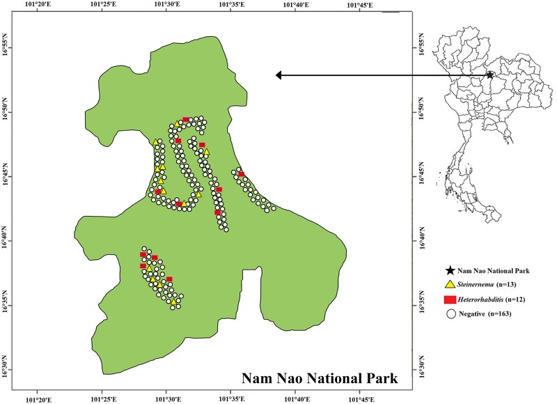
Out of the 188 soil sites from Nam Nao National Park, Phetchabun Province, Thailand, 25 tested positive for EPNs (Fig 1). Of the 940 soil samples, 27 (2.87%) were found positive for Steinernema or Heterorhabditis. This yielded 13 isolates of Heterorhabditis and 14 isolates of Steinernema. Most EPNs were isolated from loam (96.30%), with the soil pH ranging between 4.8 and 7.0, soil temperature ranging between 19 °C and 30 °C and soil moisture ranging between 1.0 and 8.0 (Table 1). One isolate of the EPNs was isolated from sandy loam and none of the EPNs were isolated from soil with a clay texture.
Fig 1. Map of sites for soil collection in Nam Nao National Park, Phetchabun Province, Thailand.
A total of 188 soil sampling sites with their statuses of positive or negative for EPNs, Steinernema and Heterorhabditis, in Nam Nao National Park, Phetchabun Province, Thailand.
Table 1. Temperature, pH and moisture from soil samples (n = 940) with and without EPNs in Nam Nao National Park, Phetchabun Province, Thailand.
| Soil parameter | Soil with EPNs (n = 27) |
Soil without EPNs (n = 913) |
P-value (Mann-Whitney test) |
|---|---|---|---|
| Temperature (°C) | 20–25 | 19–30 | 0.055 |
| pH | 6.2–7.0 | 4.8–7.0 | 0.165 |
| Moisture | 1.0–3.0 | 1.0–8.0 | 0.972 |
Identification and phylogeny of entomopathogenic nematodes
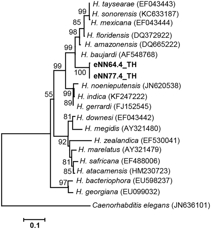
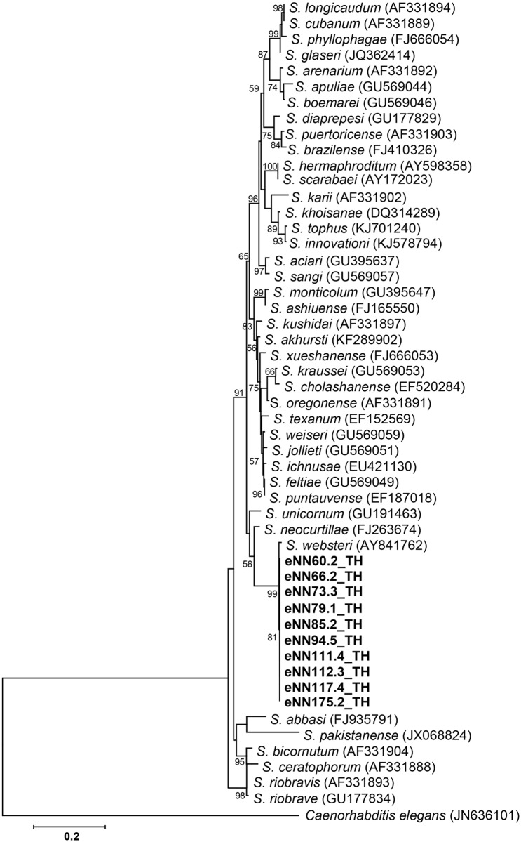
To identify Heterorhabditis and Steinernema species, PCR-based analysis and sequencing of the ITS and 28S rDNA regions were performed together with a BLASTN search of the edited sequences. Two isolates of Heterorhabditis (accession no. MG209260 and MG209261) were identified as Heterorhabditis baujardi with 99% identity after BLASTN search using 542 nucleotides of the ITS region. For the genus Steinernema (accession no. MG209262, MG209263, MG209264, MG209265, MG209266, MG209267, MG209268, MG209269, MG209270 and MG209271), 10 isolates of S. websteri were identified with 99% identity after BLASTN search using 502 nucleotides of the 28S rDNA region. S. websteri was the most recovered species in soil samples from the studied area. The remaining 15 EPN isolates were lost through protozoa and fungal contamination. A phylogenetic tree based on the maximum likelihood method revealed two isolates of the genus Heterorhabditis grouped in H. baujardi (accession no. AF548768.1) (Fig 2) and 10 isolates of the genus Steinernema grouped in S. websteri (accession no. AY841762.1) (Fig 3).
Fig 2. Phylogenetic tree of Heterorhabditis.
Maximum likelihood tree of a 542 bp of the ITS region from two Heterorhabditis isolates from Nam Nao National Park, Phetchabun Province, Thailand (codes ending with TH), together with 18 Heterorhabditis sequences downloaded from the GenBank database. Caenorhabditis elegans (accession no. JN636101.1) was used as the outgroup. Bootstrap values are based on 1,000 replicates.
Fig 3. Phylogenetic tree of Steinernema.
Maximum likelihood tree of a 502 bp of the 28S rDNA region from 10 Steinernema isolates from Nam Nao National Park, Phetchabun Province, Thailand (codes ending with TH), together with 41 Steinernema sequences downloaded from the GenBank database. Caenorhabditis elegans (accession no. JN636101.1) was used as the outgroup. Bootstrap values are based on 1,000 replicates.
Isolation, identification and phylogeny of Xenorhabdus and Photorhabdus
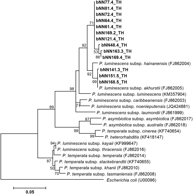
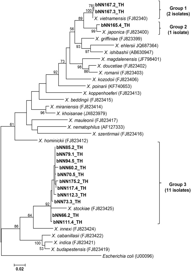
Based on colony morphology on NBTA agar, 27 isolates of symbiotic bacteria were identified as Photorhabdus (13 isolates) and Xenorhabdus (14 isolates). A partial sequence of the recA gene was amplified and sequenced to identify the Xenorhabdus and Photorhabdus species. A BLASTN search of the edited sequences was performed to find the identity. A total of 13 isolates of Photorhabdus (accession no MG209233, MG209234, MG209235, MG209236, MG209237, MG209238, MG209239, MG209240, MG209241, MG209242, MG209243, MG209244 and MG209245) were identified as P. luminescens subsp. akhurstii with 97–100% identity. Also, 14 isolates of Xenorhabdus (accession no MG209246, MG209247, MG209248, MG209249, MG209250, MG209251, MG209252, MG209253, MG209254, MG209255, MG209256, MG209257, MG209258 and MG209259) were identified as X. stockiae (11 isolates), X. vietnamensis (2 isolates) and X. japonica (1 isolate) with an identity ranging from 97–99%. Fig 4 shows the phylogenetic tree based on the maximum likelihood method, which revealed that only one group of Photorhabdus (13 isolates in the present study) was closely related to P. luminescens subsp. akhurstii (accession no. FJ862005.1) derived from the NCBI database. Two isolates of Photorhabdus (bNN64.4_TH and bNN77.4_TH) were hosted by H. baujardi (Table 2). In all, 11 isolates of P. luminescens subsp. akhurstii were associated with Heterorhabditis sp. (Table 2). Fig 5 shows the phylogenetic tree based on the maximum likelihood method of 14 Xenorhabdus isolates in the present study. The group of isolates was divided into 3 groups. Group 1 contained 2 isolates of Xenorhabdus (bNN167.3_TH and bNN167.3_TH) and the referenced X. vietnamensis (accession no. GU481051.1). Group 2 contained only 1 isolate of Xenorhabdus (bNN165.4) in the present study and X. japonica (accession no. FJ823400.1) isolated from Steinernema kushidai. Group 3 was composed of 11 isolates of Xenorhabdus, which were closely related to X. stockiae strain TH01 (accession no. FJ823425.1). Most Xenorhabdus isolates in the present study were associated with S. websteri (Table 2). Two Xenorhabdus vietnamensis isolates and one isolate of Xenorhabdus japonica were hosted by Steinernema sp. (Table 2).
Fig 4. Phylogenetic tree of Photorhabdus.
Maximum likelihood tree of a 588 bp of the recA region for 13 Photorhabdus isolates from Nam Nao National Park, Phetchabun Province, Thailand (codes ending with TH), together with 16 Photorhabdus sequences downloaded from the GenBank database. Escherichia coli (accession no. U00096.3) was used as an outgroup. Bootstrap values are based on 1,000 replicates.
Table 2. Association between EPN hosts and symbiotic bacteria in Nam in Nam Nao National Park, Phetchabun Province, Thailand.
| Association between EPN and their symbiotic bacteria | No. of Associations (isolates) | Code |
|---|---|---|
| Heterorhabditis associated with P. luminescens subsp. akhurstii | 11 | NN48.4_TH, NN61.4_TH, NN72.4_TH, NN81.4_TH, NN121.4_TH, NN141.3_TH, NN151.5_TH, NN163.3_TH, NN168.5_TH, NN169.2_TH, NN169.4_TH |
| Heterorhabditis baujardi associated with P. luminescens subsp. akhurstii | 2 | NN64.4_TH, NN77.4_TH |
| Steinernema websteri associated with Xenorhabdus stockiae | 10 | NN60.2_TH, NN66.2_TH, NN70.5_TH, NN73.3_TH, NN79.1_TH, NN85.2_TH, NN94.5_TH, NN111.4_TH, NN112.3_TH, NN117.4_TH |
| Steinernema associated with Xenorhabdus stockiae | 1 | NN175.2_TH |
| Steinernema associated with Xenorhabdus vietnamensis | 2 | NN167.2_TH, NN167.3_TH |
| Steinernema associated with Xenorhabdus japonica | 1 | NN165.4_TH |
Fig 5. Phylogenetic tree of Xenorhabdus.
Maximum likelihood tree of a 588 bp of the recA gene for 14 Xenorhabdus isolates from Nam Nao National Park, Phetchabun Province, Thailand (codes ending with TH), together with 23 Xenorhabdus sequences downloaded from the GenBank database. Escherichia coli (accession no. U00096.3) was used as an outgroup. Bootstrap values are based on 1,000 replicates.
Larvicidal activity of symbiotic bacteria against Aedes larvae
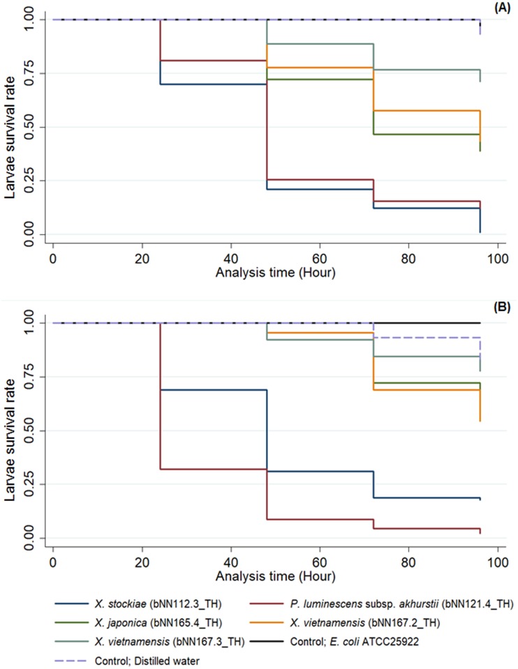
The symbiotic bacteria used for the bioassay were selected based on their locations in different groups on the phylogenetic tree. Both, the Xenorhabdus and Photorhabdus isolates in the present study were assumed to be pathogenic when ingested by Ae. aegypti and Ae. albopictus larvae. Fig 6 and Table 3 demonstrate that the highest mortality (99%) of Ae. aegypti larvae was after exposure to X. stockiae bNN112.3_TH for 96 h. A significant difference (Kaplan-Meier Estimate, P-value = 0.000) was observed when comparing the mortality rate of A. aegypti larvae between the tested group and the control groups (distilled water and E. coli ATCC® 25922). The mortality rate in the control groups was low at 0% at 24, 48, and 72 h and 7% for 96 h.
Fig 6. Kaplan-Meier overall survival curves comparing the mortality rate of Ae. aegypti larvae (A) and Ae. albopictus larvae (B) after exposure to suspensions of Xenorhabdus and Photorhabdus bacteria isolated from entomopathogenic nematodes in Nam Nao National Park, Phetchabun Province, Thailand.
Table 3. Mortality rate of Ae. aegypti and Ae. albopictus larvae after exposure to suspensions of Xenorhabdus and Photorhabdus isolated from entomopathogenic nematodes in Nam Nao National Park, Phetchabun Province, Thailand.
| Bacteria (Code) | Mortality rate (Mean ± SD) | |||||||
|---|---|---|---|---|---|---|---|---|
| Ae. aegypti | Ae. albopictus | |||||||
| 24 h | 48 h | 72 h | 96 h | 24 h | 48 h | 72 h | 96 h | |
|
X. stockiae (bNN112.3_TH) |
30 ± 26.03 | 79 ± 11.71 | 88 ± 15.40 | 99 ± 1.92 | 31 ± 53.89 | 69 ± 45.26 | 81 ± 24.11 | 82 ± 25.02 |
|
P. luminescens subsp. akhurstii (bNN121.4_TH) |
19 ± 32.72 | 74 ± 30.97 | 84 ± 26.94 | 88 ± 21.17 | 68 ± 36.63 | 91 ± 5.09 | 96 ± 5.09 | 98 ± 3.85 |
|
X. japonica (bNN165.4_TH) |
0 ± 0.00 | 28 ± 12.62 | 53 ± 21.86 | 61 ± 13.47 | 0 ± 0.00 | 8 ± 5.09 | 28 ± 7.70 | 36 ± 9.62 |
|
X. vietnamensis (bNN167.2_TH) |
0 ± 0.00 | 22 ± 1.92 | 42 ± 3.84 | 57 ± 8.82 | 0 ± 0.00 | 4 ± 1.92 | 31 ± 8.39 | 46 ± 16.78 |
|
X. vietnamensis (bNN167.3_TH) |
0 ± 0.00 | 11 ± 8.39 | 23 ± 15.28 | 29 ± 15.03 | 0 ± 0.00 | 8 ± 5.09 | 16 ± 6.94 | 22 ± 9.62 |
| E. coli ATCC® 25922 | 0 ± 0.00 | 0 ± 0.00 | 0 ± 0.00 | 7 ± 0.00 | 0 ± 0.00 | 0 ± 0.00 | 0 ± 0.00 | 0 ± 0.00 |
| Distilled water | 0 ± 0.00 | 0 ± 0.00 | 0 ± 0.00 | 7 ± 0.00 | 0 ± 0.00 | 0 ± 0.00 | 7 ± 0.00 | 17 ± 0.00 |
For the bioassay of Ae. albopictus, the highest mortality of the larvae was 98% after exposure to P. luminescens subsp. akhurstii (bNN121.4_TH) for 96 h, while the control groups (distilled water and E. coli ATCC® 25922) showed the lowest mortality at 0% at 24 and 48 h, and 7% at 72 h (Fig 6 and Table 3). Moreover, Ae. aegypti and Ae. albopictus larvae exhibited a low susceptibility to X. japonica (bNN165.4_TH) and X. vietnamensis (bNN167.2_TH and bNN167.3_TH) as indicated by the 22–61% mortality after exposure for 96 h.
Discussion
Here, we report a low recovery of EPNs (2.87%) from Nam Nao National Park of Thailand. Our findings revealed two genera of EPNs, which consisted of 10 isolates of Steinernema and two isolates of Heterorhabditis. Most EPNs were isolated from loam, which is consistent with the study by Thanwisai et al. (2012) [15]. In addition, EPNs were not found in clay in the present study.
Soil parameters including temperature, pH, and moisture are important for EPN survival and infectivity. In the present study, the soil moisture was 1.0–3.0% (average 1.8%) for the EPN-positive samples and 1.0–8.0% (average 2.0%) for the EPN-negative samples. This indicated that EPNs, especially S. websteri and H. indica (the most common species in Thailand), could live in a restricted range of moisture contents. In addition, soil moisture contents between 4.0–8.0% may not suitable for the survival of EPNs because none were recovered from this range of moisture contents. Although the difference in soil moisture contents found in the present study between the EPN-positive and EPN-negative samples was not significantly great (P-value = 0.972), soil moisture is an important factor that supports the ability of EPNs to infect hosts and its survival in different environments [43, 44]. High and low moisture content could affect the movement of EPNs to find a new host; thus, this soil parameter might affect survival rates. The appropriate moisture content for survival is 8–18% for H. indica, 6–20% for S. themophilum and 8.0–25.0% for S. glaseri [45]. This indicates that each species of EPN responds to different levels of moisture. Soil temperatures between EPN-positive samples (20–25 °C) and EPN-negative samples (19–30 °C) in the present study was not significantly great (P-value = 0.055). In general, temperatures under 0 °C and above 40°C would lead to EPN death. Temperatures between 10 and 15 °C could limit the movement of EPNs, while temperatures between 30 and 40 °C could result in low infectivity of the EPNs [46]. In the present study, the soil pH between EPN-positive samples (6.2–7.0) and EPN negative samples (4.8–7.0) was not significantly great (P-value = 0.165). Soil pH is one of the factors that is important for the infectivity and survival of EPNs. For Steinernema, infection and survival rates showed a low level of decrease when the soil pH was between 4 and 8. Rapid decreases in EPN infectivity and survival rates were observed with pH = 10 [47].
Herein, we isolated and identified two isolates of H. baujardi and 10 isolates of S. websteri. The most common species found in Nam Nao National Park was S. websteri, which has previously been isolated from soil samples collected from the roadside areas of several provinces of Thailand [15,31,32]. This EPN was also found in Mae Wong National Park, Kampheang Phet province and central Thailand [33]. This confirmed that S. websteri was abundant in natural and cultivated fields across the country. H. baujardi, a less common species that was found in the present study, is also recovered in a low number of isolates across the country [16]. In the early studies of EPNs in Thailand, Tangchitsomkid and Sontirat reported the first isolation of the Steinernema and Heterorhabditis genera from soil samples from Kanchanaburi, Kalasin, Maha Sarakham, Khon Kaen, Nong Khai, Phichit and Sa Kaeo provinces in Thailand [48]. Steinernema saimkayi, a novel species, has been isolated from a soil sample collected from the land used to cultivate tamarind in Phetchabun province [28]. Steinernema minutum, a second novel species reported in our country, was recovered from soil samples in Chumphon province, which is in south Thailand [29]. Our finding is consistent with previous studies that the most common species of EPNs in Nam Nao National Park was S. websteri. However, when comparing our findings with a previous study in Mae Wong National Park, we did not isolate H. zealandica and S. kushidai, which may be due to the low distribution of these two EPNs in the National Park.
Of the symbiotic bacteria, all 13 isolates of Photorhabdus were molecularly identified as P. luminescens subsp. akhurstii. Xenorhabdus species (14 isolates) were molecularly identified as X. stockiae (11 isolates), X. vietnamensis (2 isolates) and X. japonica (1 isolate). In the present study, we found X. stockiae was associated with S. websteri, which was consistent with the previous research in Thailand [15, 33]. However, S. websteri was also symbiotically associated with X. nematophila [49]. Two isolates of X. vietnamensis were first identified in Nam Nao National Park of Thailand. Unfortunately, the EPN host X. vietnamensis was only identified to the genus level in the present study. A previous study in Vietnam reported X. vietnamensis was associated with S. sangi [50,51]. The finding of one isolate of X. japocina in Nam Nao National Park is similar to the finding of Muangpat, who first recorded X. japonica associated with S. kushidai in Mae Wong National Park of Thailand [33]. For the genus Photorhabdus, P. luminescens subsp. akhurstii was associated with H. baujardi. Elsewhere, P. luminescens subsp. hainanensis, P. luminescens subsp. akhurstii and P. luminescens subsp. laumondii were associated with H. indica [15]. The relationship found in this study between P. luminescens subsp. akhurstii and H. baujardi is a new observation in Thailand.
We demonstrated the toxicity of Thai Xenorhabdus and Photorhabdus isolates against Ae. aegypti and Ae. albopictus larvae. Xenorhabdus stockiae bNN112.3_TH and P. luminescens subsp. akhurstii bNN121.4 isolated in the present study have potential to be control agents for Ae. aegypti and Ae. albopictus. We suppose that the symbiotic bacteria are pathogenic when ingested by Aedes larvae. In general, the diet of Aedes larvae are dead and living organic material. These are bacteria, fungi and protozoans that grow on container or are suspended in fluid [52]. These bacteria may produce toxins and secondary metabolites that kill the Aedes larvae. To support this scenario, Xenorhabdus spp. might produce insecticidal compounds including toxin complexes (Tcs), which play an important role in the pathogenicity of immunosuppression by inhibiting eicosanoid biosynthesis in the target insect [4,53]. Xenorhabdus nematophila produces at least eight bacterial metabolites that play crucial roles in suppressing the immune responses of target insects [54]. X. stockiae PB09 showed miticidal activity against mushroom mites [55]. Several species of Photorhabdus have been reported to produce several toxins including toxin complexes (Tcs), make caterpillars floppy (Mcf), Photorhabdus insect-related (Pir) proteins and Photorhabdus virulence cassettes (PVC) [56]. The Tcs destroy the epithelial cells of the insect midgut and are similar to the δ-endotoxin from Bacillus thuringiensis (Bt) [5]. The Mcf is active upon injection and disrupts the insect hemocytes by promoting their apoptosis [57]. The Photorhabdus Pir toxin is composed of PirA and PirB, which have been found to be effect against mosquito larvae especially the dengue vectors, Ae. aegypti and Ae. albopictus [8, 58]. Xenorhabdus nematophila and P. luminescens showed oral toxicity against Ae. aegypti [9]. Recently, an enhanced mortality rate of Ae. aegypti was demonstrated by mixing the Cry4Ba protein of Bacillus thuringiensis with X. nematophila and P. luminescens [10]. The PVC causes toxicities against a variety of insects such as Manduca sexta and Gallaria mellonella [59]. Moreover, culture fluids from X. nematophila caused a delay in pupation and the emergence of adults. The fluids were lethal to larvae, with a cumulative mortality higher than 90% by day 14 [11]. In addition, cell suspensions of X. ehlersii isolated from S. scarabaei from Mae Hong Son Province in northern Thailand showed a potential efficacy in killing Ae. aegypti with 100% mortality [60]. In contrast, Ae. aegypti and Ae. albopictus larvae had low mortality rates (4–61%) after exposure to X. japonica NN165.4_TH, X. vietnamensis NN167.2_TH and X. vietnamensis NN167.3_TH. This may have been due to their inability to produce very toxic metabolites to Aedes. In addition, different strains of symbiotic bacteria may produce different secondary metabolites. This may give a variable effectiveness in killing the target organisms. Also, the test of the mortality rate of the insects may be related to the variable number of bacterial cells ingested by each larva. It is possible that the symbiotic bacteria are alive in the water and may produce some secondary metabolites in the water. This may be the cause of the larval death.
At present, 2.5 billion people in 100 countries are considered living in areas with a risk infection of the Dengue virus [61,62]. It is transmitted by female mosquitoes mainly of the species Ae. aegypti and, to a lesser extent, Ae. albopictus. This mosquito also transmits chikungunya, yellow fever and Zika infection [63,64]. At present, there are no particular treatments or vaccines available to combat infections by these viruses; the only effective method to prevent infection is to avoid mosquito bites [65]. Control measures for Ae. aegypti and Ae. albopictus can be achieved using repellents, by cleaning the water source where larvae breed, or applying synthetic insecticides such as organophosphates (temephos and fenthion). The insect growth regulators (diflubenzuron and methoprene) are generally used for the control of mosquito larvae [66–68]. The constant use of such insecticides induces resistance in populations of Aedes species [69]. Furthermore, biological control might be an alternative approach to avoid these effects. The advantage of using Xenorhabdus and Photorhabdus bacteria is that they rapidly kill both Ae. aegypti and Ae. albopictus larvae within 48 h. These bacteria also may be non-toxic to humans. Xenorhabdus and Photorhabdus may thus be effective in all stages of Aedes larvae.
Conclusion
We report a diversity of EPNs and symbiotic bacteria from Nam Nao National Park, Phetchabun Province, Thailand, with the most commonly isolated EPN species being Steinernema websteri. The most commonly isolated species of symbiotic bacteria was X. stockiae. Xenorhabdus vietnamensis is the first recorded member of this species of symbiotic bacteria in Thailand. P. luminescens subsp. akhurstii associated with H. baujardi is a new observation in Thailand. Xenorhabdus stockiae and P. luminescens subsp. akhurstii are potential bio-control agents for Ae. aegypti and Ae. albopictus. Our findings provide information regarding the diversity of EPNs and symbiotic bacteria in Nam Nao National Park, Phetchabun Province, Thailand and provide a natural resource for further studies of bioactive compounds for controlling mosquitoes. Further investigations of the structure and isolation of the metabolic compounds from Xenorhabdus and Photorhabdus are needed to update the basic knowledge of the secondary metabolites found in these symbiotic bacteria.
Acknowledgments
We would like to thank Ms. Chamaiporn Fukruksa, Ms. Thatcha Yimthin and Mr. Manawat Suwannaroj for their assistance in soil collection. Many thanks are extended to Mr. Uthai Chansuk, Head of Nam Nao National Park and his staff for their soil collection facility.
Data Availability
All relevant data are within the paper.
Funding Statement
This work was supported by Naresuan University (Grant Number R2558B073) to AV. The funder had no role in study design, data collection and analysis, decision to publish, or preparation of the manuscript.
References
- 1.Boemare NE, Akhurst RJ, Mourant RG. DNA relatedness between Xenorhabdus spp. (Enterobacteriaceae), symbiotic bacteria of entomopathogenic nematodes, and a proposal to transfer Xenorhabdus luminescens to a new genus, Photorhabdus gen. nov. Int J Syst Bacteriol. 1993;43:249–255. [Google Scholar]
- 2.Dillman AR, Guillermin ML, Lee JH, Kim B, Sternberg PW, Hallem EA. Olfaction shapes host–parasite interactions in parasitic nematodes. Proc Natl Acad Sci. 2012;109(35):E2324–33. doi: 10.1073/pnas.1211436109 [DOI] [PMC free article] [PubMed] [Google Scholar]
- 3.Kaya HK, Gaugler R. Entomopathogenic nematodes. Annu. Rev. Microbiol. 1993;38:181–206. [Google Scholar]
- 4.Park Y, Kim Y. Eicosanoids rescue Spodoptera exigua infected with Xenorhabdus nematophilus, the symbiotic bacteria to the entomopathogenic nematods Steinernema carpocapsae. J Insect Physiol. 2000;11:1467–1476. [DOI] [PubMed] [Google Scholar]
- 5.Forst S, Dowds B, Boemare N, Stackebrandt E. Xenorhabdus and Photorhabdus spp. Bugsthat Kill Bugs. Annu Rev Microbiol. 1997;51:47–72. doi: 10.1146/annurev.micro.51.1.47 [DOI] [PubMed] [Google Scholar]
- 6.Bode Hb. Entomopahogenic bacteria as a source of secondary metabolites. Curr Opin Chem Biol. 2009;13(2):224–230. doi: 10.1016/j.cbpa.2009.02.037 [DOI] [PubMed] [Google Scholar]
- 7.Grundmann F, Kaiser M, Schiell M, Batzer A, Kurz M, Thanwisai A, et al. Antiparasitic Chaiyaphumines from entomopathogenic Xenorhabdus sp. PB61.4. J. Nat Prod. 2014; 77: 779–83. doi: 10.1021/np4007525 [DOI] [PubMed] [Google Scholar]
- 8.Ahantarig A, Chantawat N, Waterfield NR. PirAB Toxin from Photorhabdus asymbiotica as a larvicide against dengue vectors. J Appl Environ Microbiol. 2009;75(13):4627–4629. [DOI] [PMC free article] [PubMed] [Google Scholar]
- 9.de Silva OS, Prado GR, Silva CE, Costa M, Heermann R. Oral toxicity of Photorhabdus luminescens and Xenorhabdus nematophila (Enterobacteriaceae) against Aedes aegypti (Diptera: Culicidae). Parasitol Res. 2013;112:2891–2896. doi: 10.1007/s00436-013-3460-x [DOI] [PubMed] [Google Scholar]
- 10.Park Y. Entomopathogenic bacterium, Xenorhabdus nematophila and Photorhabdus luminescens, enhances Bacillus thuringiensis Cry4Ba toxicity against yellow fever mosquito, Aedes aegypti. J Asia Pac Entomol. 2015;18:459–463. [Google Scholar]
- 11.Park Y, Kyo JJ, Kim Y. A Mixture of Bacillus thuringiensis subsp. israelensis With Xenorhabdus nematophila-cultured broth enhances toxicity against mosquitoes Aedes albopictus and Culex pipiens pallens (Diptera: Culicidae). J Econ Entomol. 2016;109(3):tow063. [DOI] [PubMed] [Google Scholar]
- 12.de Silva JL, Schwalm UF, Silva EC, Costa-da M, Heermann R, Silva-da SO. Larvicidal and growth-inhibitory activity of entomopathogenic bacteria culture fluids against Aedes aegypti (Diptera: Culicidae). J Econ Entomol. 2017;110(2):378–385. doi: 10.1093/jee/tow224 [DOI] [PubMed] [Google Scholar]
- 13.Mráček Z, Liu QZ, Nguyen KB. Steinernema xueshanense n. sp. (Rhabditida, Steinernematidae), a new species of entomopathogenic nematode from the province of Yunnan, southeast Tibetan Mts., China. J Invertebr Pathol. 2009;102(1):69–78. doi: 10.1016/j.jip.2009.05.006 [DOI] [PubMed] [Google Scholar]
- 14.Thanwisai A. Isolation of entomopathogenic nematodes and associated Xenorhabdus/Photorhabdus spp. In Thailand. Doctural dissertation, Ph. D., Mahidol University. Bangkok. 2012.
- 15.Thanwisai A, Tandhavanant S, Saiprom N, Waterfield NR, Ke Long P, Bode HB, et al. Diversity of Xenorhabdus and Photorhabdus spp. and their symbiotic entomopathogenic nematodes from Thailand. PLoS One. 2012;7(9):e43835 doi: 10.1371/journal.pone.0043835 [DOI] [PMC free article] [PubMed] [Google Scholar]
- 16.Cimen H, Lee MM, Hatting J, Hazir S, Stock SP. Steinernema innovationi n. sp. (Panagrolaimomorpha: Steinernematidae), a new entomopathogenic nematode species from South Africa. J Helminthol. 2014;3:1–4. [DOI] [PubMed] [Google Scholar]
- 17.Cimen H, Lee MM, Hatting J, Hazir S, Stock SP. Steinernema tophus sp. n. (Nematoda: Steinernematidae), a new entomopathogenic nematode from South Africa. Zootaxa. 2014;3821(3):337–353. doi: 10.11646/zootaxa.3821.3.3 [DOI] [PubMed] [Google Scholar]
- 18.Phan KL, Mrácek Z, Puza V, Nermut J, Jarosova A. Steinernema huense sp. n., a new entomopathogenic nematode (Nematoda: Steinernematidae) from Vietnam. Nematology. 2014;16:761–775. [Google Scholar]
- 19.Nthenga I, Knoetze R, Berry S, Tiedt LR, Malan AP. Steinernema sacchari n. sp. (Rhabditida: Steinernematidae), a new entomopathogenic nematode from South Africa. Nematology. 2014;16(4):475–494. [Google Scholar]
- 20.Nikdel M, Niknam G, Ye W. Steinernema arasbaranense sp. n. (Nematoda: Steinernematidae), a new entomopathogenic nematode from arasbaran forests, Iran. Nematol Mediterr. 2011;39:17–28. [Google Scholar]
- 21.Abate AB, Malan AP, Tiedt LR, Winghfield MJ, Slippers B, Hurley B. Steinernema fabii n. sp. (Rhabditida: Steinernematidae), a new entomopathogenic nematode from South Africa. Nematology. 2016;18(2):235–255. [Google Scholar]
- 22.Cimen H, Půža V, Nermut J, Hatting J, Ramakuwel T, Hazir S. Steinernema biddulphi n. sp., a New Entomopathogenic Nematode (Nematoda: Steinernematidae) from South Africa. J Nematol. 2016;48(3):148–158. [DOI] [PMC free article] [PubMed] [Google Scholar]
- 23.Cimen H, Půža V, Nermut J, Hatting J, Ramakuwel T, Hazir S. Steinernema beitlechemi n. sp., a new entomopathogenic nematode (Nematoda: Steinernematidae) from South Africa. J Nematol. 2016;18(4):439–453. [DOI] [PMC free article] [PubMed] [Google Scholar]
- 24.Půža V, Nermut J, Maráčk Z, Gengler S, Haukeland S. Steinernema pwaniensis n. sp., a new entomopathogenic nematode (Nematoda: Steinernematidae) from Tanzania. J Helminthol. 2016;91(1):20–34. doi: 10.1017/S0022149X15001157 [DOI] [PubMed] [Google Scholar]
- 25.de Bride AL, Rosa JMO, de Oliveira CMG, de Castro e Castro BM, Serrao JD, Zanuncio JC, Leite LG, Wilcken SRS. Entomopathogenic nematodes in agricultural areas in Brazil. Sci Rep. 2017;7:45254 doi: 10.1038/srep45254 [DOI] [PMC free article] [PubMed] [Google Scholar]
- 26.Li XY, Liu Q, Nermut' J, Půža V, Mráček Z. Heterorhabditis beicherriana n. sp. (Nematoda: Heterorhabditidae), a new entomopathogenic nematode from the Shunyi district of Beijing, China. Zootaxa. 2012;3569:25–40. [Google Scholar]
- 27.Malan AP, Knoetze R, Tiedt L. Heterorhabditis noenieputensis n. sp. (Rhabditida: Heterorhabditidae), a new entomopathogenic nematode from South Africa. J Helminthol. 2014; 88(2):139–51. doi: 10.1017/S0022149X12000806 [DOI] [PubMed] [Google Scholar]
- 28.Stock SP, Somsook V, Reid AP. Steinernema siamkayai n. sp. (Rhabditida: Steinernematidae), anentomopathogenic nematode from Thailand. Syst Parasitol. 1998;41(2):105–113. [Google Scholar]
- 29.Maneesakorn P, Grewal PS, Chandrapatya A. Steinernema minutum sp. nov. (Rhabditida: Steinernamatidae): A new entomopathogenic nematode from Thailand. J Nematol. 2010;42(4):281–291. [PMC free article] [PubMed] [Google Scholar]
- 30.Maneesakorn P, An R, Grewal PS, Chandrapatya A. Heterorhabditis somsookae sp. novo (Rhabditida: Heterorhabditidae): a new entomopathogenic nematode from Thailand. Int J Nematol. 2015;ISSN:1368-8774. [Google Scholar]
- 31.Vitta A, Yimthin T, Fukruksa C, Wongpeera W, Yotpanya W, Polseela R, et al. Distribution of entomopathogenic nematodes in lower northern Thailand. Southeast Asian J Trop Med Public Health. 2015;46(4):564–573. [PubMed] [Google Scholar]
- 32.Vitta A, Fukruksa C, Yimthin T, Deelue K, Sarai C, Polseela R, Thanwisai A. Preliminary survey of entomopathogenic nematodes in upper northern Thailand. Southeast Asian J Trop Med Public Health. 2017;48(1):18–26. [PubMed] [Google Scholar]
- 33.Muangpat P, Yooyangket T, Fukruksa C, Suwannaroj M, Yimthin T, Sitthisak S, Chantratita N, Vitta A, Thanwisai A. Identification and Characterization of the Antimicrobial Activity against Drug Resistant Bacteria of Photorhabdus and Xenorhabdus Associated with Entomopathogenic Nematodes from Mae Wong National Park, Thailand. Front Microbiol. 2017;8:1142. [DOI] [PMC free article] [PubMed] [Google Scholar]
- 34.Tailliez P, Pages S, Ginibre N, Boemare N. New insight into diversity in the genus Xenorhabdus, including the description of ten novel species. Int J Syst Evol Microbiol. 2006;56:2805–2818. doi: 10.1099/ijs.0.64287-0 [DOI] [PubMed] [Google Scholar]
- 35.Maneesakorn P, An R, daneshvar H, Taylor K, Bai X, Adam BJ, et al. Phylogenetic and cophylogenetic relationships of entomopathogenic nematodes (Heterorhabditidae: Rhabditida) and their symbiotic bacteria (Photorhabdus: Enterobacteriaceae). Mol Phylogenet Evol. 2011;59(2):271–280. doi: 10.1016/j.ympev.2011.02.012 [DOI] [PubMed] [Google Scholar]
- 36.Glaeser SP, Tobias NJ, Thanwisai A, Chantratita N, Bode HB, Kämpfer P. Photorhabdus luminescens subsp. namnaonensis subsp. nov., isolated from Heterorhabditis baujardi nematodes in Nam Nao district of central Thailand. Int J Syst Evol Microbiol. 2016;67:1046–1051. [DOI] [PubMed] [Google Scholar]
- 37.Bedding RA, Akhurst RJ. A simple technique for the detection of insect parasitic rhabditid nematodes in soil. Nematologica. 1975;21(1):109–110. [Google Scholar]
- 38.White GF. A method for obtaining infective nematode larvae from cultures. Science. 1927; 66(1709):302–303. [DOI] [PubMed] [Google Scholar]
- 39.Stock SP, Campbell JF, Nadler SA. Phylogeny of Steinernema Travassos 1927 (Cephalobina: Steinernematidae) inferred from ribosomal DNA sequences and morphological characters. J Parasitol Res. 2001;87(4):877–889. [DOI] [PubMed] [Google Scholar]
- 40.Hominick WM, Briscoe BR, del Pino FG, Heng J, Hunt DJ, Kozodoy E, et al. Biosystematics of entomopathogenic nematodes: Current status, protocols and definitions. J Helminthol. 1997;71(4):271–298. [DOI] [PubMed] [Google Scholar]
- 41.Tailliez P, Laroui C, Ginibre N, Paule A, Pages S, Boemare N. Phylogeny of Photorhabdus and Xenorhabdus based on universally conserved proteincoding sequences and implications for the taxonomy of these two genera. Int J Syst Evol Microbiol. 2010;60(pt8):1921–1937. [DOI] [PubMed] [Google Scholar]
- 42.Kumar S, Steche G, Tamura K. MEGA7: Molecular Evolutionary Genetics Analysis Version 7.0 for Bigger Datasets. Mol Biol Evol. 2016;33(7):1870–1874. doi: 10.1093/molbev/msw054 [DOI] [PMC free article] [PubMed] [Google Scholar]
- 43.Kung SP, Gaugler R, Kaya HK. Effects of soil temperature, moisture, and relative humidity on entomopathogenic nematode persistence. J Invertebr Pathol. 1991;57:242–249. [Google Scholar]
- 44.Grant JA, Villani MG. Soil moisture effects on entomopathogenic nematodes. Environ Entomol. 2003;32:80–87. [Google Scholar]
- 45.Yadav AK, Lalramliana. Evaluation of the efficacy of three indigenous strains of entomopathogenic nematodes from Meghalaya, India against mustard sawfly, Athalia lugens proxima Klug (Hymenoptera: Tenthredinidae). J Parasit Dis. 2012;36(2):175–180. doi: 10.1007/s12639-012-0099-y [DOI] [PMC free article] [PubMed] [Google Scholar]
- 46.Rohde C, Moino A Jr, Silva MA, Carvalho FD, Ferreira CS, 2010. Influence of soil temperature and moisture on the infectivity of entomopathogenic nematodes (Rhabditida: Heterorhabditidae, Steinernematidae) against larvae of Ceratitis capitata (Wiedeman) (Diptera: Tephritidae). Biol Control. 2010;39:608–611. [DOI] [PubMed] [Google Scholar]
- 47.Molyneux AS. Heterorhabditis spp. and Steinernema spp. temperature and aspects of behavior and infectivity. Exp Parasitol. 1986;62(2):169–180. [DOI] [PubMed] [Google Scholar]
- 48.Tangchitsomkid N, Sontirat S. Occurrence of Entomopathogenic Nematodes in Thailand. Kasetsart J (Nat. Sci.). 1998;32:347–354. [Google Scholar]
- 49.Lee MM, Stock SP. A multigene approach for assessing evolutionary relationships of Xenorhabdus spp. (γ-Proteobacteria), the bacterial symbionts of entomopathogenic Steinernema nematodes. J Invertebr Pathol. 2010;104(2):67–74. doi: 10.1016/j.jip.2010.01.005 [DOI] [PubMed] [Google Scholar]
- 50.Ke LP, Nguyen CN, Moens M. Steinernema sangi Sp. n. (Rhabditida: Steinernematidae) from Vietnam. Russ. J. Nematol. 2001;9(1):1–7. [Google Scholar]
- 51.Tailliez P, Laroui C, Ginibre N, Paule A, Pages S, Boemare N. Phylogeny of Photorhabdus and Xenorhabdus based on universally conserved protein-coding sequences and implications for the taxonomy of these two genera. Proposal of new taxa: X. vietnamensis sp. nov., P. luminescens subsp. caribbeanensis subsp. nov., P. luminescens subsp. hainanensis subsp. nov., P. temperate subsp. khanii subsp. nov., P. temperata subsp. tasmaniensis subsp. nov., and the reclassification of P. luminescens subsp. thracensis as P. temperata subsp. thracensis comb. nov. Int J Syst Evol Microbiol. 2010;60:1921–1937. doi: 10.1099/ijs.0.014308-0 [DOI] [PubMed] [Google Scholar]
- 52.Merritt RW, Dadd RH, Walker ED. Feeding behaviour, natural food, and nutritional relationships of larval mosquitoes. Annu Rev Entomol. 1992;37:349–376. doi: 10.1146/annurev.en.37.010192.002025 [DOI] [PubMed] [Google Scholar]
- 53.Dunphy GB, Webster JM. Interaction of Xenorhabdus nematophilus subsp. nematophilus with the hemolymph of Galleria mellonella. J Insect Physiol. 1984;30:883–889. [Google Scholar]
- 54.Eom S, Park Y, Kim Y. Sequential immunosuppressive activities of bacterial secondary metabolites from the entomopahogenic bacterium Xenorhabdus nematophila. J Microbiol. 2014;529(2):161–168. [DOI] [PubMed] [Google Scholar]
- 55.Namsena P, Bussaman P, Rattanasena P. Bioformulation of Xenorhabdus stockiae PB09 for controlling mushroom mite, Luciaphorus perniciosus Rack. Bioresour Bioprocess. 2016;3(19):1–7. [Google Scholar]
- 56.Rodou A, Ankrah DO, Stathopoulos C. Toxin and secretion systems of Photorhabdus luminescens. Toxins. 2010;2(6):1250–1264. doi: 10.3390/toxins2061250 [DOI] [PMC free article] [PubMed] [Google Scholar]
- 57.Jallouli W, Zouari N, Jaoua S. Involvement of oxidative stress and growth at high cell density in the viable but nonculturable state of Photorhabdus temperata ssp. temperata strain K122. Process Biochem. 2010;45:706–713. [Google Scholar]
- 58.Ffrench-Constant RH, Dowling A, Waterfield NR. Insecticidal toxin from Photorhabdus bacteria and their potential use in agriculture. Toxin. 2006;49:436–451. [DOI] [PubMed] [Google Scholar]
- 59.Forst S, Nealson K. Molecular biology of the symbiotic-pathogenic bacteria Xenorhabdus spp. and Photorhabdus spp. Microbiol Rev. 1996;60:21–43. [DOI] [PMC free article] [PubMed] [Google Scholar]
- 60.Fukruksa C, Yimthin T, Suwannaroj M, Muangpat P, Tandhavanant S, Thanwisai A, et al. Isolation and identification of Xenorhabdus and Photorhabdus bacteria associated with entomopathogenic nematodes and their larvicidal activity against Aedes aegypti. Parasit Vectors. 2017;10(1):440 doi: 10.1186/s13071-017-2383-2 [DOI] [PMC free article] [PubMed] [Google Scholar]
- 61.Villabona-Arenas CJ, Zanotto PM. Worldwide spread of Dengue virus type 1. PLoS One. 2013;13;8(5):e62649 doi: 10.1371/journal.pone.0062649 [DOI] [PMC free article] [PubMed] [Google Scholar]
- 62.Bhatt S, Gething PW, Brady OJ, Messina JP, Farlow AW, Moyer CL, et al. The global distribution and burden of dengue. Nature. 2013;496(7446):504–507. doi: 10.1038/nature12060 [DOI] [PMC free article] [PubMed] [Google Scholar]
- 63.Benelli G, Mehlhorn H. Declining malaria, rising of dengue and Zika virus: insights for mosquito vector control. Parasitol Res. 2016;115(5):1747–1754. doi: 10.1007/s00436-016-4971-z [DOI] [PubMed] [Google Scholar]
- 64.Mint Mohamed Lemine A, Ould Lemrabott MA, Hasni Ebou M, Mint Lekweiry K, Ould Ahmedou Salem MS, Ould Brahim K, et al. Mosquitoes (Diptera: Culicidae) in Mauritania: a review of their biodiversity, distribution and medical importance. Parasit Vectors. 2017;10(1):35 doi: 10.1186/s13071-017-1978-y [DOI] [PMC free article] [PubMed] [Google Scholar]
- 65.WHO [Internet]. Zika virus. c2016- [cited 2016 Dec 27]. http://www.who.int/mediacentre/factsheets/zika/en/
- 66.Silva JJ, Mendes J. Susceptibility of Aedes aegypti (L) to the insect growth regulator diflubenzuron and methoprene in Uberlândia, State of Minas Gerais. Rev Soc Bras Med Trop. 2007;40(6):612–616. [DOI] [PubMed] [Google Scholar]
- 67.Lucia A, Harburgue L, Licastro S, Zerba E, Masuh H. Efficacy of a new combined larvicidal-adulticidal ultralow volume formulation against Aedes aegypti (Diptera: Culicidae) vector of dengue. Parasitol Res. 2009;104(5):1101–1107. doi: 10.1007/s00436-008-1294-8 [DOI] [PubMed] [Google Scholar]
- 68.Salokhe SG, Deshpands SG, Mukherjee SN. Evaluation of the insect growth regulator lufenuron (Match®) for control of Aedes aegypti by simulated field trials. Parasitol Res. 2012;111(3):1325–1329. doi: 10.1007/s00436-012-2968-9 [DOI] [PubMed] [Google Scholar]
- 69.Naqqash MN, Gokce A, Bakhsh A, Salim M. Insecticide resistance and its molecular basis in urban insect pests. Parasitol Res. 2016;115(4):1363–1373. doi: 10.1007/s00436-015-4898-9 [DOI] [PubMed] [Google Scholar]
Associated Data
This section collects any data citations, data availability statements, or supplementary materials included in this article.
Data Availability Statement
All relevant data are within the paper.


