Abstract
Persistent air leaks (PALs) are associated with increased morbidity, prolonged hospital stay, and increased treatment costs. Endobronchial 1-way valves have been recently used as a potential less invasive treatment option. We sought to investigate the effects of valve therapy in treating this condition. The patients with evidence of continuous air leak flow whose chest tubes remained in place for more than 7 days were treated with bronchoscopic closure using 1-way valves. The source of the air leak was identified by the Chartis system.
A total of 11 patients (1 woman, 10 men; mean age, 68 years) who underwent valve placement were eligible to be enrolled from January 2015 through January 2017. Six patients had postoperative PAL, and 5 had a secondary spontaneous pneumothorax. The number of used valves varied from 1 to 3 (median 1). The resolution of the leak was complete in 8 patients (72.7%), whose mean duration of air leak before and after valve deployment was 58.5 and 4.5 days, respectively. There were no complications related to the valve deployment.
Bronchoscopic placement of 1-way valves is a safe procedure that could help manage patients with prolonged PALs. A prospective randomized trial with cost-efficiency analysis is necessary to better define the role of this bronchoscopic intervention and demonstrate its effect on air leak duration.
Keywords: bronchoscopic, persistent air leaks, postoperative, valve
1. Introduction
Persistent air leaks (PALs) are a result of abnormal communication between the alveoli or bronchi and the pleural space and last more than 7 days despite continuous drainage of the thoracic cavity.[1] PALs can lead to increased morbidity, prolonged hospital stay, and increased treatment costs.[2] They may arise from spontaneous pneumothorax caused by underlying lung disease; thoracic trauma, iatrogenic, and otherwise; or pulmonary surgery, such as lung biopsy, segmentectomy, lung volume reduction, or anatomic resections when there are incomplete fissures separating the lobes of the lung.[3]
Surgical procedures are considered to be the traditional method to treat PALs. However, PALs are substantially more frequent in elder patients who are inoperable because they have underlying respiratory comorbidities and a poor general health. For this reason, minimally invasive endoscopic techniques have been developed in recent years, including one-way endobronchial valves (EBV). By reducing the flow of air into the treated lobe during inspiration and expelling the secretions and air from that region during expiration, EBVs were initially developed for bronchoscopic lung volume reduction in cases of emphysema, and later were studied in the management of PALs. The first case of a PAL successfully treated with EBV in the literature was published in 2006 by Feller-Kopmanetal.[4]
In this study, we report 11 cases of PALs that were managed by endoscopic occlusion with EBV. All the patients in our study had PAL either following secondary spontaneous pneumothorax, or following a surgical intervention.
2. Patients and methods
2.1. Patients
All the patients with PALs who underwent EBV in our hospital between January 2015 and January 2017 were included in the study. The detailed medical history, clinical presentation, indication for valve placement, and thoracostomy tube duration before and after EBV placement were recorded and analyzed. We used the length of hospital stay and the thoracostomy tube duration after the placement to evaluate the main outcomes. Successful valve deployment was defined as resolution of air leak without need for further intervention or death. The study was approved by the Ethics Committee of the Second Affiliated Hospital of Zhejiang University. Written informed consents were obtained from all the patients enrolled in this study.
2.2. Bronchoscopy and valve placement
Flexible bronchoscopy with a 2.8-mm working channel was performed with the patients under general anesthesia in an endoscopy center. All the patients had their thoracostomy tube on negative pressure suction during the entire procedure with a water-seal chamber drainage system. To identify the source of the air leak, we used the Chartis system (Pulmonx), which consists of a balloon catheter and console that houses the flow and pressure sensors. The catheter was inserted into the suspected bronchi or subsegmental bronchi, and the balloon inflated so as to completely isolate the tested segment. Airflow was measured through the sensors in the console. When assessing an airway that was exposed to the leak, due to the strong negative pressure in the pleural cavity, Chartis visually displayed an abnormal block of constant negative pressure (Fig. 1).
Figure 1.

Constant negative pressure displayed by the Chartis system revealed that the tested airway was exposed to the leak.
When the source of leakage was confirmed, and there was an absence of collateral ventilation, we used the Zephyr valves (Pulmonx), which are composed of a self-expanding framework made of nitinol. There are 2 types of valves that can be respectively applied to airways with diameter of 4 to 7 mm (4.0) and 5.5 to 8.5 mm (5.5). Once the valve size was determined by using the marker on the delivery catheter tip, the Zephyr valve was loaded into the catheter and guided into the desired airway. After valve implantation, air leaks on underwater seal were clinically monitored, and chest radiography was used to assess the status of lung inflation. When there was no evidence of air leak, the thoracostomy tube was removed. All patients who were successfully treated with valves made a follow-up visit shortly after they were discharged home. The procedure was defined as successful if the air leaks were completely stopped and the chest tube could be removed.
3. Results
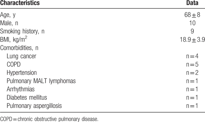
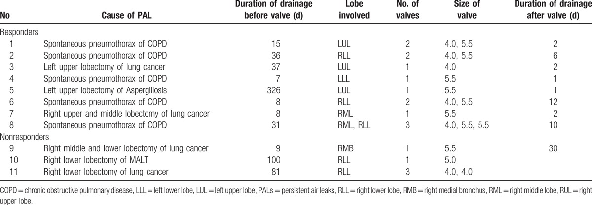
A total of 11 patients who underwent valve placement were eligible to be enrolled from January 2015 through January 2017. Six patients had postoperative PAL. Five patients had a secondary spontaneous pneumothorax, which were all caused by chronic obstructive pulmonary disease with emphysema. All of the patients were initially treated with chest tube thoracostomy. The mean patient age was 68 ± 8 years (range, 55–86 years), and 10 patients were men. Table 1 shows the patient characteristics and demographic variables. Table 2 shows the underlying causes of PALs, air leak duration of chest tube placement before and after bronchoscopy, number of valves, and treated lobe in these patients.
Table 1.
Patient characteristics.

Table 2.
Cause of PALs and outcomes of valve placement.

The mean number of implanted valves placed per patient ranged from 1 to 3, with a median of 1. The valves were placed in the left upper lobe (n = 3), right middle lobe (n = 1), right lower lobe (n = 4), left lower lobe (n = 1), right middle and right lower lobes (n = 1), and right medial bronchus (n = 1). PAL cessation occurred in all 5 patients with spontaneous pneumothorax. The mean air leak duration before valve placement was 19.4 days (range, 7–31 days). In contrast, the mean time to chest tube removal after valve placement was 6 days (range, 1–12 days).
In the surgical resection group, 3 patients were considered responders, and the mean air leak duration before and after valve placement was 123.7 days (range, 8–326 days) versus 1.7 days (range, 1–2 days). The other one patient expectorated the valve the day after the deployment and then underwent a surgical repair 1 month later. The remaining 2 patients had improvement but not resolution of their PALs and required surgical intervention ultimately. One of them (case no. 10) had fibrin glue and argon plasma coagulation, but also failed. Overall, the mean duration of air leak before and after valve deployment in those who responded was 58.5 and 4.5 days. No other adverse events such as pneumonia, infection, transient oxygen desaturation, or death associated with the implant were observed.
4. Discussion
In this article, we present that the deployment of EBVs is effective for patients with PALs following secondary pneumothorax or pneumothorax after lung resection. Complete cessation of air leak was achieved in 72.7% of our patients, with no need for other treatment after endobronchial valve implantation. The procedure was feasible, safe, and well tolerated, with few adverse events.
Air leaks, alveoli pleural, and bronchopleural fistulas can be difficult to treat, and if prolonged despite proper chest tube thoracostomy, they may increase health care cost, length of hospital stay, morbidity, and mortality. Many of them with poor performance status are caused by multiple comorbidities and impaired lung function, making surgical intervention a high-risk treatment option. Thus, the development of less invasive strategies for the management of PAL in these patients is necessary. Various bronchoscopic attempts to treat PAL have been many and include endovascular metallic coils,[5] glues,[6] spigots,[7] stents,[8] silver nitrate,[9] and gel foam.[10] But none of these methods has shown significant efficacy to replace surgical intervention in the treatment of PAL. One-way EBV placement, initially developed as a nonsurgical method of lung volume reduction for emphysema, has provided a new tool for use in the closure of persistent air leak. The valves are placed in the segmental or subsegmental airways leading to the air leak. The air is prevented from crossing the visceral pleural injury, thereby allowing it time to heal. In contrast to other blocking devices, these valves allow for expiration and clearance of bronchial secretions, therefore reducing the risk of postobstructive pneumonia, and can be easily removed when necessary.
A number of case series have reported the use of 1-way EBVs as a valid alternative for the treatment of PAL. The largest series are those published by Travaline et al.[11] Of the 40 patients with different etiologies, 93% had improvement in air leak, with 48% having complete resolution. The mean time from valve insertion to chest tube removal was 21 days, with a median of 7.5 days, and from valve procedure to hospital discharge was 19 ± 28 days, with a median of 11 days. Other series have shown similar success.[12,13] All these reports have demonstrated the safe and efficacious use of these technologies consistent with the findings of our study. Complications are rare and include pneumonia, expectoration or migration of valves, and bacterial colonization.
In our series, 3 patients were considered nonresponders and had to undergo surgery. They were all in the postsurgical group. The explanation for this is that the fistula in these patients was localized on the bronchial stump, where the valves cannot be firmly fixed; thus, the valves were either unlikely to adequately occlude the segment or were more likely to be expectorated. In these cases, the valves were used to reduce the fistula, while allowing drainage of distal secretions, to reduce the incidence of infection. A good result was the appearance of inflammation around the edges of the fistula, which leads to fibrosis and mucosal proliferation, ultimately completing closure of the leak. We can use argon plasma coagulation or fibrin glues locally to assist in sealing the defect during the treatment period.
In our study, we opted for the use of an endobronchial collateral ventilation assessment system (the Chartis system) as guidance for identifying the source of air leakage. In previously reported cases, air leaks were monitored by observing the reduction of air flow at the chest drain, whereas the Chartis system has the functions of pressure measurement and imaging. It also has been shown to be helpful in the assessment of collateral ventilation status.[14] When treating for PALs, the collateral ventilation was detected indirectly by testing adjacent lobes. The presence of collateral ventilation could be the explanation for the inability to completely resolve the identified leaks.[15]
There are few reports in the literature regarding the optimal time for the removal of valves when used in the setting of air leak control. It is well accepted that when air leaks are resolved by adequate time for tissue healing, EBVs can be removed. The removal depends on clinical judgment. In our study, all the valves were left in situ. In the medical group, considering the underlying disease of emphysema, we left the valves in situ to achieve lung volume reduction. In the postsurgical group, the patients continued follow-up in the clinic. There were no adverse events associated with the implant such as infection, granulation, or migration during the follow-up.
5. Conclusion
In conclusion, the implantation of 1-way EBVs should be considered a valuable, minimally invasive treatment option in the management of PALs in well-selected patients. The results of our series are similar to those of published articles, and the same limitations in these studies include the lack of control group, the small sample size, and the heterogeneity of the causes of PAL. To develop guidelines for the use of EBVs specifically for PAL, well-designed controlled clinical trials are needed in which valve placement is compared with other bronchoscopic techniques or surgical procedures.
Author contributions
Conceptualization: H. Xu, L. Ding.
Data curation: L. Ding.
Investigation: L. Ding.
Resources: H. Xu.
Supervision: H. Xu.
Writing – original draft: X. Huang.
Writing – review & editing: H. Xu L. Ding, X. Huang.
Footnotes
Abbreviations: EBV = endobronchial valves, LLL = left lower lobe, LUL = left upper lobe, PALs = persistent air leaks, RLL = right lower lobe, RMB = right medial bronchus, RML = right middle lobe, RUL = right upper lobe.
The authors have no conflicts of interest to disclose.
References
- [1].Shrager JB, Decamp MM, Murthy SC. Intraoperative and postoperative management of air leaks in patients with emphysema. Thorac Surg Clin 2009;19:223–31. [DOI] [PubMed] [Google Scholar]
- [2].Videm V, Pillgramlarsen J, Ellingsen O, et al. Spontaneous pneumothorax in chronic obstructive pulmonary disease: complications, treatment and recurrences. Eur J Respir Dis 1987;71:365–71. [PubMed] [Google Scholar]
- [3].Wood DE, Cerfolio RJ, Gonzalez X, et al. Bronchoscopic management of prolonged air leak. Clin Chest Med 2010;31:127–33. [DOI] [PubMed] [Google Scholar]
- [4].Fann JI, Berry GJ, Burdon TA. The use of endobronchial valve device to eliminate air leak. Respir Med 2006;100:1402–6. [DOI] [PubMed] [Google Scholar]
- [5].Sivrikoz CM, Kaya T, Tulay CM, et al. Effective approach for the treatment of bronchopleural fistula: application of endovascular metallic ring-shaped coil in combination with fibrin glue. Ann Thorac Surg 2007;83:2199–201. [DOI] [PubMed] [Google Scholar]
- [6].Fuso L, Varone F, Nachira D, et al. Incidence and management of post-lobectomy and pneumonectomy bronchopleural fistula. Lung 2016;194:299–305. [DOI] [PubMed] [Google Scholar]
- [7].Sasada S, Tamura K, Chang YS, et al. Clinical evaluation of endoscopic bronchial occlusion with silicone spigots for the management of persistent pulmonary air leaks. Intern Med 2011;50:1169–73. [DOI] [PubMed] [Google Scholar]
- [8].Andreetti C, D’Andrilli A, Ibrahim M, et al. Effective treatment of post-pneumonectomy bronchopleural fistula by conical fully covered self-expandable stent. Interact Cardiovasc Thorac Surg 2012;14:420–3. [DOI] [PMC free article] [PubMed] [Google Scholar]
- [9].Boudaya MS, Smadhi H, Zribi H, et al. Conservative management of postoperative bronchopleural fistulas. J Thorac Cardiovasc Surg 2013;146:575–9. [DOI] [PubMed] [Google Scholar]
- [10].Nicholas JM, Dulchavsky SA. Successful use of autologous fibrin gel in traumatic bronchopleural fistula: case report. J Trauma 1992;32:87–8. [PubMed] [Google Scholar]
- [11].Travaline JM, Mckenna RJ, Giacomo TD, et al. Treatment of persistent pulmonary air leaks using endobronchial valves. Chest 2009;136:355–60. [DOI] [PubMed] [Google Scholar]
- [12].Bakhos C, Doelken P, Pupovac S, et al. Management of prolonged pulmonary air leaks with endobronchial valve placement. JSLS 2016;20: e2016.00055. [DOI] [PMC free article] [PubMed] [Google Scholar]
- [13].Firlinger I, Stubenberger E, Müller MR, et al. Endoscopic one-way valve implantation in patients with prolonged air leak and the use of digital air leak monitoring. Ann Thorac Surg 2013;95:1243–9. [DOI] [PubMed] [Google Scholar]
- [14].Shah PL, Herth FJF. Current status of bronchoscopic lung volume reduction with endobronchial valves. Thorax 2014;69:280–6. [DOI] [PubMed] [Google Scholar]
- [15].Mahajan AK, Doeing DC, Hogarth DK. Isolation of persistent air leaks and placement of intrabronchialvalves. J Thorac Cardiovasc Surg 2013;145:626–30. [DOI] [PMC free article] [PubMed] [Google Scholar]


