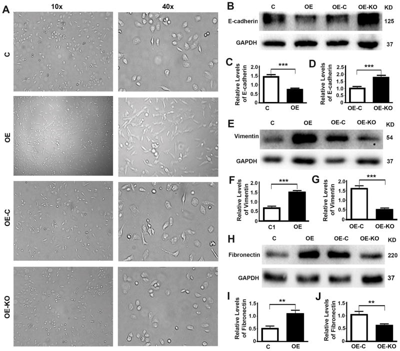
Figure 4. P62 promotes epithelial to mesenchymal transition.
(A) Representative images showing the morphology of DU145 cells (C, OE, OE-C and OE-KO). (B–J) Representative immunoblots (B,E,H) and quantification (C,D,F,G,I,J) showing levels of epithelial marker E-cadherin (B–D), Vimentin (E–G) and Fibronectin (H–J) in DU145 cells (C, OE) (C,F,I) and DU145 cells stably expressing P62 (OE-C, OE-KO) (D,G,J). **, p ≤ 0.01; and ***, p ≤ 0.001.

