Abstract
18F-Fluorocholine (FCH) PET/CT is evolving as a functional imaging modality for the preoperative imaging of abnormal parathyroid tissue(s) helping to localize eutopic and ectopic parathyroid tissue and limit the extent of surgery. FCH PET/CT may show incidental uptake in various thyroid lesions necessitating further evaluation, whereas the role of 18F-fluorodeoxyglucose (FDG) PET/CT in the detection of incidental thyroid nodules is well documented. The case of a middle-aged woman with dual pathology of parathyroid adenoma and papillary thyroid cancer detected on FCH and FDG PET/CT is presented.
Keywords: Parathyroid adenoma, Papillary thyroid carcinoma, 18F-Fluorocholine, 18F-Fluorodeoxyglucose, PET/CT
Introduction
18F-Fluorocholine (FCH) PET/CT is a promising modality for the detection of abnormal parathyroid tissue(s) [1]. FCH PET/CT may show variable tracer uptake in different incidentally detected benign and malignant lesions in the thyroid gland. FCH taken up by the thyroid gland during parathyroid imaging requires further evaluation as the uptake may be pathological [2–6]. While the incidental finding of focal FDG uptake in the thyroid gland on whole-body FDG PET/CT done for other conditions might indicate a malignant lesion requiring further work-up, the absence of FDG uptake in a large thyroid nodule nearly excludes malignancy [7–9]. We present the case of middle-aged woman in whom functional imaging studies helped detect the dual pathology of parathyroid adenoma and papillary thyroid carcinoma (PTC).
Case Report
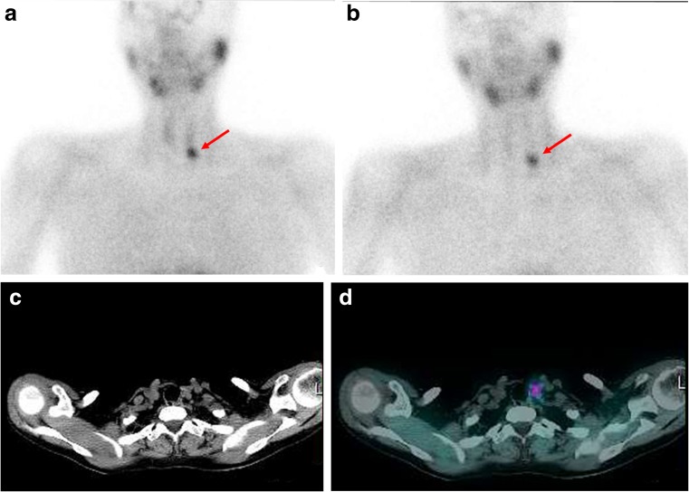
A 56-year-old woman presented with epigastric pain and vomiting (occasionally bilious) for 5 days with similar episodes in the past 2 years. Her biochemistry work-up revealed elevated calcium (12.3 mg/dL, normal range 8.8–10.2 mg/dL), alkaline phosphatase (212.94 U/L, normal range 42–128 U/L) and intact parathyroid hormone (PTH) level (167 pg/mL, normal range 15–65 pg/mL), and a low phosphate level (2.47 mg/dL, normal range 2.7–4.5 mg/dL). She was managed symptomatically with a provisional diagnosis of acute on chronic pancreatitis secondary to the raised PTH level. Neck ultrasonography showed a hypoechoic soft-tissue lesion inferior to the left thyroid lobe with multiple small hypoechoic lesions in the right upper thyroid lobe. 99mTc-Sestamibi (MIBI) planar imaging of the neck and mediastinum (Fig. 1a, b) showed a focal tracer-avid lesion below the left thyroid lobe on both early and delayed images. Transaxial CT and SPECT fused images showed a soft-tissue lesion with tracer avidity inferior to the left thyroid lobe (Fig. 1c, d) suggestive of left inferior parathyroid adenoma.
Fig. 1.
a, b 99mTc-Sestamibi dual-phase planar images show a focal tracer-avid lesion (arrows) below the left thyroid lobe on both the early (a) and delayed (b) images. c, d SPECT/CT images of the neck and mediastinum: transaxial CT (c) and fused (d) images show a small soft-tissue lesion with tracer avidity inferior to the left thyroid lobe
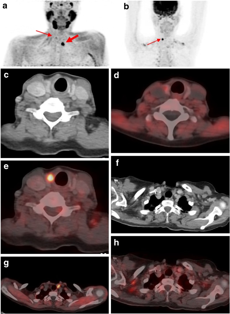
An FCH PET/CT scan performed after intravenous injection of 185 MBq of radiopharmaceutical as part of an ongoing project at our institution showed a choline-avid focus (SUVmax about 16) in the left lower neck and a faint choline-avid focus in the right upper neck on the maximum intensity projection (MIP) image (Fig. 2a) of the neck and mediastinum. Transaxial CT and fused images showed a faint choline-avid hypodense lesion in the right thyroid lobe and an intense choline-avid soft-tissue lesion inferior to the left thyroid lobe (Fig. 2c–g). The MIP image (Fig. 2b) of the FDG PET/CT scan of the neck and mediastinum showed an FDG-avid focus (SUVmax about 16) in the right upper neck. Transaxial CT and fused images showed an intense FDG-avid hypodense lesion in the right thyroid lobe and a non-FDG-avid soft-tissue lesion inferior to the left thyroid lobe (Fig. 2c–h).
Fig. 2.
a The 18F-FCH PET/CT MIP image shows an intense tracer-avid focus (thick arrow) in the left lower neck and a faint tracer-avid focus (thin arrow) in the right upper neck. c, d, f, g The transaxial CT and fused images show a right thyroid lobe hypodense lesion with faint tracer uptake and a soft-tissue lesion inferior to the left thyroid lobe with intense tracer uptake. b 18F-FDG PET/CT MIP image shows a tracer-avid focus (thin arrow) in the right upper neck. c, e, f, h Transaxial CT and fused images show a right thyroid lobe hypodense lesion with intense tracer uptake and a small soft-tissue lesion with no tracer uptake inferior to the left thyroid lobe
FDG PET/CT performed for thyroid nodule characterization and staging of the neck showed an intense FDG-avid hypodense lesion in the right thyroid lobe and no FDG uptake in the left parathyroid adenoma, while FCH PET/CT revealed mild FCH avidity in the lesion in the right upper lobe of the thyroid gland and an intense FCH-avid left inferior parathyroid adenoma. Ultrasound-guided fine-needle aspiration cytology of the right upper lobe thyroid lesion (intense FDG and mild FCH avidity) revealed the presence of PTC. The patient underwent right hemithyroidectomy for PTC and excision of the left inferior parathyroid adenoma.
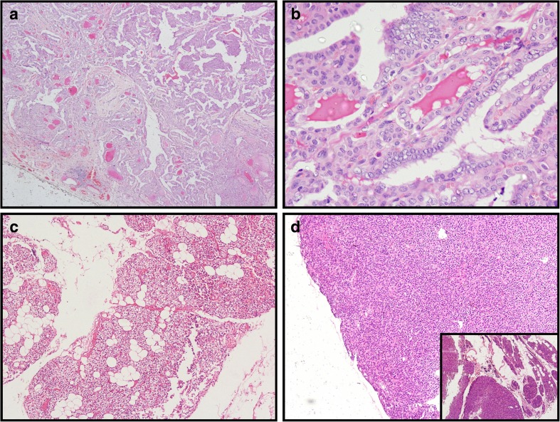
Histopathology of the surgical specimens confirmed the preoperative findings of PTC and parathyroid adenoma with histological sections showing the papillary configuration of the PTC (Fig. 3a) and the nuclear features of the PTC in the form of optical clearing, overcrowding and grooving (Fig. 3b). Figure 3c shows a histological section of the normal parathyroid gland, and Fig. 3d shows the parathyroid adenoma with the inset showing the adenoma and normal parathyroid at the periphery of the adenoma. The serum PTH level (32 pg/ml) and calcium level (8.8 ng/ml) became normal on the third postoperative day. At the time of this report, the patient was asymptomatic with a normal biochemical profile.
Fig. 3.
Histopathological examination of the surgical specimens. a, b Photomicrographs of H&E-stained sections show (a) the papillary configuration of the papillary thyroid carcinoma (×40) and (b) the nuclear features of the papillary thyroid carcinoma in the form of optical clearing, overcrowding and grooving (×400). c, d Photomicrographs of H&E-stained sections show (c) normal parathyroid gland (×100), and (d) normal parathyroid at the periphery of the adenoma (×40) and inset the adenoma
Discussion
Choline, a precursor in phosphatidylcholine biosynthesis in cellular membranes with upregulation of choline kinase in tumor, as well as its use in the evaluation of prostate cancer, shows uptake in various benign and neoplastic lesions [10]. FCH PET/CT is emerging as a novel imaging modality for the detection of parathyroid lesions [1]. However, additional pathologies including thyroid lymphoma, differentiated thyroid carcinoma, lymph node metastases from tall-cell thyroid cancer, Hürthle cell adenoma and oncocytic adenoma of the thyroid show variable FCH uptake and have been detected on FCH imaging [2–6]. FDG, a glucose metabolic marker, has demonstrated focally increased uptake in incidentally detected thyroid lesions with more than one-third of them being malignant, but performs poorly in the localization of the parathyroid gland [7, 8]. A negative FDG PET/CT study in the evaluation of suspicious thyroid nodules shows high negative predictive value for malignancy particularly in larger lesions [9].
The subject of the case presented here showed an FCH-avid, MIBI-avid and non-FDG-avid left inferior parathyroid adenoma. The incidentally detected right thyroid nodule showed intense FDG avidity and mild FCH avidity and the postoperative histopathology showed PTC. The findings reported here are different from those of a study of four newly diagnosed patients with recurrent thyroid cancer in whom the primary disease, and locoregional and lung metastases showed choline avidity (SUVmax 7.9 ± 4.9) and absent or mild FDG uptake. Similarly aggressive lymph node metastases from a thyroid malignancy have shown choline avidity without FDG avidity in a case report [4, 6]. These findings indicate that benign as well as malignant thyroid lesions may show differential FDG and FCH uptake, making the complementary use of both imaging modalities worthwhile in the situation of a patient with dual pathology.
This case report emphasizes the significance of the dual pattern of high FDG and low FCH uptake in an incidentally detected thyroid malignancy with absent FDG and high FCH uptake in parathyroid adenoma in a patient requiring appropriate evaluation.
Compliance with Ethical Standards
Conflicts of Interest
None.
Ethical Approval
All procedures performed in studies involving human participants were in accordance with the ethical standards of the institutional and/or national research committee and with the principles of the 1964 Declaration of Helsinki and its later amendments or comparable ethical standards.
Informed Consent
The institutional review board of our institution approved this retrospective study, and the requirement to obtain informed consent was waived.
Footnotes
The original version of this article was revised to correct the name of the fifth author.
An erratum to this article is available online at https://doi.org/10.1007/s13139-017-0498-1.
References
- 1.Lezaic L, Rep S, Sever MJ, Kocjan T, Hocevar M, Fettich J. 18F-Fluorocholine PET/CT for localization of hyperfunctioning parathyroid tissue in primary hyperparathyroidism: a pilot study. Eur J Nucl Med Mol Imaging. 2014;41:2083–2089. doi: 10.1007/s00259-014-2837-0. [DOI] [PubMed] [Google Scholar]
- 2.Eccles A, Challapalli A, Khan S, Barwick T, Mangar S. Thyroid lymphoma incidentally detected by 18F-fluorocholine (FCH) PET/CT. Clin Nucl Med. 2013;38:755–757. doi: 10.1097/RLU.0b013e31829f59bd. [DOI] [PubMed] [Google Scholar]
- 3.Aziz AL, Courbon F, Dierickx LO, Pascal P, Zerdoud S. Oncocytic adenoma of thyroid incidentally detected by 18F-fluorocholine PET/CT. J Nucl Med Technol. 2015;43:133–134. doi: 10.2967/jnmt.114.145433. [DOI] [PubMed] [Google Scholar]
- 4.Wu HB, Wang QS, Wang MF, Li HS. Utility of 11C-choline imaging as a supplement to F-18 FDG PET imaging for detection of thyroid carcinoma. Clin Nucl Med. 2011;36:91–95. doi: 10.1097/RLU.0b013e318203bb55. [DOI] [PubMed] [Google Scholar]
- 5.Paone G, Treglia G, Bongiovanni M, Ruberto T, Ceriani L, Giovanella L. Incidental detection of Hürthle cell adenoma by 18F-choline PET/CT scan in a patient with prostate cancer. Rev Esp Med Nucl Imagen Mol. 2013;32:340–341. doi: 10.1016/j.remn.2013.04.009. [DOI] [PubMed] [Google Scholar]
- 6.Piccardo A, Massollo M, Bandelloni R, Arlandini A, Foppiani L. Lymph node metastasis from tall-cell thyroid cancer negative on 18F-FDG PET/CT and detected by 18F-choline PET/CT. Clin Nucl Med. 2015;40:e417–e419. doi: 10.1097/RLU.0000000000000858. [DOI] [PubMed] [Google Scholar]
- 7.Soelberg KK, Bonnema SJ, Brix TH, Hegedüs L. Risk of malignancy in thyroid incidentalomas detected by 18F-fluorodeoxyglucose positron emission tomography; a systematic review. Thyroid. 2012;22:918–925. doi: 10.1089/thy.2012.0005. [DOI] [PubMed] [Google Scholar]
- 8.Hindie E, Ugur O, Fuster D, O’Doherty M, Grassetto G, Urena P, et al. 2009 EANM parathyroid guidelines. Eur J Nucl Med Mol Imaging. 2009;36:1201–1216. doi: 10.1007/s00259-009-1131-z. [DOI] [PubMed] [Google Scholar]
- 9.Vriens D, de Wilt JH, van der Wilt GJ, Netea-Maier RT, Oyen WJ, de Geus-Oei LF. The role of [18F]-2-fluoro-2-deoxy-d-glucose-positron emission tomography in thyroid nodules with indeterminate fine-needle aspiration biopsy: systematic review and meta-analysis of the literature. Cancer. 2011;117:4582–4594. doi: 10.1002/cncr.26085. [DOI] [PubMed] [Google Scholar]
- 10.Savelli G, Basile P, Muni A, Bna C, Pizzocaro C, Pagani R. Focal thyroid uptake during 18F-choline PET/CT: a case report. Case Rep Clin Med. 2015;4:345–348. doi: 10.4236/crcm.2015.411069. [DOI] [Google Scholar]


