Abstract
Background
Alterations in the structure of resistance vessels contribute to elevated systemic vascular resistance in hypertension and are linked to sympathetic hyperactivity and related lesions in target organs.
Objective
To assess the effects of exercise training on hemodynamic and autonomic parameters, as well as splenic arteriolar damages in male Wistar Kyoto (WKY) and Spontaneously Hypertensive Rats (SHR).
Methods
Normotensive sedentary (WKYS) and trained (WKYT) rats, and hypertensive sedentary (SHRS) and trained (SHRT) rats were included in this study. After 9 weeks of experimental protocol (swimming training or sedentary control), arterial pressure (AP) and heart rate (HR) were recorded in freely moving rats. We assessed the autonomic control of the heart by sympathetic and vagal autonomic blockade. Morphometric analyses of arterioles were performed in spleen tissues. The statistical significance level was set at p < 0.05.
Results
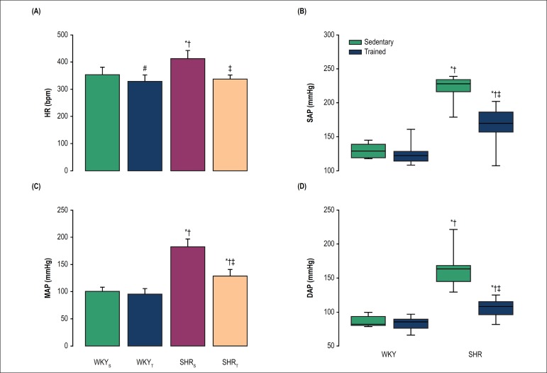
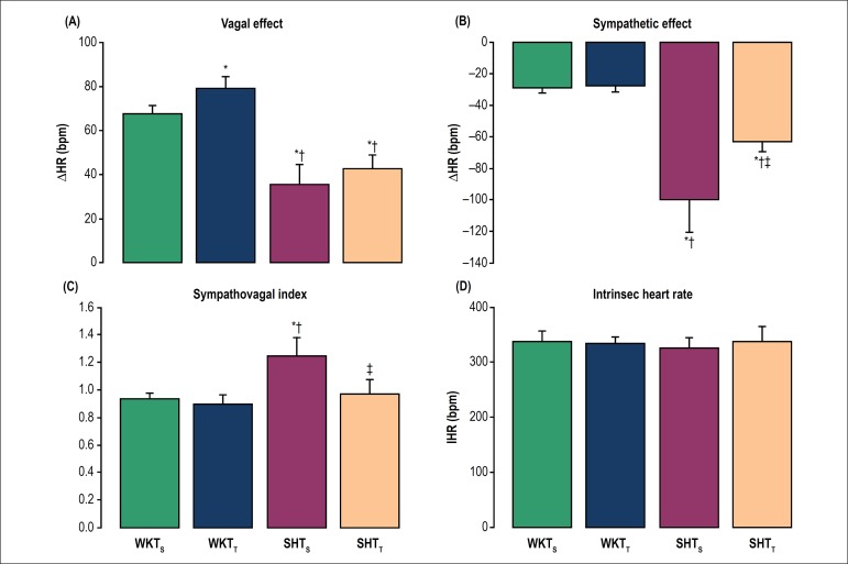
Resting bradycardia was observed in both trained groups (WKYT: 328.0 ± 7.3 bpm; SHRT: 337.0 ± 5.2 bpm) compared with their respective sedentary groups (WKYS: 353.2 ± 8.5 bpm; SHRS: 412.1 ± 10.4 bpm; p < 0.001). Exercise training attenuated mean AP only in SHRT (125.9 ± 6.2 mmHg) vs. SHRS (182.5 ± 4.2 mmHg, p < 0.001). The WKYT showed a higher vagal effect (∆HR: 79.0 ± 2.3 bpm) compared with WKYS (∆HR: 67.4 ± 1.7 bpm; p < 0.05). Chronic exercise decreased sympathetic effects on SHRT (∆HR: -62.8 ± 2.8 bpm) in comparison with SHRS (∆HR: -99.8 ± 9.2 bpm; p = 0.005). The wall thickness of splenic arterioles in SHR was reduced by training (332.1 ± 16.0 µm2 in SHRT vs. 502.7 ± 36.3 µm2 in SHRS; p < 0.05).
Conclusions
Exercise training attenuates sympathetic activity and AP in SHR, which may be contributing to the morphological improvement of the splenic arterioles.
Keywords: Exercise, Physical Exertion, Hypertension, Vascular Resistance, Arterioles, Rats
Introduction
Essential hypertension is inwardly connected to the blood vessels and is characterized by chronic increases in peripheral vascular resistance, mainly resulting from functional and structural alterations of the microcirculation. These lesions can be both the cause and the consequence of the elevation of arterial pressure (AP).1 The major pathways that interact to develop morphological changes in arteriolar vessels in hypertension may compromise the splenic vessels (arteriolar hyalinosis, fibrinoid necrosis) and the interstitial space, causing fibrosis.2-5 The arteriolar hyalinosis occurs by filtration of plasma proteins through the endothelium. It is not exclusive of any disease, being observed in arterioles of normal aging, especially in arterioles of the spleen. However, it occurs earlier and more intense in arterial hypertension.6
The autonomic nervous system plays a key role in the stabilization of AP control for maintaining homeostasis. In this respect, the literature data show that the sympathetic nervous system (SNS) can reciprocate incisively in the development of some forms of hypertension. Evidence of the participation of this system in the control of normal cardiovascular and metabolic functions and its role in the genesis and maintenance of several diseases is broad. The importance of understanding the workings of the SNS and systems related to it is essential not only to elucidate the path physiology of some diseases, but to understand how drugs that act on the adrenergic system interfere with the evolution of pathologies significantly altering the prognosis of patients.7
Experimental evidence has shown that chronic exercise produces beneficial effects on the cardiovascular system via alterations in neural control of the circulation. These effects include reductions in AP, sympathetic activity8 and vascular resistance9 concomitantly with attenuation in the target-organ damage.10 If there is relation between exercise training and decrease of vascular resistance, the mechanisms by which chronic exercise training improves splenic arteriolar morphometry are not well established. Thus, the aim of this study was to assess the effects of exercise training on sympathetic activity and arteriolar damages in spleens of spontaneously hypertensive rats (SHR).
Methods
Animal model and exercise training protocol
Forty male SHR and Wistar Kyoto rats (WKY) aged 45-50 weeks were randomly assigned into four experimental groups of 10 rats each: SHRT and WKYT (that were submitted to exercise training protocol by swimming) or SHRS and WKYS (that were kept sedentary for a similar period of time). The sample size (n) was determined based on studies that evaluated the effects of exercise training on hypertension. These studies served as the basis for the present study that investigates the cardiovascular effects of the accumulated exercise.11,12 All animals were kept in grouped cages (n = 3) at room temperature around 23ºC, humidity of 40-70% and photoperiod of 12-hour light/dark cycle. Efforts were made to avoid any unnecessary distress to the rats, in accordance to the Brazilian Council for Animal Experimentation. All animal protocols were approved by the local Experimental Animal Use Committee (#271/2013), and were performed according to the regulations set forth by the National Institutes of Health Guidelines for the Care and Use of Laboratory Animals.
The swimming exercise protocol was performed in a glass tank and ambient water temperature was kept at 30º ± 1ºC. The trained animals received a 20-min adaptation period on the first day, with increases of 10 min each day until reaching 1 hour on the fifth day.13 After this period, the rats trained 5 days/week with a gradual progression toward a 2-hour session during nine weeks. This protocol is defined as an aerobic endurance and low-intensity training, as the animals swam without additional work load, this method corresponds the intensity below the anaerobic threshold in rats.14 Sedentary animals were placed in the swimming apparatus for 10 min twice a week to mimic the water stress associated with the experimental protocol.
Surgical procedures and hemodynamic parameters recording
Twenty-four hours after the last exercise training session, all animals were anesthetized with sodium pentobarbital (40 mg/kg ip) and cannulas of polyethylene (PE-10) were implanted into the femoral artery for cardiovascular recording and into the femoral vein for drug infusion. Then, the polyethylene catheters were exteriorized at the posterior neck region of the animal. Rats received food and water ad libitum and were studied 1 day after catheter placement. Prophylactic treatment with antibiotics and anti-inflammatory drugs were performed to prevent postsurgical infections and inflammation, respectively.15 After 48 hours of recovery from the anesthesia and surgery, the arterial cannula was connected to an AP transducer and a signal amplifier (Model 8805A, Hewlett-Packard, USA) was converted by the analog-digital signal plate (sampling frequency - 1000 Hz) by a computerized system data acquisition (Aqdados, Tec Lynx. Eletron. SA, Sao Paulo, Brazil) and stored on computer. The animals were maintained in a peaceful environment for a period of 15 minutes and adaptive later pulsatile AP was continuously recorded at baseline for 30 minutes. During the experimental procedure, systolic AP (SAP), diastolic AP (DAP), mean AP (MAP) and heart rate (HR) were derived from pulsatile AP.
Cardiac autonomic tonus
To evaluate the exercise training influence on the tonic autonomic control of the heart, we also performed the sympathetic and vagal autonomic blockade after propranolol (5 mg/kg, i.v.) and atropine (4mg/kg, i.v.) injections, respectively, to calculate the sympathetic and vagal effects, as well as the intrinsic HR (iHR) and tonic sympathovagal index.14 The autonomic blockers were administered in a random sequence with a 15-min interval between them. After double blockade, the cardiovascular recordings lasted for 15 min. Briefly, the sympathetic effect was considered as the difference between the HR after sympathetic blockade and resting HR. Vagal effect was calculated as the difference between HR after vagal blockade and resting HR. The tonic sympathovagal index was obtained as the ratio between resting HR and iHR, considering that the iHR was the HR obtained after double autonomic blockade.16
Analysis of splenic arteriolar morphometry
All animals were anesthetized with sodium pentobarbital and euthanatized with a lethal dose of potassium chloride. Their spleens were excised postmortem and immersed in saline (0.9%) to remove excess blood. Shortly after, the organs were placed on foil, previously treated and weighed in a semi-analytical Gehaka BG2000®. Subsequently, the material was cut and placed inside a sterilized glass with 10% formaldehyde. Thereupon, the material was dehydrated using ethanol at concentrations of 80%, 90% and 95%. Diaphanization was performed with xylol. The material was placed in containers containing liquid paraffin at 60ºC. Then, the material was placed in blocks. Histological 2-µm cuts were performed using a microtome and then the material were mounted in glass slides and stained with Masson's Trichrome Blue. The area of the inner and outer layers of each arteriole was quantified by using common light microscope for capturing the images and the imageJ program to check the area of each layer. At the end of the procedures for quantification of the area of each layer, the thickness of each arteriole was obtained.
Statistical analysis
Shapiro-Wilks and Levene's tests were used to evaluate the normality and homogeneity of the sample. Results were expressed as mean ± SD (for normally distributed variables) or median with upper and lower quartiles (for non-normally distributed variables). For parametric data, we used two-way ANOVA (etiology vs. intervention), with the Tukey as a post hoc test. The nonparametric data were analyzed by the Mann-Whitney test. Pearson coefficient was used to test the correlation between sympathetic effect with area of outer wall thickness and total area thickness. Probability values of P < 0.05 were considered statistically significant. Analyses were performed using SigmaStat® v. 2.03 (SPSS, Chicago, IL, USA).
Results
The SHRS showed higher resting HR in comparison to WKYS (p < 0.001). As expected, both trained groups presented higher resting bradycardia compared with their respective sedentary groups (p < 0.001; Figure 1A).
Figure 1.
Baseline recording of heart rate (1A), systolic arterial pressure (1B), mean arterial pressure (1C) and diastolic arterial pressure (1D) in freely moving rats. WKYS (sedentary normotensive rats); WKYT (trained normotensive rats); SHRS (sedentary hypertensive rats); SHRT (trained hypertensive rats). Bars in figures 1A and 1C represent mean ± SD. Results in figures 1B and 1D are expressed as median (interquartile range). #p < 0.05 vs. WKYS; *p < 0.001 vs. WKYS; †p < 0.001 vs. WKYT and ‡p < 0.001 vs. SHRS.
Exercise training also was able to decrease baseline SAP (p < 0.001; Figure 1B), MAP (p < 0.001; Figure 1C) and DAP (p < 0.001; Figure 1D) in hypertensive animals compared with their respective sedentary group. The SHRS presented higher pressure levels than WKYS (p < 0.001) and WKYT (p < 0.001) groups. After the 9-week training period, the AP was similar in WKYT and WKYS.
To evaluate the influence of chronic exercise on the tonic autonomic control of the heart, we performed the vagal and sympathetic autonomic blockade with atropine and propranolol injections, respectively, to calculate the vagal (Figure 2A) and sympathetic effects (Figure 2B), as well as the tonic sympathovagal index (Figure 2C) and iHR (Figure 2D). No difference on vagal effect was observed between the hypertensive groups. However, the WKYT group evidenced a higher vagal effect than the WKYS group (p < 0.05). Both hypertensive groups presented a lower vagal effect when compared with their respective normotensive groups (p < 0.001). In addition, no difference in the sympathetic effect was observed between the normotensive groups (p = 0.563). On the other hand, the SHRT group showed a lower sympathetic effect as compared with SHRS group (p = 0.005). Both normotensive groups had a lower sympathetic effect when compared with their respective hypertensive groups (p < 0.001). The sympathovagal index was lower in SHRT than in SHRS (p < 0.05). No difference was observed between the groups regarding iHR.
Figure 2.
Effects of exercise training on the tonic autonomic control of the heart rate (HR) in non-anesthetized rats. (2A) vagal and (2B) sympathetic effects were obtained, respectively, by the difference between vagal blockade (by atropine) or sympathetic blockade (by propranolol) and resting HR. (2C) Sympathovagal balance was expressed by the tonic sympathovagal index, which is the ratio between resting and intrinsic HR (iHR). (2D) Intrinsic HR (bpm) obtained after autonomic double pharmacological blockade. Bars represent mean ± SD. *p < 0.05 vs. WKYS; †p < 0.05 vs. WKYT and ‡p < 0.05 vs. SHRS.
Morphometric analysis after histological processing revealed profound changes in microcirculatory profile of spleen circulation induced by training in hypertensive animals (Table 1). As expected, hypertensive splenic arterioles had a thicker wall than normotensive arterioles (p < 0.001). Despite this, exercise training was effective to normalize SHR arteriole wall/lumen ratio in spleen tissues analyzed when compared with that of SHRS (p < 0.001). The SHRS also presented a greater area of outer wall thickness when compared to WKYS and WKYT (p < 0.001). After exercise training protocol, the SHRT obtained a reduction in the area of the outer wall thickness compared to SHRS (p < 0.001). Similar results were observed in the total area thickness. The SHRS had a higher total area thickness of the splenic arterioles than the normotensive groups (p < 0.005). In addition, the SHRT evidenced an attenuation in total area thickness of splenic arterioles when compared with SHRS (p < 0.005).
Table 1.
Values related to morphological analysis of the area of the wall thickness of splenic arterioles.
| Thickness area | WKYS (n = 10) | WKYT (n = 10) | SHRS (n = 10) | SHRT (n = 10) |
|---|---|---|---|---|
| Inner wall (µm2) | 60.5 ± 3.4 | 58.8 ± 2.3 | 87.3 ± 3.3*† | 58.0 ± 2.6‡ |
| Outer wall (µm2) | 419.8 ± 29.3 | 405.6 ± 21.7 | 632.4 ± 29.1*† | 418.8 ± 16.4‡ |
| Total area (µm2) | 335.6 ± 44.7 | 349.7 ± 35.8 | 502.7 ± 36.3*† | 332.1 ± 16.0‡ |
Data are expressed as mean ± SD. Abbreviations: WKYS, sedentary normotensive rats; WKYT, trained normotensive rats; SHRS, sedentary hypertensive rats; SHRT, trained hypertensive rats. Data expressed as mean ± SEM
p < 0.05 vs. WKYS;
p < 0.05 vs. WKYT and
p < 0.05 vs. SHRS.
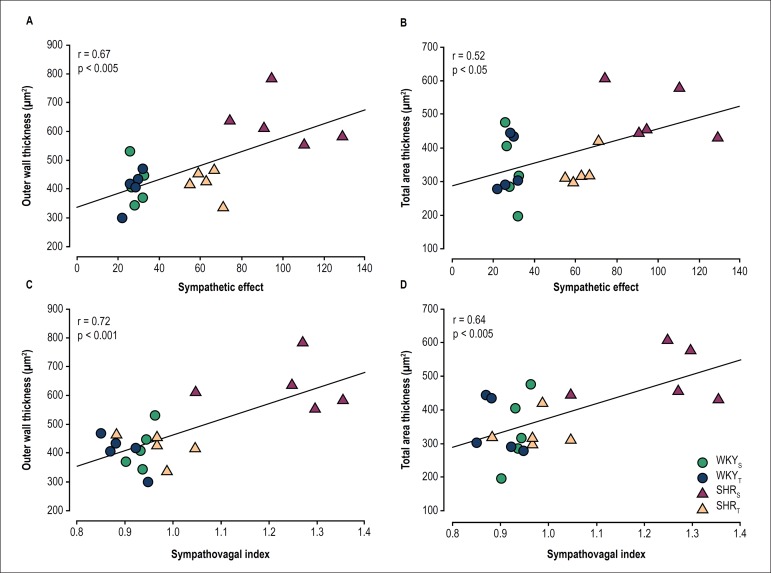
Further analysis showed a significant association between sympathetic effect and area of outer wall thickness (r = 0.67, p < 0.005; Figure 3A), sympathetic effect and total area thickness (r = 0.52, p < 0.05; Figure 3B), sympathovagal index and area of outer wall thickness (r = 0.72, p < 0.001; Figure 3C) and sympathovagal index and total area thickness (r = 0.64, p < 0.005; Figure 3D).
Figure 3.
Correlation coefficient between sympathetic effect and outer wall thickness (A), sympathetic effect and total area thickness (B), sympathovagal index and outer wall thickness (C), sympathovagal index and total area thickness (D).
Discussion
Our main findings confirmed the efficacy of exercise training to attenuate sympathetic overactivity and to lower AP in hypertensive animals, showing, in addition, that the training-induced, sympathetic-lowering effect was associated with normalization of abnormal splenic artery diameter, decreasing the degree of vascular injury in spleen. The morphometric analysis of small vessels employed in the present study revealed that the splenic vascular adjustments are specific for the SHRT. It is well documented that chronic physical exercise attenuates sympathetic hyperactivity10 and arteriolar damage on hypertension.17 To our knowledge, however, this is one of the first reports to evidence association between a reduction in splenic arteriole injury and sympathetic activity.
The cause-effect relation between hypertension and arteriolar damage (hypertrophy) is well established.18-20 In this sense, the literature evidences that an effective antihypertensive treatment should aim not only to reduce AP but also to correct injuries associated with hypertension, such as the altered vascular structure. A previous study has shown the efficacy of training to normalize arteriole wall/lumen ratio, evidencing that arteriolar response as well as vascular resistance reduction after exercise training were significantly correlated with AP reduction.21 Experimental study has found that arteriole wall/lumen ratios were reduced by increased internal and/or external diameter, which is a characteristic pattern for vascular remodeling.21 Of importance is the demonstration that exercise training, by reversing lumen encroachment, normalizes enlarged wall/lumen ratio of small arterioles in hypertensive rats. These data are in accordance with the results found in our study.
Results from studies with animal models indicate that a sustained elevation of sympathetic tonus stimulates smooth muscle cell hypertrophy, suggesting that sympathetic overactivity may contribute to changes in arterial wall thickness.22 In this way, an interesting finding in our study was a positive and significant correlation between sympathetic hyperactivity and splenic arterioles wall thickness in hypertensive rats, corroborating with results from other investigators who demonstrated that hypertension is associated with sympathetic overactivity that alters vasomotor control resulting in several abnormalities in tissue microcirculation, such as increased arteriolar wall-to-lumen ratio and decreased vessel density, which contribute to maintain an elevated total peripheral resistance.23-28 Another important finding in our research was that exercise training was able to attenuate sympathetic activity in SHR and that this effect was associated with a reduction in splenic arteriole wall thickness. Exercise training produces beneficial effects on cardiovascular system in normal and sick people via alterations (or modifications) in the neural control of circulation.29,30 These effects include reductions in AP, sympathetic outflow in humans,31,32 as well as in animal models,33,34 and vascular resistance.35,36 In addition, there is evidence that exercise training improves the conditions of the small vessels in SHR subjected to swimming protocol.37 Although this study did not address the mechanisms responsible for training-induced effects, one might speculate that arteriole adjustments are group-specific (hypertensive rats) and probably not dependent on paracrine, autocrine, metabolic, and/or myogenic factors, since similar alterations were observed in a previous study.17
It is well established that regular physical activity reduces AP in hypertensive individuals, without significant pressure changes in normotensive individuals.38-40 In fact, several studies have suggested that exercise training intensity influences the pressure-lowering effect, with larger reductions being observed with lower exercise intensities.40 We did not analyze the effect of training intensity, but our results clearly showed that the exercise protocol used caused an important AP decrease only in the SHR group. Pressure reduction was accompanied by both resting bradycardia and specific training-induced adjustment in splenic hypertensive arterioles. Resting bradycardia is considered to be an excellent hallmark for exercise training adaptation in humans and rats.39-40 Thus, the bradycardia found in trained rats clearly demonstrates the effectiveness of the exercise protocol here used.
Conclusion
Considering our findings, we can conclude that exercise training was effective in reducing AP and improving splenic arteriolar morphometry in hypertensive rats. Briefly, these data strongly suggest that this improvement was associated with decreased sympathetic nerve activity. In addition, regression of hypertrophied splenic arteriole is the anatomic response to exercise training specific to the SHR group. These compensatory adjustments, by reducing local resistance and augmenting physical capacity, contribute to the training-induced, pressure-lowering effect observed in hypertensive individuals.
Footnotes
Sources of Funding
This study was partially funded by Capes, CNPq and Fapemig.
Study Association
This study is not associated with any thesis or dissertation work.
Ethics approval and consent to participate
This study was approved by the Ethics Committee on Animal Experiments of the Universidade Federal do Triângulo Mineiro under the protocol number #271/2013.
Author contributions
Conception and design of the research: Barbosa Neto O; Acquisition of data: Lemos MP, Sordi CC; Analysis and interpretation of the data: Lemos MP, Mota GR, Marocolo Júnior M, Sordi CC, Chriguer RS, Barbosa Neto O; Statistical analysis: Lemos MP, Barbosa Neto O; Writing of the manuscript: Lemos MP; Critical revision of the manuscript for intellectual content: Mota GR, Marocolo Júnior M, Sordi CC, Chriguer RS, Barbosa Neto O.
Potential Conflict of Interest
No potential conflict of interest relevant to this article was reported.
References
- 1.Silvestre JS, Levy BI. [Hypertension: microvascular complications] Arch Mal Coeur Vaiss. 2000;93(11) Suppl:1387–1392. [PubMed] [Google Scholar]
- 2.Klag MJ, Whelton PK, Randall BL, Neaton JD, Brancati FL, Ford CE. Blood pressure and end-stage renal disease in men. N Engl J Med. 1996;334(1):13–18. doi: 10.1056/NEJM199601043340103.. [DOI] [PubMed] [Google Scholar]
- 3.Preston RA, Singer I, Epstein M. Renal parenchymal hypertension: current concepts of pathogenesis and management. Arch Intern Med. 1996;156(6):602–611. doi: 10.1001/archinte.1996.00440060016002.. [DOI] [PubMed] [Google Scholar]
- 4.Kincaid-Smith P. Clinical diagnosis of hypertensive nephrosclerosis. Nephrol Dial Transplant. 1999;14(9):2255–2256. doi: 10.1093/ndt/14.9.2255. [DOI] [PubMed] [Google Scholar]
- 5.Cuspidi C, Sala C, Zanchetti A. Metabolic syndrome and target organ damage: role of blood pressure. Expert Rev Cardiovasc Ther. 2008;6(5):731–743. doi: 10.1586/14779072.6.5.731.. [DOI] [PubMed] [Google Scholar]
- 6.Kristensen BO. Aspects of immunology and immunogenetics in human essential hypertension with special reference to vascular events. J Hypertens. 1984;2(5):571–579. doi: 10.1097/00004872-198412000-00001. [DOI] [PubMed] [Google Scholar]
- 7.Acampa M, Franchi M, Guideri F, Lamberti I, Bruni F, Pastorelli M, et al. Cardiac dysautonomia and arterial distensibility in essential hypertensives. Auton Neurosci. 2009;146(1-2):102–105. doi: 10.1016/j.autneu.2008.11.009.. [DOI] [PubMed] [Google Scholar]
- 8.Kramer JM, Beatty JA, Plowey ED, Waldrop TG. Exercise and hypertension: a model for central neural plasticity. Clin Exp Pharmacol Physiol. 2002;29(1-2):122–126. doi: 10.1046/j.1440-1681.2002.03610X.. [DOI] [PubMed] [Google Scholar]
- 9.Gando Y, Yamamoto K, Murakami H, Ohmori Y, Kawakami R, Sanada K, et al. Longer time spent in light physical activity is associated with reduced arterial stiffness in older adults. Hypertension. 2010;56(3):540–546. doi: 10.1161/HYPERTENSIONAHA.110.156331.. [DOI] [PubMed] [Google Scholar]
- 10.Barbosa Neto O, Abate DT, Marocolo Júnior M, Mota GR, Orsatti FL, Rossi e Silva RC, et al. Exercise training improves cardiovascular autonomic activity and attenuates renal damage in spontaneously hypertensive rats. J Sports Sci Med. 2013;12(1):52–59. [PMC free article] [PubMed] [Google Scholar]
- 11.Higa-Taniguchi KT, Silva FC, Silva HM, Michelini LC, Stern JE. Exercise training-induced remodeling of paraventricular nucleus (nor)adrenergic innervation in normotensive and hypertensive rats. Am J Physiol Regul Integr Comp Physiol. 2007;292(4):R1717–R1727. doi: 10.1152/ajpregu.00613.2006.. [DOI] [PubMed] [Google Scholar]
- 12.Gutkowska J, Aliou Y, Lavoie JL, Gaab K, Jankowski M, Broderick TL. Oxytocin decreases diurnal and nocturnal arterial blood pressure in the conscious unrestrained spontaneously hypertensive rat. Pathophysiology. 2016;(2):111–121. doi: 10.1016/j.pathophys.2016.03.003. [DOI] [PubMed] [Google Scholar]
- 13.Seo TB, Han LS, Yoon JH, Hong KE, Yoon SJ, Namgung UK. Involvement of Cdc2 in axonal regeneration enhanced by exercise training in rats. Med Sci Sports Exerc. 2006;38(7):1267–1276. doi: 10.1249/01.mss.0000227311.00976.68.. [DOI] [PubMed] [Google Scholar]
- 14.Gobatto CA, de Mello MA, Sibuya CY, de Azevedo JR, dos Santos LA, Kokubun E. Maximal lactate steady state in rats submitted to swimming exercise. Comp Biochem Physiol A Mol Integr Physiol. 2001;130(1):21–27. doi: 10.1016/s1095-6433(01)00362-2. [DOI] [PubMed] [Google Scholar]
- 15.Silva FC, Guidine PA, Ribeiro MF, Fernandes LG, Xavier CH, de Menezes RC, et al. Malnutrition alters the cardiovascular responses induced by central injection of tityustoxinin Fischer rats. Toxicon. 2013 Dec 15;76:343–349. doi: 10.1016/j.toxicon.2013.09.015.. [DOI] [PubMed] [Google Scholar]
- 16.Goldberger JJ. Sympathovagal balance: how should we measure it? pt 2Am J Physiol. 1999;276(4):H1273–H1280. doi: 10.1152/ajpheart.1999.276.4.H1273. [DOI] [PubMed] [Google Scholar]
- 17.Melo RM, Martinho E Jr, Michelini LC. Training-induced, pressure-lowering effect in SHR wide effects on circulatory profile of exercised and nonexercised muscles. Hypertension. 2003;42(4):851–857. doi: 10.1161/01.HYP.0000086201.27420.33.. [DOI] [PubMed] [Google Scholar]
- 18.Folkow B. Physiological aspects of primary hypertension. Physiol Rev. 1982;62(2):347–504. doi: 10.1152/physrev.1982.62.2.347. [DOI] [PubMed] [Google Scholar]
- 19.Levy BI, Ambrosio G, Pries AR, Struijker-Boudier HA. Microcirculation in hypertension: a new target for treatment? Circulation. 2001;104(6):735–740. doi: 10.1161/hc3101.091158. https://doi.org/10.1161/hc3101.091158 [DOI] [PubMed] [Google Scholar]
- 20.Mulvany MJ. Small artery remodeling and significance in the development of hypertension. News Physiol Sci. 2002 Jun;17:105–109. doi: 10.1152/nips.01366.2001.. [DOI] [PubMed] [Google Scholar]
- 21.Amaral SL, Zorn TM, Michelini LC. Exercise training normalizes wall-to-lumen ratio of the gracilis muscle arterioles and reduces pressure in spontaneously hypertensive rats. J Hypertens. 2000;18(11):1563–1572. doi: 10.1097/00004872-200018110-00006. [DOI] [PubMed] [Google Scholar]
- 22.Pauletto P, Scannapieco G, Pessina AC. Sympathetic drive and vascular damage in hypertension and atherosclerosis. Hypertension. 1991;17(4) Suppl:III75–III81. doi: 10.1161/01.hyp.17.4_suppl.iii75. [DOI] [PubMed] [Google Scholar]
- 23.Intengan HD, Schiffrin EL. Structure and mechanical properties of resistance arteries in hypertension: role of adhesion molecules and extracellular matrix determinants. Hypertension. 2000;36(3):312–318. doi: 10.1161/01.hyp.36.3.312. [DOI] [PubMed] [Google Scholar]
- 24.Laurent S, Boutouyrie P, Lacolley P. Structural and genetic bases of arterial stiffness. Hypertension. 2005;45(6):1050–1055. doi: 10.1161/01.HYP.0000164580.39991.3d.. [DOI] [PubMed] [Google Scholar]
- 25.Laurent S, Briet M, Boutouyrie P. Large and small artery cross-talk and recent morbidity-mortality trials in hypertension. Hypertension. 2009;54(2):388–392. doi: 10.1161/HYPERTENSIONAHA.109.133116.. [DOI] [PubMed] [Google Scholar]
- 26.Yasmin O’Shaughnessy KM. Genetics of arterial structure and function: towards new biomarkers for aortic stiffness? Clin Sci (Lond) 2008;114(11):661–677. doi: 10.1042/CS20070369.. [DOI] [PubMed] [Google Scholar]
- 27.Martinez-Lemus LA, Hill MA, Meininger GA. The plastic nature of the vascular wall: a continuum of remodeling events contributing to control of arteriolar diameter and structure. Physiology (Bethesda) 2009 Feb;24:45–57. doi: 10.1152/physiol.00029.2008.. [DOI] [PubMed] [Google Scholar]
- 28.Cheng C, Daskalakis C, Falkner B. Alterations in capillary morphology are found in mild blood pressure elevation. J Hypertens. 2010;28(11):2258–2266. doi: 10.1097/HJH.0b013e32833e113b.. [DOI] [PMC free article] [PubMed] [Google Scholar]
- 29.Cornelissen VA, Fagard RH. Effects of endurance training on blood pressure, blood pressure-regulating mechanisms, and cardiovascular risk factors. Hypertension. 2005;46(4):667–675. doi: 10.1161/01.HYP.0000184225.05629.51.. [DOI] [PubMed] [Google Scholar]
- 30.Zucker IH, Patel KP, Schultz HD, Li YF, Wang W, Pliquett RU. Exercise training and sympathetic regulation in experimental heart failure. Exerc Sport Sci Rev. 2004;32(3):107–111. doi: 10.1097/00003677-200407000-00006. Erratum in: Exerc Sport Sci Rev. 2004;32(4):191. [DOI] [PubMed] [Google Scholar]
- 31.Iwasaki KI, Zhang R, Zuckerman JH, Levine BD. Dose-response relationship of the cardiovascular adaptation to endurance training in healthy adults: How much training for what benefit? J Appl Physiol. 2003;95(4):1575–1583. doi: 10.1152/japplphysiol.00482.2003.. 1985. [DOI] [PubMed] [Google Scholar]
- 32.Roveda F, Middlekauff HR, Rondon MU, Reis SF, Souza M, Nastari L, et al. The effects of exercise training on sympathetic neural activation in advanced heart failure: a randomized controlled trial. J Am Coll Cardiol. 2003;42(5):854–860. doi: 10.1016/s0735-1097(03)00831-3. https://doi.org/10.1016/S0735-1097(03)00831-3 [DOI] [PubMed] [Google Scholar]
- 33.Collins HL, Rodenbaugh DW, DiCarlo SE. Daily exercise attenuates the development of arterial blood pressure related cardiovascular risk factors in hypertensive rats. Clin Exp Hypertens. 2000;22(2):193–202. doi: 10.1081/ceh-100100072. [DOI] [PubMed] [Google Scholar]
- 34.Kramer JM, Beatty JA, Plowey ED, Waldrop TG. Exercise and hypertension: a model for central neural plasticity. Clin Exp Pharmacol Physiol. 2002;29(1-2):122–126. doi: 10.1046/j.1440.1681.2002.036.10.X.. [DOI] [PubMed] [Google Scholar]
- 35.Gando Y, Yamamoto K, Murakami H, Ohmori Y, Kawakami R, Sanada K, et al. Longer time spent in light physical activity is associated with reduced arterial stiffness in older adults. Hypertension. 2010;56(3):540–546. doi: 10.1161/HYPERTENSIONAHA.110.156331.. [DOI] [PubMed] [Google Scholar]
- 36.Thijssen DH, Maiorana AJ, O’Driscoll G, Cable NT, Hopman MT, Green DJ. Impact of inactivity and exercise on the vasculature in humans. Eur J Appl Physiol. 2010;108(5):845–875. doi: 10.1007/s00421-009-1260-x.. [DOI] [PMC free article] [PubMed] [Google Scholar]
- 37.Abate DT, Barbosa Neto O, Rossi e Silva RC, Faleiros AC, Correa RR, da Silva VJ, et al. Exercise-training reduced blood pressure and improve placental vascularization in pregnant spontaneously hypertensive rats - pilot study. Fetal Pediatr Pathol. 2012;31(6):423–431. doi: 10.3109/15513815.2012.659535.. [DOI] [PubMed] [Google Scholar]
- 38.Meredith IT, Jennings GL, Esler MD, Dewar EM, Bruce AM, Fazio VA. Time-course of the antihypertensive and autonomic effects of regular endurance exercise in human subjects. J Hypertens. 1990;8(9):859–866. doi: 10.1097/00004872-199009000-00010. [DOI] [PubMed] [Google Scholar]
- 39.Negrão CE, Irigoyen MC, Moreira ED, Brum PC, Freire PM, Krieger EM. Effect of exercise training on RSNA, baroreflex control and blood pressure responsiveness. Pt 2Am J Physiol. 1993;265(2):R365–R370. doi: 10.1152/ajpregu.1993.265.2.R365. [DOI] [PubMed] [Google Scholar]
- 40.Gava NS, Véras-Silva AS, Negrão CE, Krieger EM. Low-intensity exercise training attenuates cardiac beta-adrenergic tone during exercise in spontaneously hypertensive rats. Pt 2Hypertension. 1995;26(6):1129–1133. doi: 10.1161/01.hyp.26.6.1129. https:doi.org/10.1161/01.HYP.26.6.1129 [DOI] [PubMed] [Google Scholar]


