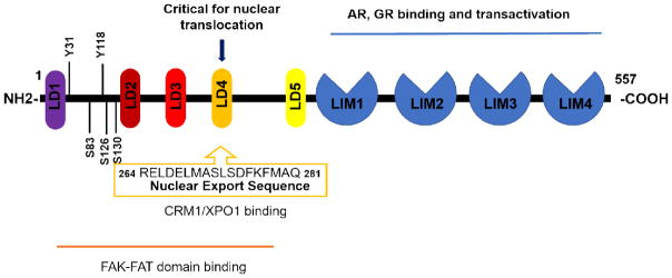
Figure 1. Schematic structure of paxillin with highlights of nuclear function related domains.
Paxillin contains five LD domains on the N-terminal half and four LIM domains on the C-terminal half of the protein. The shown N-terminal domains are critical for FAK binding and nuclear translocation, with highlights of critical serine/tyrosine phosphorylation sites as well as the NES sequence within LD4 domain. The LIM domains on the C terminal half are related to paxillin’s binding and transactivating of AR and GR.

