eLife logoLink to eLife
. 2018 Apr 16;7:e29546. doi: 10.7554/eLife.29546

Control of voluntary and optogenetically perturbed locomotion by spike rate and timing of neurons of the mouse cerebellar nuclei

Rashmi Sarnaik 1,, Indira M Raman 1,
Editor: Gary L Westbrook2
PMCID: PMC5902160  PMID: 29659351

Abstract

Neurons of the cerebellar nuclei (CbN), which generate cerebellar output, are inhibited by Purkinje cells. With extracellular recordings during voluntary locomotion in head-fixed mice, we tested how the rate and coherence of inhibition influence CbN cell firing and well-practiced movements. Firing rates of Purkinje and CbN cells were modulated systematically through the stride cycle (~200–300 ms). Optogenetically stimulating ChR2-expressing Purkinje cells with light steps or trains evoked either asynchronous or synchronous inhibition of CbN cells. Steps slowed CbN firing. Trains suppressed CbN cell firing less effectively, but consistently altered millisecond-scale spike timing. Steps or trains that perturbed stride-related modulation of CbN cell firing rates correlated well with irregularities of movement, suggesting that ongoing locomotion is sensitive to alterations in modulated CbN cell firing. Unperturbed locomotion continued more often during trains than steps, however, suggesting that stride-related modulation of CbN spiking is less readily disrupted by synchronous than asynchronous inhibition.

Research organism: Mouse

Introduction

The activity of neurons of the cerebellar nuclei (CbN) facilitates motor learning, error correction, and coordination of movements (Manto, 2008; McCormick and Thompson, 1984; Medina et al., 2002; Morton and Bastian, 2004; Raymond et al., 1996; Thach, 1968; Thach et al., 1992). CbN neurons, which form the sole output of the non-vestibular cerebellum, receive excitation from mossy fibers from multiple brain regions and convergent inhibitory input from Purkinje cells of the cerebellar cortex. Learning-related reductions of Purkinje cell activity facilitate novel motor behaviors, presumably by disinhibiting CbN cells (Albus, 1971; Gilbert and Thach, 1977; Ito et al., 1964; Jirenhed et al., 2007; Marr, 1969; Medina and Mauk, 1999). Indeed, movements can be triggered by optogenetically reducing firing by Purkinje cells, thereby elevating CbN cell activity (Heiney et al., 2014; Lee et al., 2015; Proville et al., 2014; Witter et al., 2013; Ten Brinke et al., 2017). Conversely, in behaving animals, pharmacologically inactivating regions of the CbN prolongs reaction times, implicating the CbN in controlling even simple movements (Goodkin and Thach, 2003; Mason et al., 1998). Moreover, motor control is severely disrupted by pushing CbN cell output to either extreme, that is, by silencing cerebellar output through complete ablation or by complete disinhibition through Purkinje cell loss (Glickstein, 1994; Mark et al., 2015; Vinueza Veloz et al., 2015; Yu et al., 2015). Movement disorders can also be generated by mutations that make CbN cells fire irregularly (Fremont et al., 2017; LeDoux et al., 1998; Walter et al., 2006). Together, these results suggest that both the rate and timing of CbN cell activity must be precisely regulated.

Nevertheless, how the firing patterns of CbN neurons shape ongoing, well-practiced movements such as locomotion is less well defined. The firing rates of Purkinje as well as CbN neurons increase and decrease systematically during walking (Armstrong and Edgley, 1984a, Armstrong and Edgley, 1984b) and other volitional movements (Armstrong and Rawson, 1979; Arshavsky et al., 1980; Harvey et al., 1979; Thach, 1968), consistent with the idea that modulated CbN cell firing contributes to motor behaviors; however, passive motion can induce similar modulation (Casabona et al., 2010; Cody et al., 1981). The question is complicated further by the responses of CbN cells to Purkinje cell input. Despite extensive convergent inhibition and powerful inhibitory synaptic contacts (Ito et al., 1964; Palkovits et al., 1977; Person and Raman, 2012a), the firing rates of Purkinje and CbN cells are not always anticorrelated (Armstrong and Edgley, 1984b; McDevitt et al., 1987; Thach, 1968). Predicting CbN output from Purkinje cell rates may be nontrivial in part because both cell types are excited directly (CbN) or indirectly (Purkinje, via granule cells) by mossy fibers, whose activity is also modulated during locomotion (Powell et al., 2015). In addition, CbN cells in vitro are sensitive to the temporal correlation among convergent inhibitory inputs (De Zeeuw et al., 2011; Gauck and Jaeger, 2000; Person and Raman, 2012a; Wu and Raman, 2017).

Such observations raise the questions of how CbN cells respond to modulated inhibition from Purkinje cells and how these responses relate to behavior. To explore these questions, we recorded extracellularly from stride-modulated Purkinje or CbN cells, while monitoring hind paw movement in awake, head-fixed mice running voluntarily on a cylindrical treadmill. The rate and temporal pattern of inhibition of CbN cells were briefly altered by optogenetically stimulating ChR2-expressing Purkinje cells. Perturbations of CbN cell firing rate modulation correlated well with irregularities (‘slips’) in the step cycle, and the slip probability varied with whether the light stimulus evoked asynchronous or relatively synchronous Purkinje cell firing.

Results

Targeting Purkinje and CbN cells with running-related activity

To investigate the activity of principal neurons of the cerebellum during voluntary locomotion, we recorded from Purkinje neurons and CbN cells in awake, head-fixed mice running on a freely rotating (non-motorized) cylindrical treadmill, while simultaneously tracking locomotion by video-monitoring the ipsilateral hind paw (Figure 1A). The mice were the offspring of Ai27D x Pcp2-cre crosses and expressed channelrhodopsin (ChR2) in Purkinje cells. Loose cell-attached recordings were made from either Purkinje neurons in the lobulus simplex or CbN cells in the interpositus nucleus, which are known to be modulated during walking in cats (Armstrong and Edgley, 1984a). Pilot studies identified corresponding regions of the mouse interpositus in which firing rates of CbN cells were modulated during strides (Materials and methods). In separate experiments, Purkinje cells projecting to regions of the CbN with stride-modulated cells were identified by retrograde tracing with fluorescent markers (Materials and methods). Labeled Purkinje somata were consistently found in the ventral third of lobulus simplex, always including the bottom of the primary fissure (Figure 1B), and often extending about 500 μm more posteriorly. These regions of lobulus simplex were stereotaxically targeted for Purkinje cell recordings. The angle of approach of the electrode was adjusted so that Purkinje and CbN cells in likely connected regions could be obtained in the same penetration. In 35/41 recorded Purkinje cells and 39/46 recorded CbN cells, at least one condition of stimulation via the optical fiber in the patch electrode increased firing rates of Purkinje cells or decreased firing rates of CbN cells, confirming that the dominant effect of ChR2 activation was to excite Purkinje cells, and also altered movements of the ipsilateral hindlimb (analyzed further below). Ipsilateral forelimb or trunk movements could occasionally be evoked as well, indicating that the Purkinje and CbN cells were not exclusively associated with regulation of hindlimb movement. Nevertheless, only the hindlimb was monitored, since the goal was to track continuity or discontinuity of the stride, rather than to correlate neuronal activity with actions of specific muscles.

Figure 1. Identification of Purkinje and CbN cells involved in hindlimb movement during locomotion.

Figure 1.

(A) Top, Schematic of the setup including a head-fixed mouse running on the cylindrical treadmill, with paw movement recorded with an infrared camera. The patch pipette contained an electrode wire and an optical fiber. Bottom. Side-view of the ipsilateral hindlimb (left). A cursor (green cross) tracked the X and Y positions of tip of the paw within a marked ROI (green box). The paw record (right) was captured at ~ 240 frames/s. (B) Left, AAV2/9-mediated retrograde labeling of Purkinje inputs following injection of a viral mixture (ChR2-mCherry and ChR2-eYFP) in the interpositus nucleus of Pcp2-cre mice. Sample image of a coronal section showing an overlap (white) of mCherry (magenta) and eYFP (green) label in Purkinje cells of lobulus simplex. sim, simplex, prf, primary fissure, IA, interpositus anterior, IP, interpositus posterior, IDL, interpositus dorsolateral hump, Lat, lateral nucleus, Me, medial nucleus. Upper right, higher magnification image of labeled Purkinje somata (arrows) at the bottom of lobulus simplex surrounding the primary fissure. Lower right, higher magnification image of the site of injection in the nucleus interpositus posterior, showing labeled terminals of Purkinje afferents, but not CbN cell bodies. (C) and (D), Left, Sample images of coronal sections from two different mice. Green, Alexa 488-dextran amine injected at two sites along the recording track in mice expressing ChR2 and tdTomato (magenta) in all Purkinje cells (Ai27D x Pcp2-cre). Dotted arrows, injection pipette track. (D), right. Higher magnification images of the labeled Purkinje somata (top; arrows) and CbN cell somata (bottom) along the track in D.

After recordings, Purkinje and CbN cell locations were labeled with injections of fluorescent dye. All Purkinje cells in the study were in the lobulus simplex (VIa) and spanned about 200–300 µm in the anterior-posterior axis (Figure 1C and D). CbN cell locations were usually in the posterior interpositus (Figure 1C and D), but sometimes extended to the anterior and dorsolateral divisions.

Firing rates during rest and run

To obtain baseline information for subsequent optogenetic manipulation as well as for comparison to previous work, we began by recording action potentials from either Purkinje or CbN cells while mice ran on a non-motorized treadmill. Firing rates were analyzed in the subset of mice that both rested and ran during the recording period. Traces from a sample Purkinje and CbN cell, along with instantaneous firing rate and paw position, are shown in Figure 2A for rest (upper panels) and run (lower panels). At rest, Purkinje cells fired 93 ± 6 spikes/s (N = 35) and CbN cells fired 85 ± 6 spikes/s (N = 33; Figure 2B). Both these values are somewhat higher than spontaneous firing rates recorded in juvenile to weanling rodent cerebellar slices with all synaptic transmission blocked (Purkinje cells ~50 Hz, Khaliq et al., 2003; Monsivais et al., 2005; for CbN cells of male mice, ~70 Hz, Mercer et al., 2016). During running, mean firing rates for both Purkinje and CbN cells both increased equivalently compared to rest (Figure 2B; mean run rate Pkj, 119 ± 8 spikes/s; CbN, 101 ± 7 spikes/s, rest vs. run, Pkj, p<0.001; CbN, p=0.06, paired t-test; Pkj vs. CbN run-rest difference, Pkj, 26 ± 6 spikes/s; CbN, 16 ± 8 spikes/s, p=0.3, Wilcoxon rank sum test), suggesting that both classes of cells undergo a net increase in excitation during locomotion.

Figure 2. Running related changes in Purkinje and CbN cell firing rates.

Figure 2.

(A) Sample traces of loose cell-attached recordings from a Purkinje (Pkj) cell (left) or a CbN cell (right) in awake, head-fixed mice on a non-motorized treadmill during periods of rest (top) or run (bottom). Paw, ipsilateral hind paw position in the x-domain. IFR, instantaneous firing rate; IFR hist, histogram of the IFR in 20 ms bins, overlaid for rest and run. (B, C, D) Mean, minimal, or maximal firing rates during run periods across multiple strides vs. mean firing rate during rest (≥2 s) for all cells. Open symbols, individual cells; filled symbols, means; grey lines, unity.

Firing rate modulation with stride

In both cell types, firing rates were modulated during the stride (Figure 2A, lower panels), consistent with previous work in cats on motorized treadmills moving at a fixed rate and for Purkinje cells in freely moving rats (Armstrong and Edgley, 1984a, Armstrong and Edgley, 1984b; Sauerbrei et al., 2015). Superimposing histograms of the firing rate during running and rest (20 ms bins) revealed that firing was modulated both above and below the mean rest firing rates for the same cell (Figure 2A, bottommost plots). Most Purkinje and CbN cells showed bidirectional modulation of the instantaneous firing rates, with minima below rest (Pkj, 81 ± 7 spikes/s, p=0.05, CbN, 61 ± 6 spikes/s, p=0.0014, Wilcoxon rank sum test, one-sided, Figure 2C) and maxima above rest (Pkj, 159 ± 9 spikes/s, p<0.001; CbN, 152 ± 13 spikes/s, p<0.001, Wilcoxon rank sum test, one-sided, Figure 2D).

Next, we quantified the extent of firing rate modulation and its relative phase to the stride. Since locomotion was voluntary, the stride duration of the mouse on the treadmill varied with running speed, step length, or other alterations of gait, but left-right limb alternation was preserved as in freely walking mice (Bellardita and Kiehn, 2015; Machado et al., 2015). The stride duration ranged from 140 ms for the briefest strides (i.e., fastest locomotion) to 419 ms for the longest strides (mean, 222 ms; S.D., 71 ms, N = 9091 strides). Stride durations, along with the associated firing rates of the recorded neuron, are illustrated for one Purkinje cell (Figure 3A and C) and one CbN cell (). The modulation of firing rates of Purkinje and CbN cells during different stride durations was examined by separating each stride into its four components: the lift of the paw, the swing of the paw forward, the plant of the paw, and the stance as the paw moves backward on the treadmill. Aligning strides to the lift revealed that, despite variations in stride duration, firing rates tended to rise and fall at consistent phases of the stride for both Purkinje and CbN cells (Figure 3C, 3DFigure 3C and D), indicating that the phase relationship between firing and the stride did not greatly change with speed. Therefore, to analyze the changes in firing rate over the course of the step cycle, we normalized the duration of strides aligned to the lift by dividing the stride into a total of ten bins before (stance) and after (swing) the lift (); eliminating the longest or shortest strides did not alter these plots, justifying collapsing the data across stride durations. The mean instantaneous firing rate per bin was calculated for each stride and averaged across all strides. These firing rates were plotted, along with normalized paw position, against normalized stride time (; Materials and methods). We refer to this change in instantaneous firing rate on the time scale of the stride (usually 200–300 ms) as ‘stride-related modulation'.

Figure 3. Modulation of firing rates relative to the phase of strides.

Figure 3.

(A) Sample records from a running mouse of Purkinje cell spikes, paw position, and instantaneous firing rate, illustrating stance, swing, lift, plant, component phases of a stride. (B) As in (A), for a CbN cell. (C) Top, sample strides aligned to lift. Raster plots of firing by the Purkinje cell in (A) during strides sorted by duration and aligned to the lift phase. Every third stride of 171 strides is plotted. (D) As in (C), for the CbN cell in (B). Rasters during every third stride of 159 strides. (E, F) Red, binned instantaneous firing rates averaged across all lift-aligned strides vs. normalized stride bin. Blue, paw position averaged across all normalized strides. Data from the Purkinje cell in (A, C) and the CbN cell in (B, D). (G) Modulation indices (MI) for all cells calculated from the mean IFR curves as in (E, F). Open symbols, individual cells, filled symbols, means; st, stance phase; sw, swing phase. (H) Population average of all IFR tuning curves across all Purkinje and CbN cells, superimposed on paw position. Solid line, stance; dotted line, swing. On all panels, all figures, ns, non-significant, *p<0.05, **p<0.01, ***p<0.001.

The depth of stride-related modulation on a cell-by-cell basis was estimated as a modulation index (MI), defined as the ratio of maximal change in firing rate to the sum of the maximal and minimal firing rates, (max − min)/(max + min). An MI of 0 thus indicates no modulation and an MI of 1 indicates maximal modulation. Purkinje and CbN cells showed similar degrees of modulation for the whole stride (Figure 3G, left; Pkj, 0.4 ± 0.03, N = 35; CbN, 0.4 ± 0.04, N = 39; p=0.7, Wilcoxon rank sum test). As an alternative test of modulation, we analyzed the individual spike times during the step cycle for all strides in a given recording with Kuiper’s tests. All cells demonstrated a significant deviation from a uniform distribution of spike times throughout the step cycle, confirming stride-related modulation of firing rates (Pkj, N = 35; CbN, N = 39; p<0.001, all cells). Because the extent of modulation often appeared different in the stance and the swing, as in previous studies (Armstrong and Edgley, 1984a, Armstrong and Edgley, 1984b; Orlovsky, 1972a), we compared the MI for the stance alone (StMI) to that for the swing alone (SwMI). Most Purkinje and CbN cells were modulated to a greater extent in the (hindlimb) stance than in the swing (Figure 3G, right; Pkj, StMI = 0.3 ± 0.03 vs. SwMI = 0.2 ± 0.02; CbN, StMI = 0.3 ± 0.04 vs. SwMI = 0.2 ± 0.03, p<0.001 both within-cell comparisons, Wilcoxon signed-rank test); note that hindlimb and forelimb stance and swing are out of phase with each other (Machado et al., 2015).

To investigate the phase relationship between stride-related modulation in Purkinje and CbN cells, we averaged the activity of all Purkinje neurons or all CbN neurons. Although the population of neurons was clearly heterogeneous, as described below, this measure permits comparison to related studies. The mean data showed that the Purkinje cell population had firing rates that peaked earlier in the stance phase of the stride than did the CbN cell population (Figure 3H). Since most strides were 200–300 ms long, the phase advance of approximately two bins corresponds to ~ 40–60 ms, considerably longer than a few synaptic delays. The 90° shift (i.e., one quarter of a stride) remained when the firing rates were normalized for each cell to avoid excessively weighting neurons with higher firing rates. This phase relationship differs somewhat from a similar analysis of forelimb-related Purkinje and CbN cells in cats walking at fixed rates on a motorized treadmill, which revealed an in-phase relationship for modulated Purkinje and CbN cell firing rates and greater activity on or just before the swing phase (Armstrong and Edgley, 1984a, Armstrong and Edgley, 1984b). While several differences between the studies, including species, the limb monitored, whether the stride was fixed or not, and likely the specific recording regions, might contribute to the different average phase relationships, both the present and the previous study provide evidence against a preponderance of anti-phase relationship between the averaged activity of Purkinje and CbN cells in regions that are likely to contain connected cells.

Moreover, the individual Purkinje and CbN neurons actually showed a wide range of firing patterns, with different cells showing peak firing rates at different phases of the stride. To test whether individual cells clustered into in-phase or antiphase activity patterns, we provisionally divided the cells into six discrete categories or ‘modulation classes’ based on the relative phase between the peak of activity and peak position of the ipsilateral hind paw: Class I, in phase, with activity increasing in the stance and decreasing in the swing; Class II, anti-phase, with activity decreasing in the stance and increasing in the swing; Class III, formally defined as activity leading the step cycle by 90°, but experimentally evident as activity first rising and then falling in stance; Class IV, formally defined as lagging the step cycle by 90°, but experimentally evident as first falling and then rising in stance; Class V, two peaks at ± 90° from the lift, rising then falling in both stance and swing, and Class VI, two troughs at ± 90° from the lift, falling then rising in both stance and swing. Sample data from cells in each category are illustrated in Figure 4A (Pkj) and Figure 4B (CbN). A subset of unclassifiable cells was grouped together in Class VII.

Figure 4. Multiple categories of stride-related firing rate modulation in Purkinje and CbN cells.

Figure 4.

(A, B) Instantaneous firing rates (red, IFR) and paw position (blue, paw) during normalized strides for six different Purkinje cells (A, Classes I-VI, N = 101, 208, 108, 77, 75, 118 strides) and CbN cells (B, Classes I-VI, N = 102, 67, 107, 95, 295, 54 strides). Insets, schematics of modulation pattern in each class. (C) Distribution of cells in each of the six classes. (D) Modulation indices sorted by class. Triangles, Purkinje; circles, CbN; Open symbols, individual cells, filled symbols, means; uncl., unclassified.

Neurons in each of the six categories were found in the population of both Purkinje and CbN cells (Figure 4C). Although certain patterns of activity occurred more frequently than others, for the task of well-learned, unperturbed, voluntary locomotion, there was no obvious prevalence of either in-phase relationships (i.e., similar class occupancy) or anti-phase relationships (i.e., occupancy of classes I and II, III and IV, or V and VI) for Purkinje and CbN cells. Instead, both in-phase and anti-phase categories were present. For instance, Class III was highly represented in both cell types (Pkj, 34%, CbN, 21%). In contrast, Class I was highly represented only in CbN cells (31%) with a correspondingly higher number of the anti-phase Class II Purkinje cells (17%) than in-phase Class I Purkinje cells (6%). Cells with distinct phase relationships could be located near one another; in cases in which two cells were recorded on the same penetration, different modulation categories were present in 5 of 6 Purkinje cell pairs and 3 of 5 CbN cell pairs. Additionally, the depth of stride-related modulation was comparable in most classes, for both Purkinje and CbN cells (Figure 4D). Together these data suggest that, although individual neuronal responses are distinct, the activity patterns across cells form a continuum such that a subset of Purkinje and CbN cells elevate their activity at each phase of the stride.

Alterations of gait induced by optogenetic activation of Purkinje cells

Next, we tested how perturbing CbN cell activity influenced voluntary, well-practiced locomotion. Changes in strides were monitored while ChR2-expressing Purkinje cells were optogenetically stimulated with either steps or trains of light. The choice of light patterns was informed by previous work demonstrating that CbN cells in brain slices or anesthetized mice respond differentially to inhibition from Purkinje cells activated synchronously as opposed to asynchronously (Person and Raman, 2012a). We reasoned that, if the degree of coherence of inhibitory inputs to CbN cells is a relevant variable in vivo, then differences in motor behavior might be evident when Purkinje cells are artificially stimulated to fire relatively more synchronously as compared to asynchronously. We predicted that light steps would increase firing rates of stimulated Purkinje cells, but with no systematic temporal correlation across cells, while trains would elicit spikes more or less simultaneously in the subset of illuminated cells. It is worth noting at the outset, however, that additional variables besides stimulus pattern might influence the experiments. For example, Purkinje cells that received optogenetic stimulation would also be subject to natural synaptic input, and CbN cells would in turn receive inhibition resulting from the summed synaptic and optogenetic drive to Purkinje cells as well as natural synaptic excitation. Since it was not possible to determine the effects of stimulation in advance, we tested a range of stimuli and interpreted the data in the context of these uncertainties.

Experiments were done on the same Purkinje and CbN cells characterized above. Once data from a neuron had been collected during voluntary rest or running, light was applied through an optical fiber included in the patch pipette. Each trial consisted of either a 1 s step or a 1 s train of illumination, with 3–10 trials per condition. Light stimuli were applied whether the mouse was running or stationary. The primary observation was that alterations of gait could be induced on a fraction of trials by optogenetic stimulation of Purkinje cells (by either steps or trains), whereas on other trials running was unimpeded.

Before analyzing the effects of steps and trains, we first examined the light-induced changes in strides. In trials with unimpeded locomotion, mice occasionally ran more rapidly. As long as strides remained regular, however, these were classified as ‘non-slip’ trials (Figure 5A). We quantified the attributes of these strides by automated stride detection (Materials and methods) that measured the plant-to-plant duration, the slope of the stance phase, and the slope of the swing phase of (1) the stride just before the light and (2) the stride during the light stimulus that differed most greatly from the pre-stimulus stride. A slope increase indicates a faster movement of the paw, that is, a more rapid gait. In 122 non-slip trials identified during recordings from CbN cells, the stride before vs. during the light had a duration of 288 ± 1 ms vs. 290 ± 3 ms (p=0.95, paired t-test), a swing slope of 8.0 ± 0.2 vs. 8.4 ± 0.2 AU/s (p=0.14, paired t-test, where 1 AU is the full forward-to-backward range of the paw, Materials and methods), and a stance slope of 5.4 ± 0.2 vs. 5.9 ± 0.2 AU/s (p=0.003, paired t-test, Figure 5—figure supplement 1).

Figure 5. Effects of optogenetic stimulation of Purkinje cells on locomotion.

(A-D) Sample recordings from a Purkinje (left) and CbN cell (right), with paw position, instantaneous firing rate, and stimulus pattern, for non-slip trials (A) and slip trials, including prolonged strides (B), incomplete strides (C), and arrested strides (D). Blue shading, light stimulation. Arrows in (C) indicate the time of the slip.

Figure 5.

Figure 5—figure supplement 1. Parameters of slips.

Figure 5—figure supplement 1.

(A) Stride duration during vs. before light stimulation for all automatically detected slip trials obtained during recordings from Purkinje cells (left) and CbN cells (right). Black, prolonged strides, green, incomplete strides, open symbols, values from each trial, closed symbols, mean values. Grey symbols, mean ± SEM (within the symbol) values for all non-slip trials. Solid lines, unity; dotted lines, threshold for slip classification, that is, 20% deviation from unity. (B) Stance or swing slope during vs. before light stimulation for all automatically detected arrested stride slip trials obtained during recordings from Purkinje cells (left) and CbN cells (right). Whichever phase (stance or swing) was more greatly altered is plotted. Grey symbols, mean ± SEM (within the symbol) values for all non-slip trials. Solid lines, unity; dotted lines, threshold, that is, 20% deviation from unity.

In contrast, in other trials, strides were more substantially perturbed. These trials were classified as ‘slip’ trials, although this term does not imply a literal sliding motion but a deviation from regularity. All slip trials contained at least one stride that deviated by ≥20% from the last full stride preceding the light in at least one of the following ways: an increase in duration (a prolonged stride, Figure 5B), a decrease in duration (an incomplete stride, Figure 5C), or a decrease in either swing slope or stance slope (an arrested stride, Figure 5D). Of the 368 slips automatically identified with these criteria, 295 trials (80%) exhibited more than one of these attributes (an altered stride). The parameters of all slips are shown in Figure 5—figure supplement 1. In the 293 trials with prolonged strides, durations more than doubled, increasing by 128 ± 7% (from 244 ± 6 to 522 ± 16 ms, p<0.001, paired t-test). In the 97 trials with incomplete strides, durations dropped by 40 ± 2% (from 491 ± 44 ms to 233 ± 13 ms, p<0.001, paired t-test). In the 313 trials with arrested strides, the paw transiently stopped its usual ‘sawtooth’ movement, and the swing slope fell by 60 ± 2% (from 8.3 ± 0.2 to 3.2 ± 0.2 AU/s, 173 trials, p<0.001, paired t-test) and/or the stance slope fell by 50 ± 1% (from 5.5 ± 0.1 to 2.7 ± 0.1 AU/s, 232 trials, p<0.001, paired t-test); 92/313 arrested strides had reductions in both stance and swing slopes.

Comparable measurements were made for 323 non-slips and 432 slips evoked while recordings were made from Purkinje cells. Although the optical fiber was in a different position relative to the CbN cell recordings, the effects on locomotion were similar; for non-slips, the stride duration was unchanged (from 310 ± 11 to 322 ± 19 ms, p=0.6, paired) although running again tended to accelerate (swing slope, from 7.9 to 8.6 AU/s, p<0.001 paired; stance slope from 5.2 ± 0.1 to 5.4 ± 0.1 AU/s, p=0.15, paired). For prolonged strides, stride duration increased by 133 ± 9% (N = 315, p<0.001, paired); for incomplete strides, stride duration decreased by 40 ± 2% (N = 145, p<0.001, paired); for arrested strides, swing slope decreased by 57 ± 2% (N = 263, p<0.001, paired) and stance slope decreased by 52 ± 1% (N = 215, p<0.001, paired; 368 total arrested strides; 110 with both swing and stance slope reduced). Of the 432 slips, 337 trials (78%) had changes in more than one parameter and were therefore described as altered strides.

In the 315 trials in which a clear onset of the slip could be unambiguously detected, the latency to slip was ~ 120 ms, regardless of the pattern of optogenetic stimulation or the phase of the stride (steps: 120 ± 17 ms, N = 125 trials; trains 117 ± 13 ms N = 190 trials). Along with (tracked) hindlimb deviations during slips, however, irregular movements of the (untracked) forelimb and trunk were sometimes also noted, consistent with cerebellar control of whole-body coordination, in which movements of different body regions are correlated but can be phase-shifted (Machado et al. 2015). Because the hindlimb did not necessarily initiate slips, the latency of ~ 120 ms places an upper bound on the lag between light onset and the first errant movement.

Control experiments demonstrated that slips of any kind during locomotion were rare when light stimuli were applied to two Pcp2-cre mice not expressing ChR2. Without ChR2, slips occurred on 1.3% of trials for 1.7 mW steps (1/77 trials), as compared to 93.9% for the same intensity in ChR2 expressing mice (31/33 trials, analyzed in detail below). These data indicate that slips are not visual responses to light and instead support the idea that most slips can be ascribed with confidence to the optical stimulation of Purkinje cells with ChR2.

As mentioned above, for a cell to be included in the study, a light-induced perturbation had to be observed in at least one trial with at least one step or train of any intensity or frequency (Pkj, N = 35, CbN, N = 39). In all cells, as many stimulus conditions as possible were tested. Because mice were running voluntarily, however, occasionally they would stop before or during a stimulation without resuming running afterward. These trials were excluded from the analyses, and consequently not every condition is represented in every cell. It was also necessary to control the range of the stimuli tested to ensure that slip occurrence was rare; if slips were evoked too frequently, mice tended to stop running. Therefore, if the same stimulus repeatedly evoked slips, further replications were not carried out. This constraint limited the number of slip-inducing trials that could be obtained, especially with highly effective stimuli. Often, however, slip and non-slip trials were evoked with the same stimulus condition. In these cases, slips did not cluster to the beginning or end of the series, consistent with the desired result of maintaining the stimuli near a threshold for evoking slips. The threshold nature of the response is also consistent with previous demonstrations that correlated Purkinje cell activity can vary from stride to stride (Sauerbrei et al., 2015); the light-evoked depolarization may therefore superimpose differently upon naturally occurring synaptic input on different trials. The observation that mice often continued running through the light stimulus also suggests that the array of stimuli led to spike rates that were within the natural range, rather than consistently pushing cerebellar output to an extreme of rapid firing or silencing. Subsequent analyses examined the relationship between light stimuli, neuronal activity, and behavior.

Effects of optogenetic step and train stimuli on Purkinje and CbN cell firing

We first examined how the firing rates of Purkinje and CbN cells were changed by the different patterns of optogenetic stimulation. The firing rates of recorded neurons were measured during several levels of illumination (0.56, 0.65, 0.87, 1.7, and 2.6 mW); for both Purkinje and CbN cells, responses at the lowest two levels were indistinguishable, and the data were pooled. Consistent with ChR2-mediated depolarizing drive to Purkinje cells, light steps elevated Purkinje firing rates and reduced CbN firing rates throughout the 1 s stimulus. Example traces of spikes and instantaneous firing rate plots for onset of stimulation are shown in Figure 6A and B (top panels), along with the changes in rate over the full 1 s plotted for each cell as PSTHs (50 ms bins, bottom panels). Averaging the PSTHs across all 35 Purkinje cells showed that the elevation in firing rate increased with stimulus strength (Figure 6C). The time course of the effect was similar across all stimulus levels. The mean response of all trials in all Purkinje cells showed an initial transient increase in firing rate that decayed slightly at the highest strengths to a plateau that remained above the pre-stimulus ‘baseline’ firing rate for the duration of the step (Figure 6C). Likewise, the mean response averaged across all trials in all 39 CbN cells showed that firing was strongly suppressed in the first ~100 ms, followed by a transient restoration of firing, but rates remained below baseline for the remainder of the step (Figure 6D). Overall, the stimuli evoked firing rates that covered a wide portion of the dynamic range above baseline for Purkinje cells and below baseline for CbN cells (Figure 6E). CbN cell firing rate was often elevated after the offset of light steps (rate for 200 ms before vs. after stimulation, 89 ± 2.2 spikes/s vs. 106 ± 4.5 spikes/s, N = 161 trials p<0.001, Wilcoxon signed-rank paired test), consistent with previous studies in which Purkinje cell firing was optogenetically activated (Lee et al., 2015; Witter et al., 2013).

Figure 6. Effects of light steps on Purkinje and CbN cell firing.

Figure 6.

(A, B) Top, Sample traces from a Purkinje (A) and CbN cell (B) in response to the first 400 ms of a light step (blue shading). Instantaneous firing rate (red, IFR). Bottom, PSTH of the examples in (A, B) for the full 1 s of stimulation. Light onset, 0 ms. Bin width, 50 ms; blue line, light duration, dotted line, mean pre-stimulus firing rate for that trial (1 s). (C, D Mean PSTHs across all trials for all step intensities for all 35 Purkinje (C) and all 39 CbN cells (D) Bin width, 50 ms; pre, pre-stimulus rates; dotted line, mean pre-stimulus rates in all conditions. (E) Mean firing rates over the 1 s stimulus for all cells at each stimulus strength.

The effects of light trains (50-, 100-, 150-, and 200 Hz 1 ms pulses at 1.7 or 2.6 mW) were distinct from those evoked by steps. As shown for a 100 Hz train, Purkinje cell action potentials were often time-locked to each light pulse, and CbN cell spikes tended to occur between light pulses (Figure 7A, 7BFigure 7A and B, top panels). The precision of response timing throughout the 1 s stimulation was examined by plotting spike times as PSTHs (1 ms bins) relative to the onset of each pulse in the train. The illustrated Purkinje cell showed a well-timed threefold increase in spike probability, and the CbN cell showed a well-timed drop to 0 spike probability. This temporal structure was consistent throughout the 1 s train, as shown by grouping all light pulses for the early, middle, and late phases of the stimulation window (Figure 7A, 7B, bottom panels). Averaging the PSTHs across all 35 cells and all trials showed that Purkinje spike probability peaked 2–3 ms after stimulus onset, dropping back to near pre-stimulus values by 4 ms (Figure 7C). At higher frequencies, the mean elevation of spike probability from pre-stimulus values was lower, consistent with the relatively slow ChR2 currents not evoking a spike after every light pulse, but Purkinje cell spikes continued to time lock (p<0.001 at each frequency, Rayleigh test). This time-locked pattern of response was consistent across all Purkinje cells. Since the optical stimulation was not limited to a single neuron, the light trains likely had the desired effect of increasing the degree of synchrony of Purkinje cells. In contrast to the response to trains, when the responses to steps were binned at 1 ms, the spike probability remained elevated across all bins, throughout the step (Figure 7C, grey).

Figure 7. Effects of light trains on Purkinje and CbN cell firing.

(A, B) Top, Sample traces from a Purkinje (A) and CbN cell (B) in response to the first 400 ms of a 100 Hz light train (blue shading). Instantaneous firing rate (red, IFR). Bottom, Mean PSTH across cycles for the early (1-33), middle (34-67) and late (68-100) pulses in the train, showing resetting of spike times. Bin width, 1 ms; inter-pulse interval, 10 ms; pre, mean spike probability per 1 ms over a 1 s pre-stimulus epoch. (C, D) Mean PSTHs across all 35 Purkinje and 39 CbN cells for all train frequencies for early and late cycles analyzed as in (A) and (B). Far right, inset, PSTH traces from the early group at different frequencies are offset for clarity (scale = 0.1 change in spike probability). Grey open circles, PSTH of responses to light step (0.87 mW) from Figure 6, binned at 1 ms and overlaid for comparison; dotted grey line, mean pre-stimulus firing rates for all train conditions. (E) Mean firing rates over the 1 s stimulus for all cells at each frequency.

Figure 7.

Figure 7—figure supplement 1. Local field potentials of Purkinje cell responses to optical stimulation.

Figure 7—figure supplement 1.

(A) Upper traces: Local field potentials evoked by 1 s light steps (blue square pulse) of 0.65, 1.7, and 2.8 mW, with the optical fiber positioned as it was for CbN cell recordings. Each record is the mean of ten trials. Middle traces: Expansion of the 300 ms boxed region in the upper records, illustrating the high-frequency oscillation for the first 50 ms, likely owing to coherent firing at the onset of stimulation. Bottom traces: Mean of ten pre-stimulus records, same gain as middle traces. (B, C, D, E) As in (A) for train stimulation (2.8 mW) at 50, 100, 150, and 200 Hz. On middle traces, the blue tick marks indicate the timing of light pulses to illustrate time-locking of the population spike to the stimulus. (F) FFTs of responses to steps and trains, showing frequency match between the stimulus and response for both mice tested.

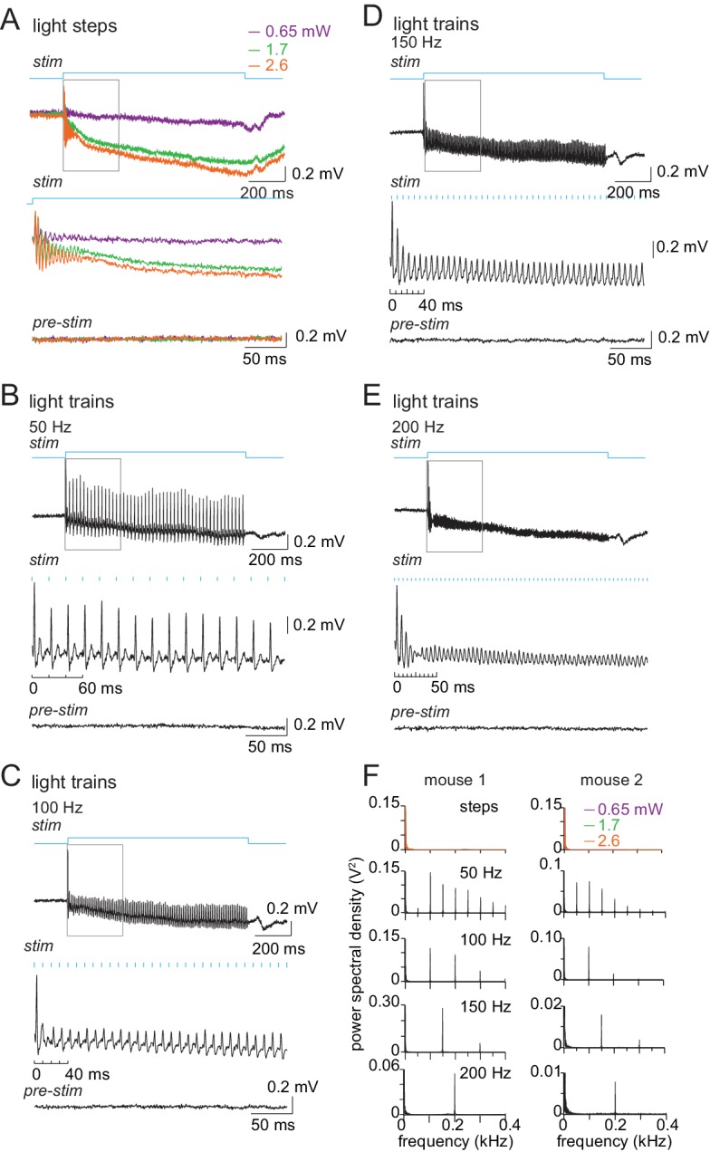
CbN cell spikes also time-locked to the stimulus at all frequencies, briefly dropping their firing rates during the window between 2 and 6 ms after light pulse onset (Figure 7D, p<0.001 at each frequency, Rayleigh test). As in Purkinje cells, spike probability deviated less from pre-stimulus values at higher frequencies, but the resetting of spike times remained evident. Since each CbN cell receives convergent input from 30 to 50 Purkinje cells (e.g., Person and Raman, 2012a), the consistent gaps in firing indicate that a measurable proportion of converging Purkinje cells must have provided coherent inhibition at regular intervals that matched the stimulus train frequency. The observation that similar response patterns were present across all 39 cells, for all trials, at each train frequency made it reasonable to conclude that the trains led to multiple Purkinje cells firing action potentials in the same 2–3 ms time window, and therefore that they increased the synchrony of Purkinje cell firing. For CbN cells, like for Purkinje cells, superimposing the responses to steps plotted in 1 ms bins confirmed that the temporal structure of spiking imposed by trains was distinct from the lack of structure imposed by steps. Specifically, after the first 10 ms, the spike probability fell below ~0.05 per 1 ms bin throughout the stimulation, consistent with steps generating asynchronous Purkinje cell firing. Trains had a smaller effect on firing rates than did steps. As train frequencies increased from 50 to 200 Hz, Purkinje cell firing rates increased from about 110 to 160 spikes/s, whereas rates of CbN cells remained at about 60–70 spikes/s at all train frequencies (Figure 7E).

Because the optical fiber was inside the recording pipette, however, the fiber was necessarily in different locations during the recordings from Purkinje and CbN cells. To obtain a measure of Purkinje cell activity with light stimulation applied in the locations associated with CbN recordings, we broke the tip of the electrode to be ~15 µm diameter, positioned the fiber at the depths that it reached during CbN cell recordings, and recorded local field potentials from Purkinje cells. This method had the added advantage of permitting simultaneous recordings from cells activated by the light stimulation, providing a direct investigation rather than just the inference of synchrony. Recordings were made in anesthetized mice to facilitate data acquisition, since testing whether high-frequency stimulation of ChR2 elicited well-timed Purkinje spikes simultaneously in multiple cells did not require awake animals. Light steps led to a brief period of coherent high-frequency firing that lasted ~50 ms; for the next ~950 ms, evidence of synchronous firing was not detectable (Figure 7 Supplement 1AFigure 7—figure supplement 1A). In contrast to the responses to steps, light trains led to coherent spiking at frequencies that matched the train and that lasted throughout the 1 s stimulus (Figure 7—figure supplement 1B–E). Consistent with the change in spike rates seen in the PSTHs from CbN cells in awake mice, the amplitude of the signal was highest for 50 Hz trains, illustrating that Purkinje cells followed the stimulus most effectively at the lowest frequency tested. Even with high-frequency trains, however, FFTs of the local field potentials in both mice showed peaks at the stimulation frequency and harmonics for all trains, verifying that some Purkinje cells fired synchronously on each cycle of the stimulus across the range of frequencies tested (Figure 7—figure supplement 1F).

Thus, the consistent time-locking of Purkinje and CbN cell firing to trains, as well as the local field potentials, confirmed that light trains could be used to restructure the temporal pattern of firing of stimulated Purkinje cells in a way that would (1) differ from firing patterns induced by steps, and (2) favor coherent firing by Purkinje cells, that is, increase the degree of synchrony of spikes. It is worth noting that the resetting of spike times in the CbN cells occurred on a time scale of 5–20 ms, a much shorter period than the duration of a stride.

Relation between firing rates and stride cycle during optogenetic stimulation

Before the recorded CbN cell activity could be related to normal and perturbed locomotion, it was necessary to test the extent to which activity of an individual CbN cell could be correlated with movement, since the firing by a single cell could not be assumed to give an accurate readout of behavior. Therefore, we first pooled results from steps and trains and compared neuronal activity during non-slip trials during stimulation (‘light on’) to control strides when no stimulation was applied (‘no light’). The same analysis, that isi.e., binning and averaging instantaneous firing rates across all strides, was applied to light-on and no light conditions. As shown for a CbN cell (Figure 8A), even though light changed the firing rates during non-slip trials, the spike rates rose and fell at the same phase of the stride as for control strides. The binned firing rates during the stride for the light-on and no-light conditions were plotted, as shown for a Purkinje and a CbN cell (Figure 8B and C, left), and the correlation between light-on and no-light binned firing rates was calculated by linear regression (Figure 8B and C, right). On non-slip trials, most Purkinje and CbN neurons had high positive correlations between the light-on and no-light conditions (Figure 8D), with ≥ 70% exceeding r = 0.4 (Purkinje cells, N = 21/30; CbN cells, N = 15/20). Thus, despite changes in mean firing rate across the duration of each stride, on non-slip trials, the phase relation between the firing rate of individual cells and the step cycle was relatively unperturbed. In other words, normal stride-related modulation was preserved on non-slip trials.

Figure 8. Correlation between firing rate modulation of individual neurons and slip/non-slip trials.

Figure 8.

(A) Comparison of CbN cell activity (black), paw position (blue), and instantaneous firing rate (red) without light (top, ‘no light’) and during light stimulation (bottom, ‘light on’) in non-slip trials. (B, C) Left, Instantaneous firing rate (IFR) and paw position during normalized strides for a Purkinje (B) and a CbN cell (C) for non-slip trials. Right, Binned IFRs from left panels for ‘light on’ vs. ‘no light’ conditions; r, correlation coefficient; dotted line, unity. (D) Histogram of correlation coefficients measured as in (B), (C) for all Purkinje (left) and CbN (right) cells. (E) As in (A) for two slip trials. Note differences in time base between ‘no light’ and ‘light on’ pairs. (F, G) As in (C), for slip trials for the illustrated CbN cells. Insets, overlay of paw traces during the pre-stimulus (grey, 'no light') stride template and the slip epoch (blue, 'light on'). (H) As in (D), for all slip trials. (I). As in (C) for prolonged stride aligned to the lift of a control stride ('no light'), for the same slip trial as shown in (G). (J) Left. Polar plots from (I) of control and prolonged lift-aligned stride. 0 indicates bin with maximal firing rate in control stride. Right. Mean of all prolonged lift-aligned stride trials, showing the peak response is near 0, which is the bin with the peak firing rate in control ('no light').

Next, we compared spiking during slip trials between light-on and no-light conditions, with a goal of testing the extent to which changes in stride-related modulation of firing in individual cells and slips were mutually predictive. Sample traces are shown for different CbN cells during an arrested stride and a prolonged stride (Figure 8E). Because each slip was distinct, these analyses were done on a trial-by-trial basis. Also, since by definition strides were disrupted during slips, firing rates could not be binned against a clearly defined concurrent stride. We reasoned, however, that if the slip and the activity of the recorded neuron were independent, then the pre-stimulus firing rate modulation should persist throughout the stimulation. We therefore tested whether firing rates during slips continued to be modulated as they were during unperturbed locomotion, by binning firing rates during the slip against a template stride just preceding light onset (Materials and methods). The binned firing rates for the light-on and no-light conditions were plotted, as illustrated for the arrested and prolonged stride (, left), and correlations were calculated (Figure 8F, 8GFigure 8F and G, right). Unlike for non-slip trials, a low proportion of perturbed strides showed correlations ≥0.4 (Purkinje cells, N = 69/340, 20% trials; CbN cells, N = 60/286, 21% trials, Figure 8H, open bars), indicating that disruption of firing rate modulation of individual cells and disruption of strides were tightly associated.

The prolonged strides provided a special case for examining this relationship, as inspection of the data suggested that firing rate modulation with respect to the unperturbed stride might be maintained but extended to match the lengthened stride. Therefore, prolonged strides were aligned to the lift and binned, and the correlation between light-on and no-light conditions was examined (Figure 8I). To quantify this effect across trials, the binned no-light and light-on firing rates were plotted on polar coordinates (Figure 8J, left), with 0° indicating the bin with the peak firing rate for the control stride (‘no light’). The distribution for all prolonged strides is shown in Figure 8J (right). The skew toward the right half of the plot suggests that the maximal firing rates were achieved in the same phase (stance or swing) of the prolonged and control strides (Pkj, N = 277, p=0.002; CbN, N = 246, p=0.002, Rayleigh test). Together, the data suggest that modulation of firing rates individual Purkinje and CbN cells correlates well with the regularity of the stride cycle: when modulation persists during optogenetic stimulation, strides continue relatively normally, whereas when modulation is disrupted, slips occur. The most plausible explanation for this result seems to be that direct stimulation of Purkinje cells evokes similar responses (of altering or failing to alter modulation) in multiple CbN cells, which together exert an influence over locomotion that is ultimately manifested in hind paw movement. The association between slips and the loss of stride-related modulation in single cells also justifies an analysis of the ability of light steps and trains to disrupt single-cell firing and perturb movements.

With this information in hand, we tested whether CbN cells responded differentially to steps and trains (asynchronous and synchronous inhibition) in a way that could be related to the measured behavior. We first compared the effects of light steps of different strengths on CbN cell firing rates during slip and non-slip trials. The mean firing rate during the 1 s light-on stimulation period was plotted against the firing rate for 1 s just before stimulation on each trial (Figure 9A–9D, every trial from N = 13, 18, 10, 7 cells). In all trials, since the mouse was running before the light-on period, the value represents the mean rate during one or more strides. As the stimulus strength increased, CbN firing rates were more likely to fall below the pre-stimulus baseline, and a larger number of slip trials were evident. Across all trials, the probability of evoking a slip with a light step was 76% with the weakest stimuli and increased to 94% with the strongest stimuli (Figure 9E, black symbols, χ2 (7, N = 155)= 7.1, p=0.07, chi-square test). The firing rates of CbN cells on slip trials but not non-slip trials showed a corresponding decrease as the step intensity was raised (Figure 9E, green symbols). Plotting the change in firing rate relative to the pre-stimulus period likewise showed greater firing rate decreases at higher intensities for slip as opposed to non-slip trials (Figure 9F, two-way ANOVA F(3,147) = 3.91, p=0.05, interaction between step intensity and slip/non-slip, p=0.08). Interestingly, almost all trials on which CbN firing rates were suppressed below ≤ 5 spikes/s resulted in slips (Figure 9A–D). The only exceptions were with the weakest stimulation, which likely recruited the fewest Purkinje cells. The observation that suppressing firing in CbN cells almost invariably correlated with discontinuities of movement (Figure 9G) suggests that even well-practiced motor behaviors, such as locomotion, relies on signals that can be disrupted by sudden and transient loss of input from CbN cells.

Figure 9. Sensitivity of smooth locomotion to optogenetic changes in CbN firing rates and patterns.

(A-D) Scatter plots for each light intensity of mean CbN cell firing rates during light steps (1 s) vs. pre-stimulus firing rates (1 s) for single trials. Filled symbols, slip; open symbols, non-slip trials; black line, unity; dashed line, five spikes/s. (E) Slip probability (black) and mean CbN cell firing rates (green) for slip (solid symbols) and non-slip (open symbols) trials. Numbers, slip/total trials (F) Change in CbN cell firing rates compared to pre-stimulus firing rates in slip and non-slip trials vs. light step strength. Symbols as in (E). (G) Slip probability vs. binned mean firing rates across all step intensities (bin = 20 spikes/s). (H–K) As in (A–D) for each light train frequency. (L) As in (E), for trains. (M) As in (F), for trains. (N) Left, Box plot of firing rates for 564 pairs of trials selected for similar mean firing rates during a step and a train of light stimuli applied to the same cell. Grey symbols, outliers; whiskers, two standard deviations. Right, Bar graph of fraction of paired trials in which a slip was evoked only by the step (dark blue) or only by the train (light blue).

Figure 9.

Figure 9—figure supplement 1. Reanalysis of data of Figure 9 with slip measurements constrained to the period within the trial when locomotion was perturbed.

Figure 9—figure supplement 1.

The trials consist of the subset of those in Figure 9 in which slip onsets and offsets were clearly identifiable. (A) Slip probability (black) and mean CbN cell firing rates (green) for slip (solid symbols) and non-slip (open symbols) trials. Steps: χ2 (7, N = 132)=3.1, p=0.4, chi-square test. (B) Change in CbN cell firing rates compared to pre-stimulus firing rates in slip and non-slip trials vs. light step strength. Symbols as in (A). Two-way ANOVA F(3,124) = 3.9, p=0.05, interaction between step strength and slip/non-slip, p=0.6. (C) As in (A), for trains. Trains: χ2 (7, N = 185)=25.8, p<0.001, chi-square test. (D) As in (B), for trains. Two-way ANOVA F(3,177) = 2.95, p=0.09, interaction between frequency and slip/non-slip, p=0.06.

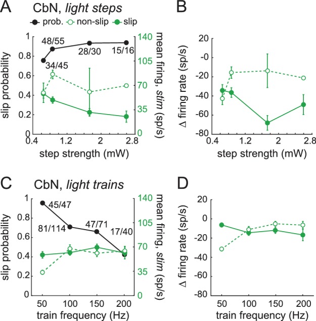
The same analysis applied to trials with light trains showed a different distribution of CbN cell firing rates before and during stimulation. The mean rates measured over the 1 s stimulus period continued to cluster around or just below the unity line, illustrating that trains were less effective at suppressing CbN cell firing rates, at all frequencies of stimulation (Figure 9H–K, trials from N = 8, 25, 13, 12 cells). Nevertheless, slip as well as non-slip trials were present, with ≥49% of trials eliciting slips at each frequency. Slip and non-slip trials could not be well distinguished on the basis of firing rate, which remained near 60 spikes/s regardless of train frequencies (except two non-slip trials with 50 Hz trains; Figure 9L), nor could they be distinguished on the basis of CV of the interspike intervals. For trials with ≥2 interspike intervals during light stimulation, CVs on slip and non-slip trials, respectively, were 0.90 ± 0.02 (N = 201) and 0.93 ± 0.05 (n = 82) for trains and 1.2 ± 0.07 (N = 111) and 1.0 ± 0.17 (N = 18) for steps. Although the CV was lower for trains than for steps (p=0.007, F(1,408) = 7.23, two-way ANOVA), the analysis revealed neither an effect of slip/non-slip on CV (p=0.4) nor an interaction between slip/non-slip and step/train (p=0.2). The data are therefore consistent with temporal restructuring of CbN cell spikes (on a millisecond time scale) by trains of synchronous inhibition, but the consequent decrease in CV was not predictive of whether or not a slip occurred.

The probability of train-evoked slips varied inversely with frequency, with 50 Hz stimulation evoking a high slip probability (0.96), and 200 Hz stimulation evoking the lowest slip probability (0.49, Figure 9L, χ2 (7, N = 285)=25.4, p<0.001, chi-square test). The slip probability therefore was not simply a direct consequence of the amount of illumination time as a readout of the magnitude of inhibition. Instead, it may have related to the efficacy of recruiting Purkinje cells to time-lock to the stimulus; both the PSTHs and the local field potentials suggested that the 50 Hz trains favored synchrony of more Purkinje cells. The larger number of Purkinje cells recruited to a perfectly regular, physiologically arbitrary stimulus pattern likely made them more effective at restructuring CbN cell spike timing (on a time scale of milliseconds) in a manner that led to deviation from the normal stride-related modulation pattern, thus favoring slips even without greatly suppressing firing rates.

Indeed, the change in firing rates compared to baseline was similar between slip and non-slip trials at the different train frequencies (Figure 9M, two-way ANOVA F(3,277) = 0.51, p=0.5, interaction between train frequency and slip/non-slip, p=0.09). These data further suggest that factors such as synaptic depression were not the primary determinants of whether a slip occurred; such a scenario predicts that strong inhibitory synaptic depression would cause high firing rates without slips, while less depression would do the opposite. Since the absolute and relative firing rates were similar in slip and non-slip trials and only slightly changed relative to no-light conditions, the simplest interpretation is that the train stimuli altered stride-related modulation on slip-trials in a way that interfered with movement.

In these analyses, firing rates were all averaged across the full 1 s light stimulus. As stated above, however, slips occurred with a lag and did not always persist throughout the stimulus window. Therefore, for the subset of trials in which slip onset and offset could be identified with certainty, the measurements were repeated with the analysis window constrained to the duration of the slip. This analysis of the data gave indistinguishable results ().

Although trains could induce slips, pooling all stimulus conditions showed that they did so with a 15% lower probability than steps (steps, 0.86; trains 0.71, Fisher’s exact test, p<0.001); as in the case of steps, however, in the few instances (six trials) when trains brought CbN cell firing rates below five spikes/s, slips always occurred (Figure 9H–9K). One possibility, therefore, is that the different slip probabilities elicited by steps and trains related simply to their differential effects on rate. To test this possibility, we selected the subset of trials in which a step and train applied to the same cell resulted in similar firing rates (within 10 spikes/s). Pairs of trials with firing rates below 10 spikes/s (which always gave high slip probabilities) were excluded. This analysis yielded 564 pairs of trials, for which the mean firing rate for both steps and trains was 71 ± 1 spike/s (Figure 9N, left, N = 13 cells, 65 distinct step trials, 141 distinct train trials).

Even with comparable firing rates elicited by comparable light intensities, the probability of evoking a slip remained higher for steps than for trains (0.89 vs. 0.72, p<0.001, McNemar’s test for paired data). Stated differently, a slip was evoked only by the step in 127 pairs (22.5% of all trials), and only by the train on 31 pairs (5.5% of all trials, Figure 9N, right); on the other trials the response to steps and trains was the same. Since the firing rates were similar in these within-cell pairs, it seems likely that similar numbers of Purkinje cells were activated and that the total inhibition during the paired trials was comparable. Therefore, the higher incidence of non-slip trials during trains suggests that trains interfered less often than did steps with normal stride-related modulation.

Indeed, when the spiking patterns during these rate-matched pairs of trials were compared, the records associated with slips showed an altered stride-related modulation compared to that seen over the time period of a single pre-stimulus stride. In contrast, the records associated with non-slips showed a stride-related modulation comparable to the pre-stimulus control (shown for two rate-matched pairs, Figure 10A and B, left and middle). These changes were evident when firing rate was plotted against paw position for the no-light and light-on conditions, regardless of whether a step or a train generated the slip (Figure 10A, 10B, right). We therefore reexamined the data of Figure 8E, that is, the correlation of firing rates during and before light stimulation for all slips associated with non-zero CbN cell firing rates, and separated the responses of steps and trains (). Again, steps gave a larger fraction of slips (83/102 trials, 81%) than did trains (203/306 trials, 66%). Thus, experimentally applied asynchronous Purkinje cell inhibition was relatively more effective than synchronous inhibition at disrupting the stride-related modulation of CbN spikes in a manner that induces irregularities of locomotion.

Figure 10. Loss of stride-related firing rate modulation during slips evoked by steps or trains.

Figure 10.

(A) Left, Rate matched trials from a single CbN cell in which a step led to a slip (top) and a train led to a non-slip (bottom). Middle, Overlaid paw records from the pre-stimulation and stimulation period (upper plots) and binned instantaneous firing rates (10 bins) during the pre-stimulus stride and during the stimulation over an equivalent period (lower plots) for the trials at left. Right, firing rate vs. paw position for the trials at left. (B) As in (A) for a different CbN cell in which both the step and train led to a slip. (C) Data from Figure 8H showing the correlation between the firing rate during the pre-stimulus stride and during the stimulus on slip trials, separated by steps and trains, and normalized to the total number of step trials (122) or train trials (286). (D) Cumulative histograms for the percentage of trials with a given correlation coefficient, for slip trials only. The maximal value of each histogram corresponds to the percentage of slip trials resulting from steps (81%) or trains (63%).

Discussion

These results provide evidence that smooth execution of well-trained, voluntary locomotion in mice is sensitive to the cyclic modulation of CbN cell firing rates associated with the stride cycle. Not only did silencing CbN cells via optogenetic activation of Purkinje cells reliably disrupt ongoing movement, but perturbing the modulation of CbN cells was also associated with irregularities in the step cycle, even when CbN cell firing was not fully suppressed by experimentally increased inhibition. Conversely, maintenance of the modulation pattern correlated well with continued walking, even when CbN firing rates were experimentally reduced. In addition, the results suggest that cerebellar output is sensitive to the degree of coherence of Purkinje cell firing, since CbN cells responded differentially to experimentally applied asynchronous or synchronous inhibition superimposed on natural activity patterns. Light steps, which increased Purkinje spike rates without consistently re-patterning spike timing on a millisecond time scale, slowed CbN cell firing and reliably elicited slips. Light trains, which increased Purkinje spike rates to a lesser extent but consistently increased the synchrony of convergent inhibition, generated slips on >50% of trials, suggesting that a resetting of the timing of inhibition could be sufficient to disrupt cerebellar output and the associated behavior. Nevertheless, relative to steps, trains were more frequently associated with non-slip trials. Since non-slip trials were associated with stride-related firing rate modulation, trains appear more permissive of continued modulation, which probably arises from direct mossy fiber excitation of CbN cells. Together, the data are consistent with the idea that simple spike synchrony can create synchronous gaps in inhibition on the millisecond time scale, during which (modulated) excitation from mossy fibers can more effectively elicit (modulated) firing in CbN cells.

Cerebellar output and the persistence of well-trained movements

Cerebellar lesions and alterations of cerebellar physiology give rise to ataxia (Manto, 2008; Morton and Bastian, 2007; Zackowski et al., 2002), and specifically reducing CbN cell output interferes with natural, ongoing behavior (Thach and Bastian, 2004). For instance, after portions of the cerebellar nuclei are inactivated with muscimol reaching and grasping movements in primates and cats show slowed reaction times, increased reach trajectory length, and general ‘clumsiness’ (Goodkin and Thach, 2003; Mason et al., 1998; Milak et al., 1997), as in the prolonged strides and other slips seen here. Similarly, in mice, silencing CbN cells by optogenetic stimulation of Purkinje cells alters the set point of whiskers without preventing whisking (Proville et al., 2014). Additionally, the sufficiency of elevated CbN cell activity to induce movements in stationary mice has been demonstrated by optogenetically altering firing exclusively of Purkinje cells (Heiney et al., 2014; Lee et al., 2015; Witter et al., 2013).

Nevertheless, dissecting how patterns of CbN cell firing relate to movements is complicated by the difficulty in determining to what extent changes in firing rate report sensory feedback and/or actively drive motor output. Passive movements like paw flexion are sufficient to alter both Purkinje and CbN cell firing patterns (Arshavsky et al., 1980; Casabona et al., 2010; Cody et al., 1981; McDevitt et al., 1987), and treadmill-induced locomotion in decerebrate animals produces modulated activity in Purkinje cells and target neurons. Transiently obstructing limb movement during locomotion in decerebrate ferrets or cats elevates simple spike rates for ~100–200 ms in Purkinje cells (Lou and Bloedel, 1992a; Lou and Bloedel, 1992b) and abolishes modulation of interpositus cell firing (Schwartz et al., 1987), but locomotion persists even with loss of cerebellar output (Orlovsky, 1972b). Such observations have led to the suggestion that such modulation normally seen in cerebellar output neurons represents sensory feedback associated with movement, which may facilitate but may not be necessary for locomotion. Additionally, despite the extensive inhibition of CbN cells by Purkinje cells, changes in gait can be subtle when exclusively Purkinje cell firing is changed (; Levin et al., 2006; Machado et al., 2015; Mullen et al., 1976; Walter et al., 2006). Thus, to what extent CbN cell firing patterns and Purkinje-mediated inhibition of CbN cell firing facilitates, produces, or reports movements may vary with the behavior and/or conditions.

The present experiments on voluntary locomotion in intact mice demonstrate that transiently suppressing modulation of CbN cell firing by directly stimulating Purkinje cells is sufficient to disrupt smooth, well-practiced locomotion. Additionally, slips were correlated with the loss of modulation rather than the absolute firing rate, suggesting that CbN cells do not simply provide an excitatory tone to downstream regions. The latency of the disruption (~120 ms) suggests either an indirect multi-synaptic signaling before the motor response becomes evident in the hind paw record or that the hindlimb slip is secondary to shorter latency disruption in other muscle groups under cerebellar regulation (e.g., Machado et al., 2015). In either case, these data provide evidence that cerebellar output can actively participate in locomotion.

Modulation of Purkinje and CbN cells during ad-lib locomotion

The repetitive nature of the step cycle makes locomotion a useful behavior for examining the activity of likely-connected Purkinje and CbN cells. Such data isare of interest since CbN cells receive concomitant synaptic excitation from mossy fibers and inhibition from Purkinje cells, which are themselves indirectly excited by mossy fibers via granule cells. Thus, excitation and inhibition may rise and fall together in CbN cells with lags of only two synaptic delays. Here we find that both Purkinje cells and CbN cells showed an elevation of mean firing rates during locomotion relative to rest. Similar firing rate increases in both cell types have been observed in walking cats (Armstrong and Edgley, 1984a, Armstrong and Edgley, 1984b) and primates during rapidly alternating arm movements (Thach, 1968), raising the question of how inhibition from Purkinje cells interacts with mossy fiber excitation.

Because the goal of the study was to collect data from as many modulated neurons as possible, we recorded from sites restricted enough to ensure that cells showed hindlimb-related activity, but broadly distributed enough to permit random sampling of the population (~400×400 x 600 µm for CbN cells). This approach revealed a wide range of stride-related modulation patterns in different cells. It remains possible that a topographic organization of phase modulation exists, but the resolution of the present work did not permit any such arrangement to be detected. Nevertheless, a reasonable prediction is that the population activity of Purkinje and CbN cells may reveal whether stride-related firing rate modulation in the two cell groups is inverted, indicating that Purkinje cell inhibition overrides excitation and sets CbN firing rates, or co-modulated, suggesting that mossy fiber excitation dominates. In cats, recordings from cells with forelimb receptive fields showed that population averages of firing rates of Purkinje and CbN cells rose and fell at the same phases of the step cycle (Armstrong and Edgley, 1984a; Armstrong and Edgley, 1984b). The corresponding analysis of the present data with respect to hindlimb movement in mice gave mean peak Purkinje firing rates that were somewhat phase-advanced to that of CbN cells. In neither study, however, did inverse firing patterns predominate. Also, in both studies, firing rates showed one or sometimes two peaks of activity at specific phases of the stride, and the peak firing rates of either Purkinje cells or CbN cells could occur at any phase of the stride. A similar range of stride-related activity patterns of Purkinje cells was reported in a study of freely moving rats, which further demonstrated that the variety of activity patterns across Purkinje cells, and even across strides within a single Purkinje cell, may result from the relation of Purkinje cell firing rates to different aspects of locomotion, including speed, acceleration, pitch, and roll (Sauerbrei et al., 2015). Here, the pattern of modulation with respect to the stride across a given Purkinje or CbN cell did not depend strongly on stride duration; one possibility is that the range of values assumed by variables such as pitch and roll was reduced by head fixation.

Responses of CbN cells to the rate and timing of Purkinje-mediated inhibition

Although the connection patterns between response classes could not be deduced from the present data, Purkinje neurons with different modulation patterns may well converge onto CbN cells. For instance, specific saccadic eye movements in primates may be encoded by convergence of Purkinje cells with different activity patterns (Herzfeld et al., 2015). Thus, several factors may govern the outcome of synaptic interactions among excitatory and inhibitory inputs to CbN cells.

A series of in vitro studies have suggested that the degree of coherent firing, or synchrony, of convergent Purkinje cell inputs may be one such factor. The intrinsic ion channels of CbN cells generate spontaneous action potential firing at 70–100 spikes/s (Mercer et al., 2016; Person and Raman, 2012a; Raman et al., 2000; Telgkamp and Raman, 2002) even with no synaptic input. CbN cells therefore require active hyperpolarization and/or shunting from Purkinje cells to prevent them from firing. About 30–50 Purkinje cells provide strong synaptic contacts to each CbN cell, but each simple spike evokes unusually brief GABAA receptor-mediated IPSCs in CbN cells, with decay kinetics of 2–2.5 ms (Person and Raman, 2012a). If converging Purkinje cells fire asynchronously, the inhibitory currents overlap in time and CbN cell firing can be effectively suppressed. Conversely, if Purkinje cells synchronize, brief periods of inhibition are followed by windows of reduced inhibition when CbN cells can fire (Person and Raman, 2012a; Wu and Raman, 2017). The degree of synchrony influences the effect of synaptic excitation: lower jitter among Purkinje inputs permits CbN cells to fire more action potentials in response to a fixed amount of excitation. In other words, the efficacy of excitation is raised by the synchrony of inhibition (Wu and Raman, 2017).

In the present experiments, synchrony could not be measured directly, since it was not possible to identify and record simultaneously from multiple converging Purkinje cells. Nevertheless, all CbN cells showed a consistent spike suppression on repeated cycles of stimulation at fixed latencies with millisecond-level precision, reflecting coherent inhibition that was interpreted as arising from increased synchrony of firing by converging Purkinje cells. Supporting this idea, Purkinje cell spike probability increased reliably with each cycle of the light train, and local field potentials in anesthetized mice demonstrated Purkinje spike-related signals at rates that matched the train stimuli. In contrast, steps did not generate well-timed inhibition of target cells. Although step-evoked local field potentials began with a ~50 ms damped oscillation of ~300 Hz, no corresponding pattern emerged in recordings of CbN cells in awake mice, possibly owing to an anesthetic-induced increased coherence in the local field potential, a lack of convergence of Purkinje cells sampled in the local field recording, or an absence of gaps in inhibition of the target cells. For the remaining 950 ms of stimulation, the millisecond-level timing of CbN cell spikes diverged for steps and trains, and the disruption of locomotion often persisted well into this later stimulus period. Taken together, it seems reasonable to infer that trains increased the synchrony of Purkinje cell firing throughout the stimulus.

These in vitro observations and in vivo analyses provide a context for interpretation of the step and train data in the present study. In many respects, the effects of steps and trains were similar: if light stimulation altered stride-related modulation, then a slip occurred. Purkinje and CbN cell firing rates, however, were reduced to a lesser extent by trains than steps, possibly owing to the shorter illumination times associated with trains and/or the limited responsiveness of ChR2 to high-frequency light pulses. Nevertheless, imposing an arbitrary, regular pattern of relatively synchronous inhibition often restructured CbN cell spike timing (on a millisecond time scale) was enough to interfere with stride-related modulation. With such disruption, the step cycle could be perturbed even without decreasing the mean rate of CbN cell firing, providing evidence that spike patterns of CbN cells on the time scale of a single stride can have behavioral consequences.

Of note, however, are the other trials, in which the natural pattern of stride-related firing modulation persisted. In these trials, the modulated excitation from mossy fibers (Powell et al., 2015) presumably contributed to the persistence of stride-related modulation in CbN cells. In other words, on about half the trials, the naturally occurring excitation successfully overcame synchronous optogenetically applied inhibition. Moreover, when steps and trains elicited the same firing rate in a given cell but different behavioral outcomes, steps were nearly four times more likely than trains were to generate slips, that is, to disrupt the modulation pattern. This observation is consistent with in vitro data demonstrating that excitation was most effective during gaps in inhibition induced by coherent Purkinje cell firing (Wu and Raman, 2017).

In vivo, synchrony of simple spikes has been repeatedly observed in recordings of neighboring and likely converging Purkinje cells, and the frequency of observation of synchrony reportedly increases during cerebellar behaviors (Bell and Grimm, 1969; Ebner and Bloedel, 1981; Heck et al., 2007; MacKay and Murphy, 1976; de Solages et al., 2008; Wise et al., 2010; reviewed by Person and Raman, 2012b), and tetrode recordings in freely moving rats illustrate correlated activity between stride-modulated Purkinje cells (Sauerbrei et al., 2015). The extent to which Purkinje cell synchrony occurs during movements remains unknown, but, given the high firing rates of Purkinje cells and the common inputs they receive from granule cells and inhibitory neurons, it seems highly improbable that inhibitory input to CbN cells will always be maximally asynchronous. An interesting possibility, yet to be tested, is that the degree of synchrony might vary systematically during each stride. Since stride-related firing rate modulation is correlated with smooth locomotion, and since excitation from mossy fibers appears sufficient to generate modulated firing in target cells (Powell et al., 2015), a plausible outcome of naturally occurring synchronous Purkinje cell activity might be to create physiologically appropriate gaps in which mossy fibers might evoke CbN cell spikes more efficiently, providing a relatively more permissive state for smooth execution of natural behaviors.

Materials and methods

Key resources table.

Reagent type (species)
or resource
Designation Source or reference Identifiers Additional information
strain, strain background
(Mus musculus)
B6.Cg-Gt(ROSA)26Sortm27.1(CAG-
COP4*H134R/tdTomato)Hze/J
Jackson Laboratory Jackson stock: 012567 -
strain, strain background
(Mus musculus)
B6.129-Tg(Pcp2-cre)2Mpin/J Jackson Laboratory Jackson stock: 004146 -
recombinant DNA reagent AAV9.CBA.Flex.ChR2(H134R)-
mCherry.WPRE.SV40
Addgene Addgene: 18916 -
recombinant DNA reagent AAV9.EF1a.DIO.hChR2(H134R)-
eYFP.WPRE.hGH
Addgene Addgene: 20298 -
software, algorithm custom paw tracking software Actimetrics from Actimetrics
www.actimetrics.com
free by contacting
company
software, algorithm irradiance calculator Deisseroth Lab
Optogenetics Resources
https://web.stanford.edu/group/dlab/ -

Animals

All procedures conformed to the NIH guidelines and all protocols were approved by Northwestern University’s Institutional Animal Care and Use Committee (Animal Welfare Assurance Number, A3283-01, protocol IS00000242, IMR). Ai27D (B6.Cg-Gt(ROSA)26Sortm27.1(CAG-COP4*H134R/tdTomato)Hze/J, stock 012567) and Pcp2-Cre (B6.129-Tg(Pcp2-cre)2Mpin/J, stock 004146) Jackson Laboratory, Bar Harbor, ME) mice were crossed to generate offspring heterozygous for channelrhodopsin-2 (hChR2 (H134R)), referred to as ChR2, expression in Purkinje cells. Mice had free access to food and water and were maintained under a reverse day-night cycle (12:12 hr) for at least two weeks before the training phase of experiments began. Home cages included running wheels (Bio-Serv, Flemington, NJ) to provide experience in running. Experiments were performed on adult mice (P45-P70). Initially, both male and female mice underwent training to run on a treadmill while head-fixed (see Behavior below); however, while 38/44 males (86%) ran consistently, only 4/13 (31%) females did. Since running was essential to the study, only male mice were used for experiments. The data presented are from 23 adult male mice.

Surgical procedures

Mice underwent surgery for a craniotomy to allow electrode access and for installation of a headplate for head-fixation. They were anesthetized with ketamine (120 mg/kg, intra-peritoneal) and xylazine (3 mg/kg, intra-peritoneal) and mounted in a stereotaxic apparatus (Stoelting Co., Wood Dale, IL). Body temperature was maintained at 37°C with a heating pad (Harvard Apparatus, Holliston, MA) and oxygen was delivered through the nose. Lidocaine (2%, topical) was applied to the skin and buprenorphine SR-LAB (1 mg/kg, subcutaneous, ZooPharm, Windsor, CO) was administered peri-operatively for analgesia. A rectangular metal headplate with a central window (Dombeck et al., 2007) was attached to the skull with Metabond (Parkell, Inc., Edgewood, NY), just posterior to the bregma. A plastic well was also cemented around the recording site to hold agarose and the ground electrode. A craniotomy was made over the paravermal region of the cerebellum (bregma −6.3 mm, 1.85 mm lateral,~0.3–0.5 mm diameter). The dura was left intact and covered with Kwik-Sil (WPI, Inc., Sarasota, FL) until the time of recording. Saline solution (0.9% NaCl, 50 ml/kg, subcutaneous) was administered to aid hydration while animals recovered from surgery.

For local field potential recordings only, experiments were done in mice (N = 2) anesthetized with intraperitoneal injection of ketamine (120 mg/kg) and xylazine (3 mg/kg). Mice were held in a stereotaxic apparatus and kept on a warming pad to maintain body temperature at 37°C and oxygen was delivered through the nose. Vital signs were monitored and the animal was re-dosed with 20–50% of the initial dose of ketamine when the toe-pinch reflex began to reappear.

Behavior

At least one week after surgery, mice began training to run on a cylindrical, freely rotating, non-motorized, 18.6 cm diameter Styrofoam treadmill (Domnisoru et al., 2013; Heys et al., 2014). The setup was located in a darkened room with white noise delivered through speakers to minimize startle from external noises. Head-fixed mice were placed on the treadmill for 30 min, increasing to 2 hr over the course of week. Most male mice (86%) ran regularly on the treadmill by days 7–10. After mice ran consistently for at least 60% of a 2 hr session, recordings were made (see Electrophysiological Recording below) during head-fixed running for up to 3 hr per day for 1–2 weeks. During recordings, the ipsilateral hind paw was monitored from the side with a high-speed, infrared video camera (240 fps, Tamron, Commack, NY). The x-position (anterior-posterior) and y-position (dorsal-ventral) of the paw was tracked (240 fps) with a custom-written program (Actimetrics, Wilmette, IL). Image contrast was aided by painting the treadmill black and illuminating the paw with a red flashlight. During running the paw position varied mostly in the x-domain, which was stored for analysis.

Injections and anatomy

To identify Purkinje cells that projected to the CbN cells associated with locomotion, tracers were pressure injected with a picospritzer (150–400 pulses, 6 psi, 5 ms pulse) into the region of the interpositus targeted for recordings (N = 3 mice). In wild-type mice, the retrograde tracer cholera-toxin subunit B conjugated to Alexa Fluor-488 (Invitrogen) was injected, and in Pcp2-cre mice, a mixture of viruses (200–500 nL) was applied to express ChR2(H134R) with a fluorescent marker (AAV2/9 conjugated with either mCherry (AAV9.CBA.Flex.ChR2(H134R)-mCherry.WPRE.SV40; Addgene 18916) or eYFP (AAV9.EF1a.DIO.hChR2(H134R)-eYFP.WPRE.hGH; Addgene 20298; U Penn viral vector core). With either technique, labeled Purkinje cells were consistently found lateral to the primary fissure in coronal sections, that isi.e., in the lobulus simplex of the cerebellar cortex, following injections in the central regions of anterior/posterior interpositus nucleus. These regions were therefore targeted for recordings.

Electrophysiological recordings and optical stimulation

Electrophysiological recordings were made from head-fixed, running mice (Harvey et al., 2009). Borosilicate micropipettes (4–6 MΩ) were pulled on an P97 Sutter micropipette puller (Novato, CA), heat polished with a microforge MF-900 (Narishige International Inc., East Meadow, NY), and filled with (mM) 125 NaCl, 5 KCl, 10 d-Glucose, 10 HEPES, 5 CaCl2, pH = 7.4, 300 mOsm). A Ag-AgCl ground electrode was placed on the surface of the skull near the craniotomy and held by 2.5% agarose, which also covered the exposed brain surface and stabilized the recording electrode. An optic fiber (240 μm, NA 0.63, Doric Lenses, Québec, Canada) was inserted in the recording pipette through the side port of the electrode holder (Warner instruments, Holliston, MA) to permit simultaneous electrophysiological recordings and optical stimulation (Katz et al., 2013). The end of the fiber was 4–5 mm from the tip of the pipette.

Recordings were made beneath the craniotomy (bregma −6.3 mm, 1.85 mm lateral,~0.3–0.5 mm diameter) at a depth of up to 1900 µm for Purkinje cells in the lobulus simplex and 1900–2500 µm for CbN cells in the posterior and less commonly the anterior or dorsolateral horn divisions of the interpositus nucleus. Light stimuli (465 nm, 8–53 mW/mm2) were generated by an LED coupled to an LED driver (Doric Lenses). Stimuli were calibrated with a photometer (PMD100, S140C, Thorlabs, Inc., Newton, NJ). The maximum measured light intensity at the pipette tip of ~ 40 mW/mm2 was estimated to attenuate to ~ 20 mW/mm2 about 50 µm from the tip (based on (Yizhar et al., 2011) https://web.stanford.edu/group/dlab/). For this fiber size, this intensity is equivalent to 0.9 mW, which corresponds to the threshold for action potential activation in pyramidal neurons in cortical slices from Ai27 mice (Madisen et al., 2012).

Loose-cell attached recordings (Rseal > 10 MΩ) were made in current clamp mode. Electrical signals were acquired at 10 kHz with an Axoclamp-2B or Axopatch-200B amplifier, a Digidata-1550, and pClamp software (Molecular Devices, Sunnyvale, CA). Action potential waveforms were identified by a voltage threshold of the electrical trace following low-pass digital filtering at 1 kHz. In trials from 8 Purkinje cells representing cells with high and low firing rates, slips and non-slips, and steps and trains, complex spikes were identified in 1 s windows around the light stimulation and found to have rates of 1.5 ± 0.4 spikes/s (before), 2.9 ± 0.7 spikes/s (during), and 1.1 ± 0.4 spikes/s (after). An elevation of complex spikes during the stimulation, which was evident in only 3 of 8 cells, is consistent with an experimentally driven increase in inhibition of nucleo-olivary cells and increase of inferior olivary firing. Because it was not possible to disambiguate the direct effect of optically raising Purkinje cell spikes from biological effects, e.g., associated with sensory or motor signals, complex spikes were not examined further. Additionally, when complex spikes were extracted from the record, the total firing rates were reduced by < 2%. Therefore, both simple and complex spikes were included in all analyses that are presented, but the values are overwhelmingly dominated by simple spikes.

Both in the cerebellar cortex and nuclei, recordings were made from regions where light stimulation evoked altered movement of the ipsilateral hindlimb (described further in Results). Each cell was recorded continuously for 10 trials of 7 s each without optical stimulation, followed by several conditions of light steps and light trains. Both steps and trains were applied for 1 s with 2 s recording before (‘pre-stim’) and after stimulation (‘post-stim’). 3–10 trials of each stimulus condition were repeated with a 10 s inter-trial interval. Steps and trains were tested in increasing order of their strength (0.56, 0.65, 0.9, 1.7 and 2.6 mW) or frequencies (50, 100, 150, 200, Hz at 1.7 or 2.6 mW), respectively. Multiple penetrations were made per session. Between sessions, the surface of the brain was sealed with Kwik-Sil (WPI, Inc.).

At the end of the final session, the recording pipette was withdrawn and a glass injection pipette (taper ~3.5 mm, tip 20–25 µm, WPI, Inc.) was inserted at the same stereotaxic coordinates. The recording locations and track were marked by pressure injection (~1–3 psi) of 1–2.5% Alexa Fluor-488 fluorescent dye conjugated with dextran (10K, anionic, fixable; Life Technologies, Carlsbad, CA). Mice were euthanized with Euthasol (100 mg/kg, intraperitoneal, Virbac, Fort Worth, TX) and transcardially perfused with paraformaldehyde (4% in PBS, ThermoScientific, Skokie, IL). Brains were kept in 4% PFA overnight, then triple-rinsed. Coronal cerebellar sections (100 µm) were cut at 4°C in PBS (0.01 M, Sigma Life Science, St. Louis, MO) on a vibratome (VT1000S, Leica Biosystems, Nussloch, Germany). Sections were imaged on a confocal microscope (Leica Biosystems, TCS SP5, Biological Imaging Facility, Northwestern University) and processed with the Fiji version of ImageJ software (Schindelin et al., 2012). The injection location was taken as the site of the maximum intensity (laser power and gain held constant for all imaged sections).

For local field potential recordings from Purkinje cells, the pipette tip was broken to give an electrode resistance of 0.8–1 MΩ, which permitted the fiber to be positioned as it was for direct recordings from CbN cells. Each optical stimulation condition was applied ten times. The ten sweeps in each condition were averaged and the signal was bandpass filtered from 50 Hz to 1 kHz. FFTs were done on the resulting sweeps and spectral power density was plotted. The power of the unfiltered residual electrical noise at 60 Hz was blanked for clarity.

Data analyses

Data are presented as mean ± SEM except as noted. All analyses including statistical tests were performed in Matlab (Mathworks). For inclusion in the study, Purkinje and CbN cells had to show appropriate firing responses to light (increases for Purkinje and decreases for CbN) that were statistically significant (one-sided paired t-test, p<0.05), as well a behavioral ‘slip’ (described in Results) of the ipsilateral hindlimb in any light stimulus condition.

Identification of strides

For quantification of paw movement, a region of interest (ROI) was set to cover the maximal range of hind paw movements during locomotion. Tracked paw position was normalized to the ROI window and therefore has arbitrary units of magnitude (Figure 1A, green box). ‘Rest’ periods, that is, when the mouse was not running, were evident in the paw-tracking record as a minimal change in paw position (<10% of that during strides). Neuronal activity during rest periods was measured from cells in which the total duration of all the rest epochs (in the absence of light stimulation) exceeded 2 s.

‘Run’ periods were identified as sawtooth-like sections of the paw-tracking record indicative of the hind paw moving forward and backward as it completed each stride (plant-stance-lift-swing-plant). Occasionally the tracking system would lose the paw, resulting in instantaneously rising transients in the tracking record that resembled brief square pulses. Tracking usually resumed within a few samples, so these transients were viewed as recording noise and were disregarded during analyses. Strides were identified from smoothed paw traces (sliding-average filter, 50 ms duration) as a trough-to-trough cycle. At least 30 strides, each 100–500 ms in duration were required for a cell to be included in the study. All strides were aligned to their peak (the lift). The ratio of the stance phase (trough-to-peak) to the swing phase (peak-to-next-trough) was calculated for each stride. The median value of this ratio for all strides associated with a recording was used to normalize strides by dividing each stride into stance and swing bins, for a total of 10 bins. Specifically, if the stance accounted for 70% of the stride, seven bins would be allotted to the period before the lift and three bins to the period after the lift. The stance phase generally accounted for 60–70% of the whole stride, so 6–7 bins were usually stance bins. In this way, the stance and swing bins would be of comparable (though not identical) durations, and firing rates in stance and swing could be examined separately. Reanalysis of the data with 10 equal bin widths yielded similar results and did not alter classification schemes, confirming that the differences in bin widths were minimal.

Instantaneous firing rates were calculated for each neuron. Mean firing rates during a stride were calculated from averaging the instantaneous firing rates within each bin. Binned firing rates and the binned paw displacement were averaged across all strides for a cell. The depth of modulation was quantified using a modulation index (MI) defined as (FRmax - FRmin)/(FRmax + FRmin), where FRmax and FRmin are the maximal and minimal binned firing rates during the normalized stride.

Classification of firing rate modulation patterns

Binned firing rates of Purkinje and CbN cells relative to paw position during the stride were classified independently by both investigators, as described in Results. Relative firing rates rose and fell at consistent phases of the stride for both Purkinje and CbN cells across stride durations, which justified normalizing strides (Armstrong and Edgley, 1984a, Armstrong and Edgley, 1984b). Nevertheless, because absolute firing rates could be variable (e.g., Sauerbrei et al., 2015), firing rates were plotted against paw velocity (derived from paw position) and analyzed by linear regression in separate analyses. 40% of Purkinje cells (14/35) and 21% of CbN cells (8/39) showed a significant correlation of firing rate and velocity. This relationship was not investigated further, but is encompassed in Class III, IV, V, and VI categorizations.

Analysis of optogenetically induced slips

Optogenetic stimulation during locomotion could generate disruptions of movement that were evident in the paw-tracking record. Because the treadmill was not motorized, the running speeds of mice were not held constant, giving rise to variability in the duration of strides as well as the rate of paw movement in the stance and swing. Consequently, it was not possible to use a fully automated analysis to identify slips. We therefore used the following approach: The paw record associated with each trial (1 s pre-stim, 1 s light stimulation, 1 s post-stim) was first inspected and classified manually, blind to stimulus condition, to identify potential slip trials. Trials in which running rate changed greatly just before the stimulus or in which running ceased during the light stimulus were discarded. If strides proceeded regularly throughout the 1 s, the trial was categorized as a ‘non-slip’. If at any point in the 1 s light stimulus, however, at least one stride showed (1) a greatly lengthened or shortened duration relative to pre-stimulus strides, (2) a strongly reduced slope of the stance or swing phase relative to pre-stimulus strides, or (3) a deviation from a sawtooth pattern, the trial was categorized as a potential ‘slip.’ These potential slip trials were then evaluated quantitatively. The last complete pre-stimulus (control) stride was detected automatically (as above) and strides during optogenetic stimulation were compared to the control stride. For a trial to remain classified as a slip trial, it had to have at least one stride with either a duration ≥20% longer than control (prolonged stride), a duration ≥20% shorter than control (incomplete stride), or a slope of the stance or swing phase ≥20% lower than control (arrested stride). Several trials fit into more than one of these slip categories (altered strides). Of the 971 putative slip trials, the automated analysis successfully detected a complete stride before and during stimulation on 859 trials; 800 (93%) fulfilled the criteria of at least one category and were confirmed as slips, whereas 59 (7%) did not reach threshold for slips but did not look like non-slips so were discarded. For the other 112 strides, the automated analysis was unsuccessful owing to factors such as a pre-stimulus stride extending for some tens of ms into the stimulus period, or the lack of an identifiable stride at any point during the stimulation. We counted these trials as slips, with an awareness that 7% (~8 trials) might not have been true slips. For a total of 912 slip trials, the misclassification error for slips would therefore be <1%. With this approach, although small optogenetically induced alterations of gait may have been ignored and treated as non-slips, slip trials are highly likely to represent real, experimentally generated changes in movement. The analysis therefore errs on the side of incorrect rejections but avoids false positives. In cells included in the study, a light-induced change in gait (including the onset of locomotion from rest) occurred on at least 50% of all trials in at least one stimulus condition.

The activity of each cell with and without optogenetic stimulation was examined with respect to paw movement. For non-slip trials, binned firing rates averaged across all strides during light stimulation were compared with binned firing rates from strides without light stimulation. For slip trials, the last stride before the light stimulus served as a template, and the firing rate of the cell during the slip period was compared to that during the template. Binned firing rates during the template stride were compared to those during the slip epochs using the same histogram bins as the template. For the special case of the ‘prolonged stride’ slip category, prolonged strides were aligned to the lift and analyzed as for undisrupted strides and binned firing rates between normal and slip strides were directly compared.

Statistical testing

The normality of each data set was assessed by a Shapiro-Wilk test. For normal data, t-tests (paired or unpaired) were performed. For non-normal data, signed-rank (paired) or Wilcoxon rank sum tests (unpaired) were performed. Tests were two-tailed, except when testing for optogenetically induced increases or decreases in firing rates, in which case the tests were one-sided. Rayleigh tests for circular non-uniformity or Kuiper’s tests (as noted) were used to test for non-uniform distributions of data, e.g., over a stride cycle or following successive pulses in light trains (toolbox for circular statistics with Matlab, Berens, 2009). Pearson’s correlation coefficients were used to evaluate the linear correlation between firing rates in two conditions. Two-way ANOVAs were performed to assess the relation among light stimuli, behavioral responses, and changes in firing rates. Fisher’s exact tests and χ2 tests for independence were used to assess dependence of slip probability on light pattern or light parameters. McNemar’s tests for paired nominal data were used to test for slip probability in pairs of trials with matched firing rates. All p-values are reported. In figures, one, two, or three asterisks indicate p<0.05,<0.01, or < 0.001.

Acknowledgements

We are grateful to Dr. Daniel Dombeck for advice on recording from awake head-fixed running mice, Dr. David Ferster for the paw-tracking program and comments on the manuscript, Dr. Izumi Sugihara (Tokyo Medical and Dental University) for consultation on anatomy, Dr. Mitra Hartmann for advice on local field potentials, as well as members of the Raman lab for helpful discussions. Confocal imaging was done in the Biological Imaging Facility, Northwestern University.

Funding Statement

The funders had no role in study design, data collection and interpretation, or the decision to submit the work for publication.

Contributor Information

Indira M Raman, Email: i-raman@northwestern.edu.

Gary L Westbrook, Vollum Institute, United States.

Funding Information

This paper was supported by the following grant:

  • National Institute of Neurological Disorders and Stroke R37-NS39395 to Indira M Raman.

Additional information

Competing interests

Reviewing editor, eLife.

No competing interests declared.

Author contributions

Conceptualization, Formal analysis, Validation, Investigation, Visualization, Writing—original draft, Writing—review and editing.

Conceptualization, Formal analysis, Supervision, Funding acquisition, Validation, Visualization, Writing—original draft, Project administration, Writing—review and editing.

Ethics

Animal experimentation: All procedures conformed to the NIH guidelines and all protocols were approved by Northwestern University's Institutional Animal Care and Use Committee (Animal Welfare Assurance Number, A3283-01, protocol IS00000242, IMR).

Additional files

Transparent reporting form
DOI: 10.7554/eLife.29546.015

References

  1. Albus JS. A theory of cerebellar function. Mathematical Biosciences. 1971;10:25–61. doi: 10.1016/0025-5564(71)90051-4. [DOI] [Google Scholar]
  2. Alviña K, Khodakhah K. KCa channels as therapeutic targets in episodic ataxia type-2. Journal of Neuroscience. 2010;30:7249–7257. doi: 10.1523/JNEUROSCI.6341-09.2010. [DOI] [PMC free article] [PubMed] [Google Scholar]
  3. Armstrong DM, Edgley SA. Discharges of nucleus interpositus neurones during locomotion in the cat. The Journal of Physiology. 1984a;351:411–432. doi: 10.1113/jphysiol.1984.sp015253. [DOI] [PMC free article] [PubMed] [Google Scholar]
  4. Armstrong DM, Edgley SA. Discharges of Purkinje cells in the paravermal part of the cerebellar anterior lobe during locomotion in the cat. The Journal of Physiology. 1984b;352:403–424. doi: 10.1113/jphysiol.1984.sp015300. [DOI] [PMC free article] [PubMed] [Google Scholar]
  5. Armstrong DM, Rawson JA. Activity patterns of cerebellar cortical neurones and climbing fibre afferents in the awake cat. The Journal of Physiology. 1979;289:425–448. doi: 10.1113/jphysiol.1979.sp012745. [DOI] [PMC free article] [PubMed] [Google Scholar]
  6. Arshavsky YI, Orlovsky GN, Pavlova GA, Perret C. Activity of neurons of cerebellar nuclei during fictitious scratch reflex in the cat. II. Interpositus and lateral nuclei. Brain Research. 1980;200:249–258. doi: 10.1016/0006-8993(80)90917-8. [DOI] [PubMed] [Google Scholar]
  7. Bell CC, Grimm RJ. Discharge properties of Purkinje cells recorded on single and double microelectrodes. Journal of Neurophysiology. 1969;32:1044–1055. doi: 10.1152/jn.1969.32.6.1044. [DOI] [PubMed] [Google Scholar]
  8. Bellardita C, Kiehn O. Phenotypic characterization of speed-associated gait changes in mice reveals modular organization of locomotor networks. Current Biology. 2015;25:1426–1436. doi: 10.1016/j.cub.2015.04.005. [DOI] [PMC free article] [PubMed] [Google Scholar]
  9. Berens P. CircStat: A MATLAB toolbox for circular statistics. Journal of Statistical Software. 2009;31:1–21. [Google Scholar]
  10. Casabona A, Bosco G, Perciavalle V, Valle MS. Processing of limb kinematics in the interpositus nucleus. The Cerebellum. 2010;9:103–110. doi: 10.1007/s12311-009-0149-x. [DOI] [PubMed] [Google Scholar]
  11. Cody FW, Moore RB, Richardson HC. Patterns of activity evoked in cerebellar interpositus nuclear neurones by natural somatosensory stimuli in awake cats. The Journal of Physiology. 1981;317:1–20. doi: 10.1113/jphysiol.1981.sp013810. [DOI] [PMC free article] [PubMed] [Google Scholar]
  12. de Solages C, Szapiro G, Brunel N, Hakim V, Isope P, Buisseret P, Rousseau C, Barbour B, Léna C. High-frequency organization and synchrony of activity in the purkinje cell layer of the cerebellum. Neuron. 2008;58:775–788. doi: 10.1016/j.neuron.2008.05.008. [DOI] [PubMed] [Google Scholar]
  13. De Zeeuw CI, Hoebeek FE, Bosman LW, Schonewille M, Witter L, Koekkoek SK. Spatiotemporal firing patterns in the cerebellum. Nature Reviews Neuroscience. 2011;12:327–344. doi: 10.1038/nrn3011. [DOI] [PubMed] [Google Scholar]
  14. Dombeck DA, Khabbaz AN, Collman F, Adelman TL, Tank DW. Imaging large-scale neural activity with cellular resolution in awake, mobile mice. Neuron. 2007;56:43–57. doi: 10.1016/j.neuron.2007.08.003. [DOI] [PMC free article] [PubMed] [Google Scholar]
  15. Domnisoru C, Kinkhabwala AA, Tank DW. Membrane potential dynamics of grid cells. Nature. 2013;495:199–204. doi: 10.1038/nature11973. [DOI] [PMC free article] [PubMed] [Google Scholar]
  16. Ebner TJ, Bloedel JR. Correlation between activity of Purkinje cells and its modification by natural peripheral stimuli. Journal of Neurophysiology. 1981;45:948–961. doi: 10.1152/jn.1981.45.5.948. [DOI] [PubMed] [Google Scholar]
  17. Fremont R, Tewari A, Angueyra C, Khodakhah K. A role for cerebellum in the hereditary dystonia DYT1. eLife. 2017;6:e22775. doi: 10.7554/eLife.22775. [DOI] [PMC free article] [PubMed] [Google Scholar]
  18. Gauck V, Jaeger D. The control of rate and timing of spikes in the deep cerebellar nuclei by inhibition. The Journal of Neuroscience. 2000;20:3006–3016. doi: 10.1523/JNEUROSCI.20-08-03006.2000. [DOI] [PMC free article] [PubMed] [Google Scholar]
  19. Gilbert PF, Thach WT. Purkinje cell activity during motor learning. Brain Research. 1977;128:309–328. doi: 10.1016/0006-8993(77)90997-0. [DOI] [PubMed] [Google Scholar]
  20. Glickstein M. Cerebellar agenesis. Brain. 1994;117 ( Pt 5:1209–1212. doi: 10.1093/brain/117.5.1209. [DOI] [PubMed] [Google Scholar]
  21. Goodkin HP, Thach WT. Cerebellar control of constrained and unconstrained movements. I. Nuclear inactivation. Journal of Neurophysiology. 2003;89:884–895. doi: 10.1152/jn.00114.2002. [DOI] [PubMed] [Google Scholar]
  22. Harvey CD, Collman F, Dombeck DA, Tank DW. Intracellular dynamics of hippocampal place cells during virtual navigation. Nature. 2009;461:941–946. doi: 10.1038/nature08499. [DOI] [PMC free article] [PubMed] [Google Scholar]
  23. Harvey RJ, Porter R, Rawson JA. Discharges of intracerebellar nuclear cells in monkeys. The Journal of Physiology. 1979;297:559–580. doi: 10.1113/jphysiol.1979.sp013057. [DOI] [PMC free article] [PubMed] [Google Scholar]
  24. Heck DH, Thach WT, Keating JG. On-beam synchrony in the cerebellum as the mechanism for the timing and coordination of movement. Proceedings of the National Academy of Sciences. 2007;104:7658–7663. doi: 10.1073/pnas.0609966104. [DOI] [PMC free article] [PubMed] [Google Scholar]
  25. Heiney SA, Kim J, Augustine GJ, Medina JF. Precise control of movement kinematics by optogenetic inhibition of Purkinje cell activity. Journal of Neuroscience. 2014;34:2321–2330. doi: 10.1523/JNEUROSCI.4547-13.2014. [DOI] [PMC free article] [PubMed] [Google Scholar]
  26. Herzfeld DJ, Kojima Y, Soetedjo R, Shadmehr R. Encoding of action by the Purkinje cells of the cerebellum. Nature. 2015;526:439–442. doi: 10.1038/nature15693. [DOI] [PMC free article] [PubMed] [Google Scholar]
  27. Heys JG, Rangarajan KV, Dombeck DA. The functional micro-organization of grid cells revealed by cellular-resolution imaging. Neuron. 2014;84:1079–1090. doi: 10.1016/j.neuron.2014.10.048. [DOI] [PMC free article] [PubMed] [Google Scholar]
  28. Ito M, Yoshida M, Obata K. Monosynaptic inhibition of the intracerebellar nuclei induced rom the cerebellar cortex. Experientia. 1964;20:575–576. doi: 10.1007/BF02150304. [DOI] [PubMed] [Google Scholar]
  29. Jirenhed DA, Bengtsson F, Hesslow G. Acquisition, extinction, and reacquisition of a cerebellar cortical memory trace. Journal of Neuroscience. 2007;27:2493–2502. doi: 10.1523/JNEUROSCI.4202-06.2007. [DOI] [PMC free article] [PubMed] [Google Scholar]
  30. Katz Y, Yizhar O, Staiger J, Lampl I. Optopatcher--an electrode holder for simultaneous intracellular patch-clamp recording and optical manipulation. Journal of Neuroscience Methods. 2013;214:113–117. doi: 10.1016/j.jneumeth.2013.01.017. [DOI] [PubMed] [Google Scholar]
  31. Khaliq ZM, Gouwens NW, Raman IM. The contribution of resurgent sodium current to high-frequency firing in Purkinje neurons: an experimental and modeling study. The Journal of Neuroscience : The Official Journal of the Society for Neuroscience. 2003;23:4899–4912. doi: 10.1523/JNEUROSCI.23-12-04899.2003. [DOI] [PMC free article] [PubMed] [Google Scholar]
  32. LeDoux MS, Hurst DC, Lorden JF. Single-unit activity of cerebellar nuclear cells in the awake genetically dystonic rat. Neuroscience. 1998;86:533–545. doi: 10.1016/S0306-4522(98)00007-4. [DOI] [PubMed] [Google Scholar]
  33. Lee KH, Mathews PJ, Reeves AM, Choe KY, Jami SA, Serrano RE, Otis TS. Circuit mechanisms underlying motor memory formation in the cerebellum. Neuron. 2015;86:529–540. doi: 10.1016/j.neuron.2015.03.010. [DOI] [PMC free article] [PubMed] [Google Scholar]
  34. Levin SI, Khaliq ZM, Aman TK, Grieco TM, Kearney JA, Raman IM, Meisler MH. Impaired motor function in mice with cell-specific knockout of sodium channel Scn8a (NaV1.6) in cerebellar purkinje neurons and granule cells. Journal of Neurophysiology. 2006;96:785–793. doi: 10.1152/jn.01193.2005. [DOI] [PubMed] [Google Scholar]
  35. Lou JS, Bloedel JR. Responses of sagittally aligned Purkinje cells during perturbed locomotion: relation of climbing fiber activation to simple spike modulation. Journal of Neurophysiology. 1992a;68:1820–1833. doi: 10.1152/jn.1992.68.5.1820. [DOI] [PubMed] [Google Scholar]
  36. Lou JS, Bloedel JR. Responses of sagittally aligned Purkinje cells during perturbed locomotion: synchronous activation of climbing fiber inputs. Journal of Neurophysiology. 1992b;68:570–580. doi: 10.1152/jn.1992.68.2.570. [DOI] [PubMed] [Google Scholar]
  37. Machado AS, Darmohray DM, Fayad J, Marques HG, Carey MR. A quantitative framework for whole-body coordination reveals specific deficits in freely walking ataxic mice. eLife. 2015;4:e07892. doi: 10.7554/eLife.07892. [DOI] [PMC free article] [PubMed] [Google Scholar]
  38. MacKay WA, Murphy JT. Integrative Versus Delay Line Characteristics of Cerebellar Cortex. Canadian Journal of Neurological Sciences / Journal Canadien Des Sciences Neurologiques. 1976;3:85–97. doi: 10.1017/S031716710002583X. [DOI] [PubMed] [Google Scholar]
  39. Madisen L, Mao T, Koch H, Zhuo JM, Berenyi A, Fujisawa S, Hsu YW, Garcia AJ, Gu X, Zanella S, Kidney J, Gu H, Mao Y, Hooks BM, Boyden ES, Buzsáki G, Ramirez JM, Jones AR, Svoboda K, Han X, Turner EE, Zeng H. A toolbox of Cre-dependent optogenetic transgenic mice for light-induced activation and silencing. Nature Neuroscience. 2012;15:793–802. doi: 10.1038/nn.3078. [DOI] [PMC free article] [PubMed] [Google Scholar]
  40. Manto M. The cerebellum, cerebellar disorders, and cerebellar research--two centuries of discoveries. The Cerebellum. 2008;7:505–516. doi: 10.1007/s12311-008-0063-7. [DOI] [PubMed] [Google Scholar]
  41. Mark MD, Krause M, Boele HJ, Kruse W, Pollok S, Kuner T, Dalkara D, Koekkoek S, De Zeeuw CI, Herlitze S. Spinocerebellar ataxia type 6 protein aggregates cause deficits in motor learning and cerebellar plasticity. Journal of Neuroscience. 2015;35:8882–8895. doi: 10.1523/JNEUROSCI.0891-15.2015. [DOI] [PMC free article] [PubMed] [Google Scholar]
  42. Marr D. A theory of cerebellar cortex. The Journal of Physiology. 1969;202:437–470. doi: 10.1113/jphysiol.1969.sp008820. [DOI] [PMC free article] [PubMed] [Google Scholar]
  43. Mason CR, Miller LE, Baker JF, Houk JC. Organization of reaching and grasping movements in the primate cerebellar nuclei as revealed by focal muscimol inactivations. Journal of Neurophysiology. 1998;79:537–554. doi: 10.1152/jn.1998.79.2.537. [DOI] [PubMed] [Google Scholar]
  44. McCormick DA, Thompson RF. Cerebellum: essential involvement in the classically conditioned eyelid response. Science. 1984;223:296–299. doi: 10.1126/science.6701513. [DOI] [PubMed] [Google Scholar]
  45. McDevitt CJ, Ebner TJ, Bloedel JR. Relationships between simultaneously recorded Purkinje cells and nuclear neurons. Brain Research. 1987;425:1–13. doi: 10.1016/0006-8993(87)90477-X. [DOI] [PubMed] [Google Scholar]
  46. Medina JF, Mauk MD. Simulations of cerebellar motor learning: computational analysis of plasticity at the mossy fiber to deep nucleus synapse. The Journal of Neuroscience : The Official Journal of the Society for Neuroscience. 1999;19:7140–7151. doi: 10.1523/JNEUROSCI.19-16-07140.1999. [DOI] [PMC free article] [PubMed] [Google Scholar]
  47. Medina JF, Nores WL, Mauk MD. Inhibition of climbing fibres is a signal for the extinction of conditioned eyelid responses. Nature. 2002;416:330–333. doi: 10.1038/416330a. [DOI] [PubMed] [Google Scholar]
  48. Mercer AA, Palarz KJ, Tabatadze N, Woolley CS, Raman IM. Sex differences in cerebellar synaptic transmission and sex-specific responses to autism-linked Gabrb3 mutations in mice. eLife. 2016;5:e07596. doi: 10.7554/eLife.07596. [DOI] [PMC free article] [PubMed] [Google Scholar]
  49. Milak MS, Shimansky Y, Bracha V, Bloedel JR. Effects of inactivating individual cerebellar nuclei on the performance and retention of an operantly conditioned forelimb movement. Journal of Neurophysiology. 1997;78:939–959. doi: 10.1152/jn.1997.78.2.939. [DOI] [PubMed] [Google Scholar]
  50. Monsivais P, Clark BA, Roth A, Häusser M. Determinants of action potential propagation in cerebellar Purkinje cell axons. Journal of Neuroscience. 2005;25:464–472. doi: 10.1523/JNEUROSCI.3871-04.2005. [DOI] [PMC free article] [PubMed] [Google Scholar]
  51. Morton SM, Bastian AJ. Cerebellar control of balance and locomotion. The Neuroscientist. 2004;10:247–259. doi: 10.1177/1073858404263517. [DOI] [PubMed] [Google Scholar]
  52. Morton SM, Bastian AJ. Mechanisms of cerebellar gait ataxia. The Cerebellum. 2007;6:79–86. doi: 10.1080/14734220601187741. [DOI] [PubMed] [Google Scholar]
  53. Mullen RJ, Eicher EM, Sidman RL. Purkinje cell degeneration, a new neurological mutation in the mouse. Proceedings of the National Academy of Sciences. 1976;73:208–212. doi: 10.1073/pnas.73.1.208. [DOI] [PMC free article] [PubMed] [Google Scholar]
  54. Orlovsky GN. Activity of rubrospinal neurons during locomotion. Brain Research. 1972a;46:99–112. doi: 10.1016/0006-8993(72)90008-X. [DOI] [PubMed] [Google Scholar]
  55. Orlovsky GN. The effect of different descending systems on flexor and extensor activity during locomotion. Brain Research. 1972b;40:359–372. doi: 10.1016/0006-8993(72)90139-4. [DOI] [PubMed] [Google Scholar]
  56. Palkovits M, Mezey E, Hámori J, Szentágothai J. Quantitative histological analysis of the cerebellar nuclei in the cat. I. Numerical data on cells and on synapses. Experimental Brain Research. 1977;28:189–209. doi: 10.1007/BF00237096. [DOI] [PubMed] [Google Scholar]
  57. Person AL, Raman IM. Purkinje neuron synchrony elicits time-locked spiking in the cerebellar nuclei. Nature. 2012a;481:502–505. doi: 10.1038/nature10732. [DOI] [PMC free article] [PubMed] [Google Scholar]
  58. Person AL, Raman IM. Synchrony and neural coding in cerebellar circuits. Frontiers in Neural Circuits. 2012b;6:97. doi: 10.3389/fncir.2012.00097. [DOI] [PMC free article] [PubMed] [Google Scholar]
  59. Powell K, Mathy A, Duguid I, Häusser M. Synaptic representation of locomotion in single cerebellar granule cells. eLife. 2015;4:e07290. doi: 10.7554/eLife.07290. [DOI] [PMC free article] [PubMed] [Google Scholar]
  60. Proville RD, Spolidoro M, Guyon N, Dugué GP, Selimi F, Isope P, Popa D, Léna C. Cerebellum involvement in cortical sensorimotor circuits for the control of voluntary movements. Nature Neuroscience. 2014;17:1233–1239. doi: 10.1038/nn.3773. [DOI] [PubMed] [Google Scholar]
  61. Raman IM, Gustafson AE, Padgett D. Ionic currents and spontaneous firing in neurons isolated from the cerebellar nuclei. The Journal of Neuroscience : The Official Journal of the Society for Neuroscience. 2000;20:9004–9016. doi: 10.1523/JNEUROSCI.20-24-09004.2000. [DOI] [PMC free article] [PubMed] [Google Scholar]
  62. Raymond JL, Lisberger SG, Mauk MD. The cerebellum: a neuronal learning machine? Science. 1996;272:1126–1131. doi: 10.1126/science.272.5265.1126. [DOI] [PubMed] [Google Scholar]
  63. Sauerbrei BA, Lubenov EV, Siapas AG. Structured Variability in Purkinje Cell Activity during Locomotion. Neuron. 2015;87:840–852. doi: 10.1016/j.neuron.2015.08.003. [DOI] [PMC free article] [PubMed] [Google Scholar]
  64. Schindelin J, Arganda-Carreras I, Frise E, Kaynig V, Longair M, Pietzsch T, Preibisch S, Rueden C, Saalfeld S, Schmid B, Tinevez JY, White DJ, Hartenstein V, Eliceiri K, Tomancak P, Cardona A. Fiji: an open-source platform for biological-image analysis. Nature Methods. 2012;9:676–682. doi: 10.1038/nmeth.2019. [DOI] [PMC free article] [PubMed] [Google Scholar]
  65. Schwartz AB, Ebner TJ, Bloedel JR. Responses of interposed and dentate neurons to perturbations of the locomotor cycle. Experimental Brain Research. 1987;67:323–338. doi: 10.1007/BF00248553. [DOI] [PubMed] [Google Scholar]
  66. Telgkamp P, Raman IM. Depression of inhibitory synaptic transmission between Purkinje cells and neurons of the cerebellar nuclei. The Journal of Neuroscience. 2002;22:8447–8457. doi: 10.1523/JNEUROSCI.22-19-08447.2002. [DOI] [PMC free article] [PubMed] [Google Scholar]
  67. Ten Brinke MM, Heiney SA, Wang X, Proietti-Onori M, Boele HJ, Bakermans J, Medina JF, Gao Z, De Zeeuw CI. Dynamic modulation of activity in cerebellar nuclei neurons during pavlovian eyeblink conditioning in mice. eLife. 2017;6:e28132. doi: 10.7554/eLife.28132. [DOI] [PMC free article] [PubMed] [Google Scholar]
  68. Thach WT, Bastian AJ. Role of the cerebellum in the control and adaptation of gait in health and disease. Progress in Brain Research. 2004;143:353–366. doi: 10.1016/s0079-6123(03)43034-3. [DOI] [PubMed] [Google Scholar]
  69. Thach WT, Goodkin HP, Keating JG. The cerebellum and the adaptive coordination of movement. Annual Review of Neuroscience. 1992;15:403–442. doi: 10.1146/annurev.ne.15.030192.002155. [DOI] [PubMed] [Google Scholar]
  70. Thach WT. Discharge of Purkinje and cerebellar nuclear neurons during rapidly alternating arm movements in the monkey. Journal of Neurophysiology. 1968;31:785–797. doi: 10.1152/jn.1968.31.5.785. [DOI] [PubMed] [Google Scholar]
  71. Vinueza Veloz MF, Zhou K, Bosman LW, Potters JW, Negrello M, Seepers RM, Strydis C, Koekkoek SK, De Zeeuw CI. Cerebellar control of gait and interlimb coordination. Brain Structure and Function. 2015;220:3513–3536. doi: 10.1007/s00429-014-0870-1. [DOI] [PMC free article] [PubMed] [Google Scholar]
  72. Walter JT, Alviña K, Womack MD, Chevez C, Khodakhah K. Decreases in the precision of Purkinje cell pacemaking cause cerebellar dysfunction and ataxia. Nature Neuroscience. 2006;9:389–397. doi: 10.1038/nn1648. [DOI] [PubMed] [Google Scholar]
  73. Wise AK, Cerminara NL, Marple-Horvat DE, Apps R. Mechanisms of synchronous activity in cerebellar Purkinje cells. The Journal of Physiology. 2010;588:2373–2390. doi: 10.1113/jphysiol.2010.189704. [DOI] [PMC free article] [PubMed] [Google Scholar]
  74. Witter L, Canto CB, Hoogland TM, de Gruijl JR, De Zeeuw CI. Strength and timing of motor responses mediated by rebound firing in the cerebellar nuclei after Purkinje cell activation. Frontiers in Neural Circuits. 2013;7:133. doi: 10.3389/fncir.2013.00133. [DOI] [PMC free article] [PubMed] [Google Scholar]
  75. Wu Y, Raman IM. Facilitation of mossy fibre-driven spiking in the cerebellar nuclei by the synchrony of inhibition. The Journal of Physiology. 2017;595:5245–5264. doi: 10.1113/JP274321. [DOI] [PMC free article] [PubMed] [Google Scholar]
  76. Yizhar O, Fenno LE, Davidson TJ, Mogri M, Deisseroth K. Optogenetics in neural systems. Neuron. 2011;71:9–34. doi: 10.1016/j.neuron.2011.06.004. [DOI] [PubMed] [Google Scholar]
  77. Yu F, Jiang QJ, Sun XY, Zhang RW. A new case of complete primary cerebellar agenesis: clinical and imaging findings in a living patient. Brain. 2015;138:e353. doi: 10.1093/brain/awu239. [DOI] [PMC free article] [PubMed] [Google Scholar]
  78. Zackowski KM, Thach WT, Bastian AJ. Cerebellar subjects show impaired coupling of reach and grasp movements. Experimental Brain Research. 2002;146:511–522. doi: 10.1007/s00221-002-1191-9. [DOI] [PubMed] [Google Scholar]

Decision letter

Editor: Gary L Westbrook1

In the interests of transparency, eLife includes the editorial decision letter and accompanying author responses. A lightly edited version of the letter sent to the authors after peer review is shown, indicating the most substantive concerns; minor comments are not usually included.

Thank you for submitting your article "Control of free and optogenetically perturbed running by spike rate and timing of neurons of the mouse cerebellar nuclei" for consideration by eLife. Your article has been reviewed by three peer reviewers, and the evaluation has been overseen by Gary Westbrook as the Senior Editor. The reviewers have opted to remain anonymous. The reviewers have discussed the reviews with one another and the Senior Editor has drafted this decision to help you prepare a revised submission.

Summary:

All the reviewers thought the topic was very important and that the quality of the work is high. However, there were concerns as to the extent to which the results prove causality between the neural activity and locomotion, as most clearly stated in reviews 2 and 3. Overall there was broad agreement among the reviewers concerning the specific comments raised by all reviewers regarding required changes to analysis and interpretation. Because the reviews include substantial amounts of text, we have included the reviews verbatim rather than try to summarize subtleties. We expect that addressing the comments will require additional analysis and perhaps some additional experiments. A few issues also arose in the discussion that we would like you to address:

1) Due to the lack of measurement of front paw movement and the previous recordings from other species, the focus of the paper might be best directed to the question of synchronous/asynchronous activity. However, synchrony vs asynchrony with the different optogenetic stimulation paradigms has not been demonstrated, and it needs to be. The authors could address this with LFP recordings from cerebellar cortex during continuous and pulsed stimulation (at different frequencies).

2) An additional concern is the possibility of direct stimulation of Purkinje cell axon terminals, particularly in the experiments recording from CbN neurons as it appears that the distance between fiber and pipette tip was fixed in the experiments). We recognize that the recordings were done at a max depth of 2.5 mm with the fiber tip 4-5 mm above the pipette tip, thus there would have been a lot of scatter through the 2.5 mm of brain above the DCN. Nonetheless the targets of stimulation (Purkinje cells) were at a systematically different distance between the DCN recordings and Purkinje cell recordings.

Reviewer #1:

The authors made recordings from Purkinje cells (PCs) and neurons in the interpositus deep cerebellar nucleus (CbNs) in locomoting mice. They used optogenetic stimulation of PCs to alter activity in these neurons and their targets in the deep nuclei and attempt to correlate these perturbations in activity with alterations in locomotor behavior. The electrophysiological recordings in this study are of high quality, and the experiments have the potential to describe a causal relationship between cerebellar activity patterns and the execution of ongoing motor programs.

These findings build on previous work showing that PC and CbN firing patterns are periodically modulated with step cycle – described by Armstrong and colleagues over 30 years ago – and that cerebellar perturbations of various kinds (with lesions, microstimulation, or optogenetics) can alter ongoing motor programs. The potential novelty of the present study lies in its ability to draw causal relationships between the types of perturbations induced in PC activity (steps vs. trains), and specific alterations in locomotor behavior. In this regard, the authors could dig deeper into their data in order to strengthen the conclusions drawn and enhance the novelty of the findings.

Major comments:

1) A main piece of analysis that is lacking is a description of any systematic relationship between the timing and types of optogenetic perturbations and the resulting defects in the animals' gait. Here are a few questions that could be addressed:

- Does some feature of the underlying activity explain why the optogenetic stimulus is effective at causing 'slips' on some trials and not others?

- Is there a particular phase of the step cycle which was more 'vulnerable' than others to the optogenetic perturbation?

- Is the stimulation more likely to cause a 'slip' at certain running speeds than others?

- Did trains or steps induce greater numbers of different types of slips over the course of single recordings (i.e. the same cell)? In general, it is unclear if trains, steps and different powers were applied over the course of single recordings or whether each cell was only subjected to a subset of stimuli. Comparing across stimuli within cells, when the optogenetic stimulus was constant, would be a useful group of analyses.

- Is there a systematic relationship between the type of 'slip' – prolonged, altered, or incomplete stride – and any of the abovementioned criteria?

These analyses would substantially improve our understanding of how cerebellar activity can affect ongoing motor programs. Otherwise, this study could be seen as being largely confirmatory, validating a set of previously documented findings with contemporary methods (optogenetics).

2) It is somewhat worrying that 'slip' events were detected manually and (apparently) without blinding. It would be good if the authors could develop automated criteria for what they call a 'slip'. Paw tracking is admittedly a difficult analysis challenge, but formalizing it in order to remove potential bias is important. This is particularly worrying in a few of the examples shown, which seem to contain high frequency events (e.g. the paw trace in 5A, left panel just at the end of the optogenetic step) that look aberrant. Are these errors in the paw tracking itself or actual movements?

3) The authors should show explicitly that their optogenetic perturbations (light flash + ChR2) induce stride perturbations at higher rates than in baseline (no light flash) and negative control (light flash but no ChR2) conditions. Description of these measurements is mentioned deep in the Materials and methods section, but no data are shown. Adding these comparisons in various places throughout the paper (for example in Figure 8, or as a supplement) would contextualize interpretation, since variability in step-cycle PC firing is related to other behavioural parameters (see Sauerbrei et al., 2015).

4) Complex spiking in Purkinje cells appears to be completely neglected in this study. The authors should at least attempt to extract complex spikes from their recordings and analyze their response properties during the optogenetic stimulation. Is complex spiking increased during 'slips' to a greater extent than in non-slip trials? Related to the point above – does the flashing of the optogenetic stimulus induce complex spiking (this is likely a novel/unexpected sensory stimulus)?

5) The authors mention in the Materials and methods that a subset of their neurons were modulated by running speed. Was this consistent across individual step cycles? If so, it would be a departure from the findings of Armstrong and colleagues, who showed that firing rates, when normalized to step cycle, were invariant to different speeds of walking on a motorized treadmill. It would be valuable to know if during free running, the depth of modulation varies with speed (consistent with the findings of Sauerbrei et al., 2015).

Reviewer #2:

How the cerebellum contributes to already learned, ongoing movement is an important and much debated question. While most studies have focused on the role of cerebellar circuits in motor learning, the idea that the cerebellum plays a significant role in ongoing movement has only recently become more widely accepted. However, the same modern optogenetic tools that have permitted causal demonstrations that Purkinje cell firing can drive motor output have also suggested an overly simplified relationship between Purkinje cell output, cerebellar nuclear cell (CbN) firing, and movement by implying a strict inverse relationship between Purkinje cell firing and CbN firing. In contrast, in vivo recordings from CbN neurons and Purkinje cells have suggested a more complex relationship between cerebellar output and movement. Here, Sarnaik and Raman seek to address the important question of how the cerebellum contributes to ongoing movement by combining in vivo recordings from both the CbN and cerebellar cortex in combination with optogenetic manipulations designed to test how the firing patterns of neurons in each location are causally related to motor output. By tailoring optogenetic stimulation of Purkinje cells in a manner intended to either promote or reduce population synchrony, in combination with high-speed measurements of hind limb position during locomotion, they conclude that Purkinje cell synchrony is favorable for normal movement, likely by creating widows of excitability for ongoing mossy fiber excitation. If true, this conclusion is significant, and I would be in favor of publication. However, I am skeptical of the central conclusion, as I am not yet convinced that the data supports the claim that enhanced synchrony is favorable for ongoing movement. Moreover, the relationship between movement, CbN spiking and synchrony overall is not fully explored, and paper would benefit greatly from further analysis regarding the relationship between these parameters.

Major comments:

1) Movement errors may actually be highest when PC synchrony is highest for light pulse trains. Based on Figure 7, it is challenging to interpret the relationship between Chr2 train frequency and PC synchrony. Because PCs were recorded one at a time, it is unclear how light spreads from the stimulation/recording pipette to the surrounding tissue activate nearby PCs. A nearby LFP electrode could have been useful to assess relative population synchrony in absence of distance dependent paired PC recordings. However, from the current measurements, it is clear that peak PC spike probability is inversely proportional to train frequency, strongly suggesting the likelihood of decreased synchrony in the population with increasing stimulation frequency. This hypothesis is consistent with the CbN recordings. Here, the largest suppression (peak transient reduction in spike probability) happens for the lowest stimulation frequency of 50 Hz. This could suggest PC synchrony is highest for the 50 Hz stimulation, because CbN neurons integrate 50-80 PC inputs and suppression should be largest when synchrony is highest. It seems unlikely that the difference in CbN suppression is purely due to short-term depression from PC synapses, because the spike probability in PCs at 100 Hz is nearly half that at 50 Hz, which should normalize for depression between these frequencies. Hence, though the measurements are indirect, both PC and CbN recordings suggest highest PC synchrony at 50 Hz stimulation. Comparing these data with Figure 9, the highest probability of movement error occurs for 50 Hz stimulation. More specifically, in Figure 9L, though mean spike rate is invariant, motor errors decrease dramatically with simulation frequency. In combination with the data shown in Figure 7, these results suggest the possibility that with the same type of stimulation (pulse trains), motor errors occur with higher probability as PC synchrony increases, conflicting with the authors' current interpretation of the data.

2) The relationship between movement, movement errors, and CbN spiking is unclear. Figure 8 seems intended to show that locomotion can proceed normally as long as PC and CbN neuron firing maintains the same phase relationship with respect to movement during light stimulation (modulated in the same way, with only a change in spike rates). Conversely, the authors suggest that movement is preferentially disrupted when the relationship between spiking and the step cycle is impaired. However, in panels F and G, the movements being compared are totally different. Hence, I don't understand the meaning of the firing rate correlations, which are no longer paired in a meaningful way with respect to movement. How is this different from a random shuffle of movement and spiking prior to correlation measurements? Moreover, if I understand panel I correctly, when prolonged movement traces are corrected by aligning with normal movement, the firing rates then show a similar relationship to movement as for non-slip/control strides. Do these data suggest that the relationship between spiking and movement is actually the same for slip and control trials, and that the only difference lies in the altered kinematics of movement, which is definitionally different for slips? From this analysis it is not clear how disrupted Purkinje cell spiking is causing the slip as opposed to reflective of the change in motor program. The authors should justify why it is reasonable to correlate firing rates across distinct movements, and explain more clearly how this informs the role of CbN spiking in motor output.

3) How do light steps vs light trains differentially affect the relationship between CbN spiking and movement across the step cycle? Figure 8 implies that disrupting the specific phase relationship between PC/CbN spiking and movement is a central cause for movement errors. Figure 9 shows that light trains are less likely to cause movement errors than light steps for equivalent mean changes in spike rates. The authors suggest that together these observations imply that light steps are more likely to disrupt the spiking/movement phase relationship. This is a central point, but it is not demonstrated. To make sense of how light steps and trains differ in causing movement errors, it is necessary to show how they differentially affect the relationship between spiking and the step cycle. This should also be shown for different light train frequencies as well, as it remains unclear why different frequencies produce differential motor error probabilities despite producing the same net effect on spiking.

4) It is unclear whether movement errors relate to PC synchrony, or just irregularity in firing. Rodent models of cerebellar ataxia have suggested that mean PC firing rates in ataxic animals are not different from control animals with normal coordinated gait, but instead differ in regularity of firing (as measured by CV). It seems that a major difference between the pulse and train stimuli delivered here might simply be a change in firing regularity instead of a difference in population synchrony, but this is never measured. In particular, Figure 9N shows that for the same mean firing rates, light steps are more likely to produce movement errors than pulse trains. Could this simply be due to differences in firing CV? Analysis of firing regularity across conditions should be considered as an explanation for the effects on movement.

Reviewer #3:

1) This is a well-written, high quality study investigating how the temporal organization of Purkinje cell activity affects CbN activity and locomotor behavior. Unfortunately, weaknesses in experimental design undermine the main conclusions of the paper. The paper is motivated in two ways – both as examining the role of Purkinje cells/cerebellar nucleus neurons in locomotion and as examining the physiological relevance of synchronous vs. asynchronous Purkinje cell activation. Most of the conclusions are really about the latter, but the Title and Introduction, as currently written, are overly focused on the former.

2) The authors use optogenetics, with continuous vs. pulsed illumination, to generate 'asynchronous' vs. 'synchronous' responses in Purkinje cells, respectively. But, only single unit responses are shown, and the assumption that continuous stimulation will give asynchronous responses across the Purkinje cells population is not validated. The authors only claim that Purkinje cells spike rates are increased without 'consistently affecting spike timing' – but this is across trials, not across neurons. Any Purkinje cell synchronicity that exists in vivo would be largely due to shared inputs, which as the authors acknowledge, are still there in the optogenetics experiments. So even if an individual Purkinje cell fires with different temporal patterns in response to continuous light stimulation on subsequent trials it is still very possible that a set of Purkinje cells will respond synchronously with each other on those same trials. Establishing whether the stimulus conditions used here result in synchronous or asynchrounous activity across Purkinje cells on individual trials – which is crucial to support the authors' conclusions – would likely require recording or imaging across multiple Purkinje cells (or CbN) simultaneously.

3) A related point: it was interesting that the relative inefficacy of trains of stimuli in changing CbN firing rates or evoking slips was frequency dependent. That made me wonder whether differences in short term plasticity could be responsible. If so, it may not be the synchronous nature of the response to the trains that matters, so much as the fact that it is extremely regular. Would trains with more natural temporal patterns (drawn from a typical distribution of Purkinje cell interspike intervals) be more effective? With the current experimental design, synchronicity and regularity are conflated.

4) Given these caveats, I have a general worry that the findings about continuous vs. pulsed stimulation may be a consequence of the relatively unnatural case of optogenetic stimulation, rather than telling us anything fundamental about the role of physiological synchrony, which is what the authors want to investigate.

5) The authors state that "Since the goal was to track continuity or discontinuity of the stride, rather than to correlate neuronal activity with actions of specific limb muscles, only the hindlimb was monitored." For the experiments that aimed to look at synchronous vs. asynchronous activation, this may be OK. However, given 1) that the stance phase for a front paw is (to a first approximation, and depending on walking speed) the swing phase for the ipsilateral hindpaw, and 2) the important role of the cerebellum in interlimb coordination in mice (Machado et al. 2015, Vinueza Veloz et al. 2014), only having measurements of the ipsilateral hindpaw is limiting for any analysis of stride phasing of recorded responses (e.g. all of Figures 3,4). Moreover, given the nature of the treadmill used, if one paw's movement is altered by optogenetic stimulation, the treadmill movement will change, and all paws will appear to slip. This could be one reason for the seemingly long (160 ms) latency between stimulation and slips that the authors report. Careful latency comparisons of front vs. hind paw kinematics, or EMG recordings, would be necessary to conclude that any slips (or neural responses) observed were due to changes in the ipsilateral hindpaw (measured) and not secondary consequences of primary direct effects on ipsilateral front paws.

6) The authors are fairly careful in their wording, to say 'modulated CbN firing is necessary for normal, ongoing locomotion,' but I am concerned that most readers will read this as 'modulation of CbN firing is necessary…', which has not been shown. Acute shutdown of spontaneously active neurons cannot reveal whether tonic activity or modulated activity was required. The authors clearly know this, but they should address it explicitly, and may want their main conclusions (Impact statement, Abstract, first line of Discussion) to be less susceptible to confusion on this point.

[Editors' note: further revisions were requested prior to acceptance, as described below.]

Thank you for sending your article entitled "Control of voluntary and optogenetically perturbed locomotion by spike rate and timing of neurons of the mouse cerebellar nuclei" for peer review at eLife. Your revised article has been evaluated by two of the original peer reviewers, and the evaluation has been overseen by Gary Westbrook as the Senior Editor.

Summary:

The revised manuscript was seen by two reviewers, both of whom have substantial remaining concerns. Under usual circumstances, eLife would not consider a manuscript further at this point. However, the reviewers recognize that the raw data are very high quality and the results are interesting, but the interpretation and presentation fall short. The study has evolved its focus to understand how the level of coherence of Purkinje cell inhibition influences the way neurons in the cerebellar nuclei support locomotion. This is an interesting point, but what is missing is direct evidence for a relationship between these phenomena in awake animals.

The reviewers discussed whether the authors could conduct a single experiment, involving simultaneous LFP recording in the cerebellar cortex and optogenetic stimulation of Purkinje cells during locomotion. This experiment would allow for single trial analysis that would relate the presence or absence of behavioral slips to the degree of coherence in the cortical LFP, which if successful could provide support for a direct relationship.

We also suggest that the authors restrict their analyses to the period of time (<120 ms) preceding the slips rather than the whole 1-second optogenetic stimulations (both steps and trains). The stated claim in the manuscript is that steps produce less synchronous inhibition, and this is true over the whole second stimulation. However, there are very clear oscillations in the anesthetized LFP recordings during the first ~60 ms of the recordings in both conditions, which confuses the claim that optogenetic steps produce a lower degree of synchronous firing than trains (over the time interval relevant for behavior). This is why single trial analysis during behavior is a crucial experiment, and the inherent trial-by-trial variability of spiking may be a useful 'bug' that can be exploited to provide the necessary support for the authors' claims.

Before we make a final decision on the manuscript, we would like the authors to address whether they think this approach is something they wish to pursue. If so, please provide an action plan and a time frame for us to consider. The original comments of the reviewers are also included below.

Reviewer #1:

The authors have added new experiments and analyses to their manuscript, which is now significantly improved. Specifically, they have added some additional analyses as well as LFP recordings from the cerebellar cortex in anaesthetized mice during their optogenetic stimulation protocols.

I have the following comments about the new experiments:

1) The LFP recordings demonstrate – when viewed over the whole 1 s stimulation time window – that there are obvious differences in the coherence of Purkinje cells between step and train stimuli. What is worrying, however, is that in the initial ~60 ms, there is a clear oscillatory response present in their recordings (most probably reflecting synchronous firing). Given that the average latency to a slip is ~120 ms (reduced from 160 ms in the initial manuscript), this could be an important time frame for consideration, especially if the hindlimbs are 'followers' of a forelimb slip.

Related to this point, the authors bin across the PC and CbN firing rates in Figure 6 over 50 ms, which prevents the assessment of short term synchronous dynamics. In Figure 7, meanwhile, they assess spiking over the relevant (single millisecond) timescales.

2) It is a little disappointing (though understandable given the technical challenge) that the authors did not perform these LFP recordings under the same conditions as the rest of the experiments in the manuscript – i.e. in awake, running mice. While the conditions for the requested LFP recordings were not explicitly stated in the "essential reviews", it would obviously have been better to have these results obtained under the same conditions, especially considering the comments of Reviewer 3, Major point 3. Nevertheless, having seen the data from anaesthetized mice, it would be helpful to know whether:

(a) Varying the duration of light steps (say down to the 60 ms range) produces similar results to the 1 second stimulation, and

(b) Whether non-slip trials show a greater degree of LFP oscillation (Purkinje cell synchrony) than slip trials.

If the authors have this data it would be helpful to include it in the final version of the manuscript.

3) The methodological description of the LFP recordings is a little confusing. The authors state that (1) the fiber was normally ~4-5 mm from the tip of the pipette, (2) the CbN recordings were made 1.9-2.5 mm below the cerebellar surface, and (3) for LFP recordings, the pipette tip was broken at the level of the optic fiber and placed as if for CbN recordings, implying a 1.5-3.1 mm distance (4-5 minus 1.9-2.5) between the recording pipette and cerebellar surface. Is this correct?

Reviewer #3:

While the authors have added helpful new experimental data, I continue to have major difficulties with this paper. I am unconvinced that the revisions have resolved the serious issues raised by all three reviewers in the previous round. I outline my major concerns below. In summary, in my view, once the caveats with the experimental approach that must be considered are taken into account, the conclusions left do not constitute a major advance in our understanding of cerebellar circuit processing and locomotion.

1) Here is the logic as it is laid out by the authors in the abstract (and elsewhere):a. 'Trains suppressed CbN firing less effectively' than steps;b. Trains 'altered spike timing';c. Steps or trains that perturbed 'stride related modulation' (which the authors confusingly use to refer to timing) correlated well with irregularities of movement;d. However, 'Unperturbed locomotion continued more often during trains than steps'.

In other words, trains altered spike timing more effectively, but didn't cause as many slips, yet the authors' conclusion is that modulation of spike timing is what matters for inducing slips. As the authors acknowledge with the 'however', this is fundamentally contradictory. If trains truly altered spike timing more effectively, and spike timing cause irregularities in movement, then trains simply should not allow locomotion to continue unperturbed more often than during steps. The authors speculate that 'the modulation of CbN spiking required for locomotion may persist more easily during synchronous inhibition' and that this could account for the apparent discrepancy, but this is highly speculative, not to mention extremely confusing for me as an expert reviewer, and presumably even more so for a broad readership. Insufficient data is presented to support this idea and in my view, it cannot be used to explain the apparently contradictory findings, given the necessary caveats in interpreting the experiments as they were done (using sudden disruption of neural activity with optogenetics).

2) I strongly disagree with the repeated claims in the manuscript and the rebuttal that the finding that ongoing locomotion is perturbed by optogenetic stimulation provides any evidence at all that 'modulated CbN cell firing is necessary for normal, ongoing locomotion.' I pointed this problem out in my previous review, at which time I thought that the authors had simply not been careful with their language. However, now they are insisting on this point. To show that modulated CbN firing is necessary for normal, ongoing locomotion it would be necessary to effectively clamp CbN firing at a certain level and prevent it from modulating with the stride. This is not currently possible with optogenetic stimulation in spontaneously active neurons and the kind of acute perturbation of spiking presented here is particularly unsuited to making any claims about what natural patterns of spike timing may be doing during locomotion.

(See for example:

1. From the rebuttal: 'we can safely conclude that transient disruption of CbN output rapidly induces discontinuities of gait' [Ok] and 'we can begin to infer that cerebellar output, and indeed modulated cerebellar output, is necessary for smooth, learned locomotion'. [No, we cannot; this has not been shown and is presented as a primary result of the manuscript].

2. From the rebuttal, 'These observations… permit us to conclude that cerebellar output consisting of modulated CbN activity is required even for well-trained ongoing repetitive movements like locomotion.' Again, I fundamentally disagree that they do.)

3) There are many statements throughout the paper and rebuttal (including the statement quote in point #2 above) relating to the idea that one important contribution of this paper is to demonstrate that sudden optogenetic perturbations of CbN neuron activity perturbs even well-practiced, ongoing locomotion. But this is entirely expected. It has been shown repeatedly, for example by the Lena, Medina, and Otis groups that optogenetic perturbation of Purkinje cell activity affects movement acutely in naïve animals. Disrupting movement of a limb of a mouse on a freely rotating cylinder will obviously, necessarily, induce 'slips', particularly given that the authors are not even claiming that the slips they observe are a direct result of perturbation of control of the hindlimb (this is in fact their main defense for not analyzing forelimb movements.) Moreover, 'To what extent CbN cell activity is assistive, predictive, reactive, or generative remains open to discussion' is used to motivate this study. But this study does not speak to those questions. CbN neurons are deep within the brain. They are not purely sensory or motor. Disturbing their activity with a sudden optogenetic perturbation of many CbN neurons at once perturbs movement, as expected, because they project directly to motor output areas. But this does not inform what signals drive their normal activity under physiological conditions.

4) According to the authors, after the primary demonstration that disruptions of CbN activity perturb movement, their second main result is the demonstration that 'steps and trains of light that disfavor or favor Purkinje cell synchrony evoke different responses in CbN cells and different behavioral outputs.' Although the authors have added LFP data to support the idea that the optogenetic stimulation resulted in more synchrony with trains than steps (though steps also induced synchrony for the first ~100ms or so), I am still not convinced that the paper presents results that speak to the question of how physiological PC/CbN synchrony might affect locomotion. In some places in the rebuttal for example, the authors admit that they cannot speak to physiological synchrony. But sometimes they say things like: 'The results provide a framework for interpreting systems-level observations in light of observations from cellular physiology.' The link here between the two levels is too tenuous and statements like this are unsupported by actual data. Moreover, if their claims are limited to claims about optogenetic synchrony (which I think they necessarily are), I do not see this as a meaningful advance.

5) The first part of the paper is meant to be a description of PC/CbN activity in mouse cerebellum during locomotion, which the authors argue, and I agree, is a necessary step in addressing their later questions. However, this analysis is incomplete and basic aspects of this characterization are lacking. For this part of the paper to represent an advance on its own, given the many similar published studies from other species, the authors would need to thoroughly analyze the speed dependence of the physiological responses they observe, for example. Given that they cannot speak to whether they were in a fore or hindlimb region, they would also need to at least consider changes in interlimb coordination and timing across speeds, to assess whether those could account for changes in neural responses, either across speeds in the same cells, or even possibly across cells, if the animals were locomoting at different speeds in different recordings. For example, in Figure 4, could changes in interlimb phasing across speeds account for these different categories? How common are the different categories of responses across speeds? Are these even distinct categories of responses? Similarly, in Figure 3, fifth paragraph of the Results, Figures 3E, F do not show that across speeds/stride durations 'firing rates tended to rise and fall at consistent phases of the stride cycle for both Pkj and CbN cells'. 3E, F are each one representative neuron, and there is no analysis of the relative phase modulation at different stance durations. The fact that there is still an average modulation when normalizing for stride duration does not speak to whether there is a consistent dependence of phase on stride duration. This is relevant, again, because fore/hind limb phasing also depends on speed.

6) In the sixth paragraph of the Results and throughout the paper: The modulation index and the subsequent use of the word 'modulation' to refer both to changes in spike rate and timing is extremely confusing. In the rebuttal the authors try to explain that they are using modulation to refer to changes only in temporal patterns but the definition itself is spike-rate based. This needs to be fixed.

[Editors' note: further revisions were requested prior to acceptance, as described below.]

Thank you for resubmitting your work entitled "Control of voluntary and optogenetically perturbed locomotion by spike rate and timing of neurons of the mouse cerebellar nuclei". Your revised article and responses have been evaluated by Gary Westbrook (Senior editor) in consultation with two reviewers. Following this revision, the conclusions of the paper are now more in harmony with the data with respect to interpretation. However the reviewers had several comments that we would like you to consider and address so that the Senior editor can make a final decision on the manuscript. These are included below. Some are primarily for your information, but some will require adjustments in the text.

1) Interpretationsa) The changes reflected in the rewriting of the first and the last sentences of the first paragraph of the Discussion, and the results are major improvements over their predecessors, which attempted to make larger claims about what was "required" during normal locomotion. These findings are now presented in a way that is adequately supported by the data.

b) The changes in the Discussion (which now reads "In either case, these data provide evidence that cerebellar output can actively participate in locomotion") and elsewhere are similarly crucial.

c) Synchrony: The authors have taken increased care in discussing physiological synchrony throughout the paper.

d) Terminology: Clarifications of the use of terms such as spike rate, spike modulation, and spike timing/synchrony are a major improvement. The authors could go further in this regard, perhaps using "depth of modulation" more consistently, and avoiding the term "spike timing" if at all possible (because "timing" alone still could refer to time relative to the stride). These suggested changes may be helpful for eLife's broad readership.

2) Beyond interpretation, the major technical issue was to what extent synchronous vs. asynchronous responses had been demonstrated in response to trains vs. steps. The reviewers asked for a demonstration in awake animals that steps did not elicit synchrony because recordings in the anesthetized animal may not reflect the situation in awake animals. Without such a direct comparison, the conclusions need to be adjusted accordingly. Much of the toning down in the revised text helps here, but this caveat (anesthetized vs. awake LFP recordings, and the issue of the synchrony at the start of the steps) should be explicit in the Discussion.

3) Proville et al. did show that stimulating Purkinje cells during whisking caused whisker deflections (see Figure 6C of their paper). They call this a change in setpoint, and argue that it did not interfere with ongoing whisking. However, the stimulation did in fact induce a movement. If one changes the position (setpoint) of whiskers, the mouse can keep whisking (as Proville et al. found). However, if one changes, for example, the position (setpoint) of a front limb during ongoing locomotion, it will necessarily "disrupt locomotion" at the level measured in the current study – because it will prevent that limb from touching down at the expected time (if the paw is in the air) or it will directly perturb the wheel motion (if the paw is on the surface when the stimulation comes). The consequent change in support pattern or wheel motion will cause compensatory changes in the movement of all limbs, which will resemble hind limb "slips" as measured in the current study. Until now, the authors seemed to be assuming that their stimulations were directly disrupting locomotion itself – whereas it is entirely possible that stimulation simply induces movement of a limb, and that locomotor patterns are altered as a result. For example, the same slips in the hindpaws and the same changes in CbN activity might occur if one physically halted the mouse's front limb while it was in the air during a step. The authors now acknowledge this possible alternative interpretation at several points in the manuscript.

4) In response to the concerns about the completeness of the description of neural responses during locomotion, the authors state in their rebuttal, "Pilot analyses verified that the phase relationship between firing and the stride cells did not change with speed, and that eliminating especially long or especially short strides did not significantly alter the plots of stride-related modulation, justifying collapsing the data across stride durations." This is quite useful information that we recommend should be incorporated in the Results section.

5) Introduction, paragraph two: "Consequently, to what extent and during which behaviors CbN cell activity may be assistive, predictive, reactive, or generative remains open to discussion." This sentence should be deleted as its placement gives a misleading impression of what the paper is about. The question in the third paragraph asks the same thing in a way that is more relevant for the paper, and the same point is made elsewhere, including in the Discussion.

6) Paragraph beginning "The present experiments on voluntary locomotion in intact mice demonstrate that transiently suppressing modulation of CbN cell firing by directly altering Purkinje cell activity is sufficient to disrupt smooth, well-practiced locomotion”: Because the authors highlight the effects of stimulation (as opposed to inhibition) of Purkinje cell activity, they could be more specific here and say "stimulation of" instead of "altering." At the end of this paragraph, "In either case, these data provide evidence that cerebellar output can actively participate in locomotion" is a major improvement, as it does not infer causality that has not been demonstrated. The authors could also say something like "and cerebellar output is tightly linked to locomotor performance," if they wanted to strengthen this statement.

7) Subsection “Modulation of Purkinje and CbN cells during ad-lib locomotion”, passage "Here, we found that the pattern of modulation with respect to the stride across a given Purkinje or CbN cell did not depend strongly on stride duration; possibly the range of values assumed by variables such as pitch and roll was reduced by head fixation.": This is an interesting point, but it is also possible that this could be a species difference, perhaps resulting from speed-dependent interlimb phasing differences in rats vs mice.

8) Second paragraph subsection “Cerebellar output and the persistence of well-trained movements”: Suggestion: "Such observations suggest that such modulation normally seen in cerebellar output neurons… may facilitate but may not be necessary for locomotion." Delete: "represents sensory feedback associated with movement, which", because the paper does not address this issue.

9) In the same paragraph: Replace "may vary depending on the behavior and the conditions" with "is unclear".

10) The x axis label for the bottom (histogram) panels of 6A, B should be seconds not milliseconds.

eLife. 2018 Apr 16;7:e29546. doi: 10.7554/eLife.29546.018

Author response


Summary:

All the reviewers thought the topic was very important and that the quality of the work is high. However, there were concerns as to the extent to which the results prove causality between the neural activity and locomotion, as most clearly stated in Reviews 2 and 3. Overall there was broad agreement among the reviewers concerning the specific comments raised by all reviewers regarding required changes to analysis and interpretation. Because the reviews include substantial amounts of text, we have included the reviews verbatim rather than try to summarize subtleties. We expect that addressing the comments will require additional analysis and perhaps some additional experiments. A few issues also arose in the Discussion that we would like you to address:

1) Due to the lack of measurement of front paw movement and the previous recordings from other species, the focus of the paper might be best directed to the question of synchronous/asynchronous activity. However, synchrony vs asynchrony with the different optogenetic stimulation paradigms has not been demonstrated, and it needs to be. The authors could address this with LFP recordings from cerebellar cortex during continuous and pulsed stimulation (at different frequencies).

We have made and included the requested LFP recordings, which are shown as Figure 7—figure supplement 1 and discussed in the associated text.

2) An additional concern is the possibility of direct stimulation of Purkinje cell axon terminals, particularly in the experiments recording from CbN neurons as it appears that the distance between fiber and pipette tip was fixed in the experiments). We recognize that the recordings were done at a max depth of 2.5 mm with the fiber tip 4-5 mm above the pipette tip, thus there would have been a lot of scatter through the 2.5 mm of brain above the DCN. Nonetheless the targets of stimulation (Purkinje cells) were at a systematically different distance between the DCN recordings and Purkinje cell recordings.

Below, we clarify the comparisons that were made in the response to reviewers as well as in the text of the revised manuscript. The point was to stimulate Purkinje cells at any position (somata or axons). Comparisons were not made between data obtained from positions used for Purkinje stimulation and positions used for CbN cell stimulation. The LFP data directly illustrate the pattern of activity of Purkinje cells with stimulation positions used for recordings of CbN cells.

Reviewer #1:

[…] Major comments:

1) A main piece of analysis that is lacking is a description of any systematic relationship between the timing and types of optogenetic perturbations and the resulting defects in the animals' gait. Here are a few questions that could be addressed:

- Does some feature of the underlying activity explain why the optogenetic stimulus is effective at causing 'slips' on some trials and not others?

Yes. Identifying the features of CbN cell activity that correlate with slips and non-slips is a major point of the manuscript. The first factor is firing rate. When the firing rate (measured in single CbN cells) falls below ~5 Hz, the slip probability is very high. This is a key point: it indicates that silencing cerebellar output disrupts even well-trained locomotion. These data are quantified throughout Figure 9. When firing rates are not reduced, if the stride-related modulation pattern is disrupted, slips are induced (measured and quantified in Figure 8). This is another key result: it shows that seeing a disruption of stride related modulation in a single CbN cell is indeed predictive of the slip, which one could not have anticipated with certainty before the experiments and analyses were done. This idea is now explained more fully in the text associated with Figure 8. A slip-inducing decrease in firing rate or disruption of modulation can be achieved with either a step or a train, but because trains are less effective at silencing cells, slips associated with trains more often show disrupted modulation than suppressed rate. This has been explained more fully in the text associated with Figure 9.

Regarding the questions below, please note that it was necessary to ensure that slips were relatively infrequent, since voluntary running by mice tended to cease when slips were evoked too often. Therefore, stimuli were at a threshold level, necessarily limiting the data set. We have inserted text, which addresses many of the points below, in the Results to clarify the stimuli used.

- Is there a particular phase of the step cycle which was more 'vulnerable' than others to the optogenetic perturbation?

No. The latency of the slips was 120 ± 17 ms for steps and 117 ± 13 ms for trains. We clarify that these latencies were independent of the step phase.

- Is the stimulation more likely to cause a 'slip' at certain running speeds than others?

Slips did not correlate with speed but with attributes of the firing patterns in the CbN. This is now directly evident as the distribution of slope of stance (rate of paw backward movement) and slope of swing (rate of paw forward movement) overlaps for trials that led to slips and non-slips, now shown in Figure 5—figure supplement 1.

- Did trains or steps induce greater numbers of different types of slips over the course of single recordings (i.e. the same cell)?

No. Each stimulus was repeated 3-10 times and slips did not occur earlier or later in the series of repeated stimuli, clarified in the Results.

In general, it is unclear if trains, steps and different powers were applied over the course of single recordings or whether each cell was only subjected to a subset of stimuli.

Multiple replications of each stimulus condition were applied to each cell, clarified in the Results.

Comparing across stimuli within cells, when the optogenetic stimulus was constant, would be a useful group of analyses.

The analysis of Figure 9N compares across stimuli within cells (with rate-matched trials).

- Is there a systematic relationship between the type of 'slip' – prolonged, altered, or incomplete stride – and any of the abovementioned criteria?

The descriptors “prolonged,” “incomplete” and “arrested” do not separate slips into three different categories or types. They are attributes that were indicative of a slip (now quantified and illustrated in Figure 5—figure supplement 1), but most slips (632/800, or 79%) had more than one of these attributes (“altered”), so a separation among slip “types” would not be appropriate or meaningful. This is clarified in the description of slips (paragraph five, subsection “Alterations of gait induced by optogenetic activation of Purkinje cells”).

These analyses would substantially improve our understanding of how cerebellar activity can affect ongoing motor programs. Otherwise, this study could be seen as being largely confirmatory, validating a set of previously documented findings with contemporary methods (optogenetics).

As indicated above, a number of the requested analyses were in fact done, and our textual revisions clarify these points. Nevertheless, independently of these finer grain analyses, the work is not simply confirmatory. Reversible manipulations of neuronal activity during locomotion have not previously been made. The first half of the present work is intentionally descriptive, since a baseline of measurements is necessary to the investigative part of the paper, and since the results from cats on motorized treadmills from Armstrong and Edgley 1984a and 1984b cannot be extrapolated to the voluntarily running head-fixed mouse. The second half of the work manipulates Purkinje cell activity, and brings us toward the realm of causality. The slips are certainly caused by the optogenetic stimulation of Purkinje cells (see quantification of light without ChR2 below and the new automated validation of slips); the question remains of how many intervening steps are involved, but since trained mice rarely slip on the treadmill, and since slips only occurred consistently with measurable disruptions of CbN activity, we can safely conclude that transient disruption of CbN output rapidly induces discontinuities of gait. From these results, we can begin to infer that cerebellar output, and indeed modulated cerebellar output, is necessary for smooth, learned locomotion. This has not been shown and is the primary result of the manuscript. The secondary result is the demonstration that steps and trains of light that disfavor or favor Purkinje cell synchrony evoke different responses in CbN cells and different behavioral outputs. These points have been re-emphasized by textual revision throughout the manuscript.

2) It is somewhat worrying that 'slip' events were detected manually and (apparently) without blinding. It would be good if the authors could develop automated criteria for what they call a 'slip'. Paw tracking is admittedly a difficult analysis challenge, but formalizing it in order to remove potential bias is important. This is particularly worrying in a few of the examples shown, which seem to contain high frequency events (e.g. the paw trace in 5A, left panel just at the end of the optogenetic step) that look aberrant. Are these errors in the paw tracking itself or actual movements?

We developed an automated analysis method and re-analyzed all trials, described in the Materials and methods as well as Results. The attributes of all slips are now illustrated in Figure 5—figure supplement 1. The automated analysis extracts stride duration, stance slope and swing slope and compares these values for strides before the stimulus to those during the stimulus. The method provides a stringent identification of slips; as described in the Materials and methods, false positives are low. The analysis led to a rejection of only 7% of the slips identified by eye (which was in fact blinded, now stated, in subsection “Analysis of optogenetically induced slips”). Consequently, although a few numbers have been updated, no changes in results or interpretation come from the reanalysis. We also clarify that the brief transients are noise from when the tracker transiently lost the paw signal and do not reflect actual movement (subsection “Identification of strides”).

3) The authors should show explicitly that their optogenetic perturbations (light flash + ChR2) induce stride perturbations at higher rates than in baseline (no light flash) and negative control (light flash but no ChR2) conditions. Description of these measurements is mentioned deep in the Materials and methods section, but no data are shown. Adding these comparisons in various places throughout the paper (for example in Figure 8, or as a supplement) would contextualize interpretation, since variability in step-cycle PC firing is related to other behavioural parameters (see Sauerbrei et al., 2015).

As the reviewer noted, we had described these control experiments in the Materials and methods. We moved the text to the Results to incorporate it better into the narrative, and added a sentence to stress that the data provide evidence that the optical stimulation of Purkinje cells is responsible for most slips. Because these slip events in control mice were so rare (1 in 77 trials, 1.3%, without ChR2, compared to 31/33 trials, 93.9%, with ChR2), and because the investment in surgery and training mice was quite high, it did not make sense to run the complete set of experiments on many control mice.

Like Sauerbrei et al., 2015, who studied freely moving rats, we see modulated Purkinje cell firing across the step cycle as well as variability of stride duration. Sauerbrei et al. indicate that some of the variability comes from acceleration, roll, and pitch parameters as the animal moves freely. Sauerbrei et al. also illustrate that the activity simultaneously recorded Purkinje cells co-vary, so the variability is not just noise. This piece of data informs the present study in two ways: (1) they help account for why the optical stimulation may be effective on some trials and not on others, and (2) they provide evidence that synchronized Purkinje cell activity may be a real aspect of cerebellar function during locomotion. These points are now included in the Results and the Discussion.

4) Complex spiking in Purkinje cells appears to be completely neglected in this study. The authors should at least attempt to extract complex spikes from their recordings and analyze their response properties during the optogenetic stimulation. Is complex spiking increased during 'slips' to a greater extent than in non-slip trials? Related to the point above – does the flashing of the optogenetic stimulus induce complex spiking (this is likely a novel/unexpected sensory stimulus)?

Complex spikes were intentionally not studied because the questions under investigation are about firing patterns in CbN cells and their relation to simple spike mediated inhibition from Purkinje cells and to ongoing practiced movement. We originally provided information on complex spike rates in the Materials and methods, and we now added measurements from additional Purkinje cells and separated firing rates into windows before, during and after the light step. As expected, complex spike rate is low (1-2 Hz) and can go up during the light-induced activation of Purkinje cells (2-3 Hz), which is predicted to inhibit nucleoolivary cells, leading to disinhibition of the inferior olive. Because it is not possible to disambiguate the direct effects (olivary disinhibition by increase Purkinje cell firing) from biological effects (e.g., sensory or motor input to the inferior olive), further analysis is not likely to be informative. We have therefore left this as incidental information in the Materials and methods for completeness but have not pursued the studies further.

5) The authors mention in the Materials and methods that a subset of their neurons were modulated by running speed. Was this consistent across individual step cycles? If so, it would be a departure from the findings of Armstrong and colleagues, who showed that firing rates, when normalized to step cycle, were invariant to different speeds of walking on a motorized treadmill. It would be valuable to know if during free running, the depth of modulation varies with speed (consistent with the findings of Sauerbrei et al., 2015).

As indicated in the Results, aligning strides to the lift revealed that relative firing rates rose and fell at consistent phases of the stride for both Purkinje and CbN cells across stride durations. Nevertheless, for completeness we tested whether firing rates varied with velocity. As reported in 8 CbN cells (21%), firing rate correlated with velocity. Because these were a minority of cells targeted in our recordings and because this question was tangential to the questions at hand, we reported the data but did not pursue it further.

Reviewer #2:

How the cerebellum contributes to already learned, ongoing movement is an important and much debated question. While most studies have focused on the role of cerebellar circuits in motor learning, the idea that the cerebellum plays a significant role in ongoing movement has only recently become more widely accepted. However, the same modern optogenetic tools that have permitted causal demonstrations that Purkinje cell firing can drive motor output have also suggested an overly simplified relationship between Purkinje cell output, cerebellar nuclear cell (CbN) firing, and movement by implying a strict inverse relationship between Purkinje cell firing and CbN firing. In contrast, in vivo recordings from CbN neurons and Purkinje cells have suggested a more complex relationship between cerebellar output and movement. Here, Sarnaik and Raman seek to address the important question of how the cerebellum contributes to ongoing movement by combining in vivo recordings from both the CbN and cerebellar cortex in combination with optogenetic manipulations designed to test how the firing patterns of neurons in each location are causally related to motor output. By tailoring optogenetic stimulation of Purkinje cells in a manner intended to either promote or reduce population synchrony, in combination with high-speed measurements of hind limb position during locomotion, they conclude that Purkinje cell synchrony is favorable for normal movement, likely by creating widows of excitability for ongoing mossy fiber excitation. If true, this conclusion is significant, and I would be in favor of publication. However, I am skeptical of the central conclusion, as I am not yet convinced that the data supports the claim that enhanced synchrony is favorable for ongoing movement. Moreover, the relationship between movement, CbN spiking and synchrony overall is not fully explored, and paper would benefit greatly from further analysis regarding the relationship between these parameters.

The reviewer has laid out the questions clearly but has overstated our conclusions. We have no data on physiological synchrony during locomotion. The actual conclusions of the manuscript are derived directly from the results, which indicate that alterations of Purkinje cell activity that (a) suppress CbN cell firing rates or (b) disrupt temporal modulation of CbN cell firing rates on the time scale of a stride are associated with slips. These observations, which have to our knowledge have never been made explicitly before, permit us to conclude that cerebellar output consisting of modulated CbN cell activity is required even for well-trained ongoing repetitive movements like locomotion. As part of the series of manipulations, we compare different types of activation of Purkinje cells, namely asynchronous and synchronous firing. These can both induce slips, but influence CbN cells differently, with asynchronous inhibition producing lower firing rates. To control for rate (and other variables), we made within-cell comparisons of pairs of trials in which a step and a train led to comparable changes in firing rate. In these rate-matched trials, steps and trains often elicited a common response (either a slip or a nonslip). However, in 29% of these trials, a step but not a train elicited a slip, or vice versa—and step-slip/train-nonslip trials were nearly four times more likely than step-nonslip/train-slip. Since we know that slips correlate with loss of modulation and non-slips correlate with continued modulation, we can conclude that modulation can persist more easily with trains (synchrony) than with steps.

To illustrate this point more clearly, we have added Figure 10. The raw data for rate matched step and train response pairs from individual cells and all subsequent transformations are shown, to illustrate that, for either steps or trains of light, when slips occurred, stride-related modulation was disrupted, and when nonslips occurred, modulation persisted. In addition, the correlation between firing rate modulation during the slip and before the slip (shown for all slips from Figure 8H) is parsed by step or train as stimulus. The plots show directly that a higher fraction of step trials than train trials were non-modulated.

In the Discussion, we bring up previous observations (by others) of synchrony occurring under physiological conditions of normal movements. Given the evidence that CbN cell modulation is required for locomotion, and that modulation is less often disrupted by applied synchronous inhibition than applied asynchronous inhibition, we propose the idea that a consequence of physiological synchrony might by to permit modulation of CbN firing (e.g. by mossy fiber inputs) to proceed. We mention that the conclusions raise this idea as a possibility because it serves to put the experimental results into context and (we hope) direct future research. We have edited the text throughout to distinguish our interpretations relating to the artificial synchrony tested here and our proposals regarding physiological synchrony that may occur in vivo.

Major comments:

1) Movement errors may actually be highest when PC synchrony is highest for light pulse trains. Based on Figure 7, it is challenging to interpret the relationship between Chr2 train frequency and PC synchrony. Because PCs were recorded one at a time, it is unclear how light spreads from the stimulation/recording pipette to the surrounding tissue activate nearby PCs. A nearby LFP electrode could have been useful to assess relative population synchrony in absence of distance dependent paired PC recordings. However, from the current measurements, it is clear that peak PC spike probability is inversely proportional to train frequency, strongly suggesting the likelihood of decreased synchrony in the population with increasing stimulation frequency. This hypothesis is consistent with the CbN recordings. Here, the largest suppression (peak transient reduction in spike probability) happens for the lowest stimulation frequency of 50 Hz. This could suggest PC synchrony is highest for the 50 Hz stimulation, because CbN neurons integrate 50-80 PC inputs and suppression should be largest when synchrony is highest. It seems unlikely that the difference in CbN suppression is purely due to short-term depression from PC synapses, because the spike probability in PCs at 100 Hz is nearly half that at 50 Hz, which should normalize for depression between these frequencies. Hence, though the measurements are indirect, both PC and CbN recordings suggest highest PC synchrony at 50 Hz stimulation. Comparing these data with Figure 9, the highest probability of movement error occurs for 50 Hz stimulation. More specifically, in Figure 9L, though mean spike rate is invariant, motor errors decrease dramatically with simulation frequency. In combination with the data shown in Figure 7, these results suggest the possibility that with the same type of stimulation (pulse trains), motor errors occur with higher probability as PC synchrony increases, conflicting with the authors' current interpretation of the data.

Everything the reviewer states is correct, except the last phrase. We have revised the text throughout to remove the mistaken impression that we have claimed that extreme artificially induced synchrony will facilitate movement. Based on the data, this claim would indeed be incorrect. The data indicate that what facilitates movement is stride-related modulation of CbN cell spiking. Stride-related modulation of spiking can be disrupted by (1) inhibition that reduces the rate of firing enough to suppress CbN cell output altogether or (2) inhibition that alters the stride-related timing of spikes whether or not the rate is suppressed. As the reviewer correctly points out, compared to the other train stimuli tested here, 50-Hz stimulation of Purkinje cells is an unusually strong pattern of artificial regular inhibition. We have added LFP recordings, which confirm this statement. It effectively disrupts stride-related modulation by retiming the spikes. Nevertheless, as described in the response to the reviewer’s general comments, when pairwise comparisons are made, controlling for rate changes, even artificially regular trains with no relation to the stride cycle are less likely to induce slips, i.e., they are less likely to interfere with the CbN cell firing rate modulation. These ideas are now emphasized still more strongly in the manuscript in the text associated with Figure 9L and 9M, as well as Figure 10. These observations raise the possibility that if Purkinje cell activity during stepping were to be correlated across cells (as suggested by data of Sauerbrei), then inhibition might be interspersed with physiologically appropriate gaps in which mossy fiber activity would be able to generate spikes more effectively. We bring up this possibility in the last paragraph of the manuscript, as it is a rational inference from the data, which might be helpful in future experimental design, even though it is not tested in the present study.

2) The relationship between movement, movement errors, and CbN spiking is unclear. Figure 8 seems intended to show that locomotion can proceed normally as long as PC and CbN neuron firing maintains the same phase relationship with respect to movement during light stimulation (modulated in the same way, with only a change in spike rates). Conversely, the authors suggest that movement is preferentially disrupted when the relationship between spiking and the step cycle is impaired. However, in panels F and G, the movements being compared are totally different. Hence, I don't understand the meaning of the firing rate correlations, which are no longer paired in a meaningful way with respect to movement. How is this different from a random shuffle of movement and spiking prior to correlation measurements? Moreover, if I understand panel I correctly, when prolonged movement traces are corrected by aligning with normal movement, the firing rates then show a similar relationship to movement as for non-slip/control strides. Do these data suggest that the relationship between spiking and movement is actually the same for slip and control trials, and that the only difference lies in the altered kinematics of movement, which is definitionally different for slips? From this analysis it is not clear how disrupted Purkinje cell spiking is causing the slip as opposed to reflective of the change in motor program. The authors should justify why it is reasonable to correlate firing rates across distinct movements, and explain more clearly how this informs the role of CbN spiking in motor output.

The underlying assumption that the reviewer is making is firing rate in an individual cell is correlated with movement, but that is an assumption that cannot be validated without these experiments and analyses. Only afterward can we be confident that the single-cell firing rates and movement are linked. The text associated with Figures 8-10 has been edited to stress these points. The key ideas are as follows:

In panels 8F and 8G, what is being correlated is not movement but firing rates before and after stimulation. To interpret the rest of the data, we must verify that firing patterns of individual neurons can be used to make inferences about movements. What we observe (Figures 8B, 8C, 8D) is that when movements are not perturbed by light stimulation (non-slips), firing rate modulation continues as it does in the unstimulated condition (although rates can change). This observation provides evidence against the idea that movement persists normally even when firing rates in the recorded cell are altered.

Then we check the converse: does modulation of firing rates in the recorded cell continue even when movement is disrupted? Please refer to Figures 8F and 8G. We test the correlation between the firing rate during the normal stride (no light) with the firing rate over an identical time period during the slip. If the slip is independent of the activity in the recorded cell, firing rate modulation might persist regardless of the movement. But instead, we see that modulation is disrupted (as is the movement). Like the measurement of modulation during non-slips, this is a crucial control that permits us to say that modulation in the CbN cell is predictive of whether or not there is a slip. This is a necessary baseline for the analyses of Figure 9 (and 10).

The prolonged stride is a special case because we can identify strides clearly, and see that firing rate modulation now occurs on a longer time scale. This is interesting because it strengthens the relationship between the modulation in a single CbN cell and the locomotor output. Regarding causality, it is reasonable to say that the optogenetic activation of Purkinje cells (whose output is to CbN cells) caused the slip, but we cannot (and do not) make claims about whether it is a proximal or distal cause. Nevertheless, what matters here is to test whether a single-cell readout can be used to make inferences about movement. The text associated with Figures 8-10 has been edited to re-emphasize these points.

3) How do light steps vs light trains differentially affect the relationship between CbN spiking and movement across the step cycle? Figure 8 implies that disrupting the specific phase relationship between PC/CbN spiking and movement is a central cause for movement errors. Figure 9 shows that light trains are less likely to cause movement errors than light steps for equivalent mean changes in spike rates. The authors suggest that together these observations imply that light steps are more likely to disrupt the spiking/movement phase relationship. This is a central point, but it is not demonstrated. To make sense of how light steps and trains differ in causing movement errors, it is necessary to show how they differentially affect the relationship between spiking and the step cycle. This should also be shown for different light train frequencies as well, as it remains unclear why different frequencies produce differential motor error probabilities despite producing the same net effect on spiking.

We have added Figure 10 which shows paired examples of steps and trains that elicited similar firing rates and illustrates the disruption of modulation during slips but not non-slips (for both steps and trains), as well as the higher fraction of slip trials with non-zero firing rates, which were non-modulated, for trains vs. steps (from data in Figure 8).

4) It is unclear whether movement errors relate to PC synchrony, or just irregularity in firing. Rodent models of cerebellar ataxia have suggested that mean PC firing rates in ataxic animals are not different from control animals with normal coordinated gait, but instead differ in regularity of firing (as measured by CV). It seems that a major difference between the pulse and train stimuli delivered here might simply be a change in firing regularity instead of a difference in population synchrony, but this is never measured. In particular, Figure 9N shows that for the same mean firing rates, light steps are more likely to produce movement errors than pulse trains. Could this simply be due to differences in firing CV? Analysis of firing regularity across conditions should be considered as an explanation for the effects on movement.

We measured CV in all step and train trials. As expected, the CV is lower in the trains than in the steps, consistent with regularization by the optically induced regular synchronous inhibition, but was not predictive of whether a slip or non-slip would occur. We have included this information but this question seems to be predicated on a misunderstanding. The rodent studies that the reviewer is presumably referring to show regular/irregular firing in slices without synaptic input, not regularity or irregularity in vivo, and as such are not directly comparable or relevant to the present work. In the present experiments, regular trains of synchronous inhibition would be the mechanism underlying the outcome of a reduced CV.

Reviewer #3:

1) This is a well-written, high quality study investigating how the temporal organization of Purkinje cell activity affects CbN activity and locomotor behavior. Unfortunately, weaknesses in experimental design undermine the main conclusions of the paper. The paper is motivated in two ways – both as examining the role of Purkinje cells/cerebellar nucleus neurons in locomotion and as examining the physiological relevance of synchronous vs. asynchronous Purkinje cell activation. Most of the conclusions are really about the latter, but the Title and Introduction, as currently written, are overly focused on the former.

We tried to be systematic in building up to the key experiments and explicit about the multiple variables that contribute to the phenomena under study. Therefore, the manuscript begins with a descriptive portion (Figures 1-4) that lays the foundation of Purkinje and CbN cell activity during locomotion before the experimental portion that manipulates Purkinje cell activity optogenetically. After illustrating the effects of the manipulation (Figure 5), Figures 6-8 provide tests, assessments, and validation of the manipulations (Purkinje and CbN activity during optogenetic stimulation; correlation of non-slips with continued modulation, hence interpretability of individual CbN cells). Only then do we proceed with the detailed analysis of slips (Figure 9 and now the new Figure 10), of which the comparison of steps vs. trains is a subset. We think this approach makes the experimental design quite solid, especially given the multiple variables that make investigative (rather than exclusively descriptive) experiments on the relation between neuronal firing patterns and behavior quite challenging.

The manuscript is actually not only (or even primarily) about synchrony vs. asynchrony, although previous work on this topic certainly helped motivate the study. As stated in other responses above, we think that the strongest experiments and results of the paper are as follows. We present some of the first recordings of Purkinje and CbN cell activity in awake, running mice both during voluntary locomotion and during transient, reversible, experimental elevations of inhibition to the cerebellar nuclei. These results show definitively that direct manipulations of Purkinje activity that either (a) suppress the activity of CbN cells, or (b) disrupt the stride-related modulation of CbN cells, produce measurable disruptions of locomotion. These results matter for the following reasons: The experimental manipulation is new and direct. The demonstration that tracking an individual CbN cell’s activity is highly correlated with behavioral output could not have been predicted without direct measurement. The observation that silencing the output of a CbN cell (which likely indicates comparable silencing of multiple CbN cells) is sufficient to alter movement provides evidence that cerebellar activity is required for persistence even of a well-trained motor task. The observation that disruption of modulation of a CbN cell (which likely indicates disrupted modulation of multiple CbN cells) is sufficient to alter movement provides evidence that modulated cerebellar output is required for persistence even of a well-trained motor task. None of these points could have been stated before the experiments were done. We have edited throughout to ensure that the emphases are appropriate to make these points clear.

2) The authors use optogenetics, with continuous vs. pulsed illumination, to generate 'asynchronous' vs. 'synchronous' responses in Purkinje cells, respectively. But, only single unit responses are shown, and the assumption that continuous stimulation will give asynchronous responses across the Purkinje cells population is not validated. The authors only claim that Purkinje cells spike rates are increased without 'consistently affecting spike timing' – but this is across trials, not across neurons. Any Purkinje cell synchronicity that exists in vivo would be largely due to shared inputs, which as the authors acknowledge, are still there in the optogenetics experiments. So even if an individual Purkinje cell fires with different temporal patterns in response to continuous light stimulation on subsequent trials it is still very possible that a set of Purkinje cells will respond synchronously with each other on those same trials. Establishing whether the stimulus conditions used here result in synchronous or asynchrounous activity across Purkinje cells on individual trials – which is crucial to support the authors' conclusions – would likely require recording or imaging across multiple Purkinje cells (or CbN) simultaneously.

First, we have made local field potential recordings from Purkinje cells with the optical fiber placed as it was for CbN cell recordings. These data confirm that trains but not steps evoke responses whose FFTs have power at the optical stimulation frequency (and harmonics), consistent with multiple cells firing simultaneously at the applied stimulation frequency. These data are now included as Figure 7—figure supplement 1.

Second, we edited the text to clarify that the data in Figure 7 show the spike timings (in 1-ms bins) per condition averaged across all trials in all Purkinje cells (7C) and all CbN cells (7D). Although the cells are not recorded simultaneously, they are signal averaged with respect to the optical stimulus, so the data really are likely to reflect the response “across neurons.” If there were a consistent temporal relationship across Purkinje cell spikes, it would emerge in the mean PSTH for Purkinje cells and for CbN cells. Such patterns do emerge for trains but not for steps (grey mean PSTHs). Steps evoke comparatively constant spike probabilities in each 1-ms bin throughout the early and late windows, distinct from the patterns seen with trains, in which the spike probabilities consistently rise and fall (across Purkinje cells) or fall and rise (across CbN cells) in the same windows.

3) A related point: it was interesting that the relative inefficacy of trains of stimuli in changing CbN firing rates or evoking slips was frequency dependent. That made me wonder whether differences in short term plasticity could be responsible. If so, it may not be the synchronous nature of the response to the trains that matters, so much as the fact that it is extremely regular. Would trains with more natural temporal patterns (drawn from a typical distribution of Purkinje cell interspike intervals) be more effective? With the current experimental design, synchronicity and regularity are conflated.

We, too, wondered whether higher frequency trains are ineffective because inhibition by Purkinje cells is reduced by synaptic depression, but this idea is unlikely. The depth of modulation of Purkinje cells (Figure 7C, Figure 7—figure supplement 1) was lowest at high train frequencies, suggesting that fewer Purkinje cells fire on each cycle of the stimulus (even though every cycle has at least some Purkinje cells firing). Indeed, the mean firing rates of Purkinje cells were comparable at low and high frequencies (Figure 7E) giving rise to comparable firing rates of CbN cells across the range of frequencies tested (Figure 7E), suggesting that the total inhibition provided by Purkinje cells was not much lower for the high-frequency trains. Still more direct evidence comes from the analysis of rates in Figure 9. If inhibitory synaptic depression or failures of Purkinje cell spikes to follow the stimulus were the primary determinant of slips, then slips and non-slips would segregate either by absolute firing rate or by the size of the reduction in firing rate, which is not the case (Figures 9H-9M). This is in fact the reason why we did the analyses in Figures 9H-9M. These explanations have been added more explicitly to the manuscript (Results).

4) Given these caveats, I have a general worry that the findings about continuous vs. pulsed stimulation may be a consequence of the relatively unnatural case of optogenetic stimulation, rather than telling us anything fundamental about the role of physiological synchrony, which is what the authors want to investigate.

It is important to stress again here that this manuscript neither tests nor makes claims about physiological synchrony, either in the original or revised version. Optogenetic stimulation is in fact completely unnatural; it is an experimental manipulation that is being used to test how and with what probability movements are altered by interference with normal Purkinje cell firing patterns. We have edited throughout to try to clarify what we are actually studying (summarized below).

The question under investigation is what patterns and strengths of inhibition are likely to be most effective (and ineffective) at changing CbN cell firing in a manner that has behavioral consequences? Or as is stated more broadly in the Introduction, we are testing “how CbN cells respond to modulated inhibition from Purkinje cells and how these responses relate to behavior.” The choice of stimulus parameters is influenced by the awareness that biophysical studies of the Purkinje-CbN synapse indicate that firing by CbN cells is sensitive to the relative timing (the degree of coherence) of inhibitory inputs as well as the rate of inhibition, as well as the fact that both Purkinje cell simple spike synchrony and asynchrony has been repeatedly reported in vivo. We therefore manipulated two variables: the rate of Purkinje cell firing (with steps of different intensities, which keeps relative timing asynchronous) as well as the relative timing of Purkinje cell firing (with trains of different frequencies, which keeps firing rate relatively constant) to observe the effect on CbN cell firing (rates and patterns) as well as on behavior.

By doing so, we provide answers to the question under investigation: movement is altered when the firing rates of CbN are fully suppressed. Movement can also be disrupted when CbN cell firing rates are relatively unchanged. In the latter case, two explanations are possible. Either the cell’s firing pattern has changed and may correlate with the slip, or its firing pattern is unchanged and a single-cell readout is uninformative. We consistently find that the CbN cell modulation pattern changes during slips, and not during non-slips. Regardless of steps and trains, if modulation persists, locomotion is unimpeded even when CbN cell firing rates are strongly reduced (Figure 8C, 8D); conversely, when modulation is lost, locomotion is disrupted even when CbN firing rates are not reduced (Figure 8F, 8G, 8H).

Further analysis indicates that normal locomotion persists somewhat more during trains than steps (Figure 9N, new Figure 10). This means that trains (even artificial ones) are less effective at suppressing modulation. These experimental results let us think about biophysics, which indicates that mossy fiber excitation is (not surprisingly) most effective when it is not impeded by inhibition, i.e. during gaps in inhibition, which occur between synchronous IPSPs (Wu and Raman, 2017). In other words, if Purkinje cells synchronize, then their inhibition will be less likely to shunt modulated excitation. This lets us consider the physiological scenario. We know that under normal (unmanipulated) conditions, CbN cells are modulated. This raises the possibility, which we present in the discussion, that physiological Purkinje cell synchrony, which almost certainly is not a regular train of spikes if/when it happens, could be more permissive of modulation of CbN cell firing rates by incoming mossy fiber excitation. The present experiments do not purport to test physiological synchrony, but provide data that permit us to think about what would happen.

5) The authors state that "Since the goal was to track continuity or discontinuity of the stride, rather than to correlate neuronal activity with actions of specific limb muscles, only the hindlimb was monitored." For the experiments that aimed to look at synchronous vs. asynchronous activation, this may be OK. However, given 1) that the stance phase for a front paw is (to a first approximation, and depending on walking speed) the swing phase for the ipsilateral hindpaw, and 2) the important role of the cerebellum in interlimb coordination in mice (Machado et al., 2015, Vinueza Veloz et al., 2014), only having measurements of the ipsilateral hindpaw is limiting for any analysis of stride phasing of recorded responses (e.g. all of Figures 3,4). Moreover, given the nature of the treadmill used, if one paw's movement is altered by optogenetic stimulation, the treadmill movement will change, and all paws will appear to slip. This could be one reason for the seemingly long (160 ms) latency between stimulation and slips that the authors report. Careful latency comparisons of front vs. hind paw kinematics, or EMG recordings, would be necessary to conclude that any slips (or neural responses) observed were due to changes in the ipsilateral hindpaw (measured) and not secondary consequences of primary direct effects on ipsilateral front paws.

Yes, the reviewer is exactly right that multiple limbs may be affected and that the stride phasing cannot be determined. We expanded our statement of this idea and state paragraph four of subsection “Firing rate modulation with stride” that the phase relationship is shifted relative to Armstrong and Edgley’s tracking of forelimb (which brings the two studies much closer into alignment); the same idea is now stated earlier in the text. The most important part for the questions at hand, however, is that the Purkinje and CbN cells that we recorded from are in regions that project to one another, so the relative phase of modulation with respect to the hindlimb step cycle can be compared. The point of Figure 3-4 is not to relate the activity to a specific limb, but to look at the distribution of modulation patterns with respect to the stride and for Purkinje vs. CbN cells.

Regarding the optogenetics, which paw initiates the irregularity of movement is actually immaterial to the study and its conclusions. Pilot studies indicated that the hindlimb was the most reliably tracked during normal and altered locomotion. All slips included a change of hindlimb movement and correlated with a loss of modulation in the recorded Purkinje or CbN cell. The recording regions were consistent, as were the slip latencies. Therefore, it seems reasonable to conclude that the data can be analyzed to address the desired question:what alterations of CbN cell activity as a consequence of experimental manipulation of Purkinje cell activity give rise to disruptions of gait? We are not trying to say that the hind paw movement is causal of the slip, but it is a readout of the slip. We have added text (in addition to the places where it already was stated) to clarify these points. We also edited the sentences on latency in the Discussion.

6) The authors are fairly careful in their wording, to say 'modulated CbN firing is necessary for normal, ongoing locomotion,' but I am concerned that most readers will read this as 'modulation of CbN firing is necessary…', which has not been shown. Acute shutdown of spontaneously active neurons cannot reveal whether tonic activity or modulated activity was required. The authors clearly know this, but they should address it explicitly, and may want their main conclusions (Impact statement, Abstract, first line of Discussion) to be less susceptible to confusion on this point.

We edited the Abstract to define “modulated,” in addition to the definition that already was present in the Introduction. The data support the idea that slips occur when modulation is disrupted, even without suppression of firing. This is shown in Figure 8D: non-slips remain modulated, i.e. there is a high correlation between firing rate modulation before and during the light stimulus; 8H: slips become unmodulated, i.e., the correlation between firing rate modulation before and during the stimulus is lost. It is shown in another way in Figure 9ABCD; 9HIJK, note slips on unity line, i.e. with unmodulated (tonic) output. It is not really the case that it remains ambiguous whether tonic output is sufficient. The text associated with Figures 8 and 9 has been edited to make these points clearer.

[Editors' note: further revisions were requested prior to acceptance, as described below.]

Summary:

The revised manuscript was seen by two reviewers, both of whom have substantial remaining concerns. Under usual circumstances, eLife would not consider a manuscript further at this point. However, the reviewers recognize that the raw data are very high quality and the results are interesting, but the interpretation and presentation fall short. The study has evolved its focus to understand how the level of coherence of Purkinje cell inhibition influences the way neurons in the cerebellar nuclei support locomotion. This is an interesting point, but what is missing is direct evidence for a relationship between these phenomena in awake animals.

The reviewers discussed whether the authors could conduct a single experiment, involving simultaneous LFP recording in the cerebellar cortex and optogenetic stimulation of Purkinje cells during locomotion. This experiment would allow for single trial analysis that would relate the presence or absence of behavioral slips to the degree of coherence in the cortical LFP, which if successful could provide support for a direct relationship.

We also suggest that the authors restrict their analyses to the period of time (<120 ms) preceding the slips rather than the whole 1-second optogenetic stimulations (both steps and trains). The stated claim in the manuscript is that steps produce less synchronous inhibition, and this is true over the whole second stimulation. However, there are very clear oscillations in the anesthetized LFP recordings during the first ~60 ms of the recordings in both conditions, which confuses the claim that optogenetic steps produce a lower degree of synchronous firing than trains (over the time interval relevant for behavior). This is why single trial analysis during behavior is a crucial experiment, and the inherent trial-by-trial variability of spiking may be a useful 'bug' that can be exploited to provide the necessary support for the authors' claims.

Before we make a final decision on the manuscript, we would like the authors to address whether they think this approach is something they wish to pursue. If so, please provide an action plan and a time frame for us to consider. The original comments of the reviewers are also included below.

Thank you for this summary and the opportunity to respond to it. The requests for further revisions apparently stem from the desire for some key data and concerns about novelty and interpretation, centering on the following requests:

(a) Evidence for increases in synchrony of Pkj cells with trains and not steps,

(b) Shifting of the analysis window,

(c) Analyses of single trials,

(d) Curtailment of inferences, and

(e) Distinction from previous work.

We have edited the full text based on the editor’s and reviewers’ comments to address the points listed above. Below, we explain why the experiments and analyses already in the manuscript provide the answers they seek and why the reviewers’ newly suggested experiments and analyses would fail to answer the central questions. We also clarify key points of the work and its distinction from previous studies.

We hope that the edits and these explanations will clarify why we think the key points have been addressed.

(a) Evidence for increases in synchrony of Purkinje cells with trains and not steps:

The reviewers’ key question is whether Purkinje (Pkj) cells synchronize in response to trains.

Answers provided by existing experiments: In our original study, we provided evidence that synchrony was elevated by trains, from direct recordings from individual Pkj cells as well as from direct recordings from individual CbN cells. Since each CbN cell is targeted by a few dozen Pkj cells, the response of each cell reflects the combination of convergent inhibitory inputs. If inhibition (assessed as the decrease of spike probability in a 1-ms bin) of a CbN cell is measurably time-locked to the stimulus train applied to ChR2-expressing Pkj cells, the most reasonable interpretation is that the inhibition is coherent (relatively synchronous). This offers a direct measure of the activity of Pkj cells that are (1) stimulated by light and (2) converging. We found that individual CbN cells and the population of CbN cells indeed showed evidence of Pkj cell synchrony in the form of inhibition time-locked on the time scale of ms. This was true for trains of all frequencies, but not for steps (Figure 6B, 6D for steps, Figure 7B, 7D for trains).

Explanation of shortcomings of LFP recordings: Nevertheless, the reviewers requested LFP data from Pkj cells stimulated by steps and trains of light. To comply with reviewer requests, we obtained the data requested. The problem with LFP recordings, however, is that there is no way to know how many Purkinje cells contribute to the measurement or whether they converge in the CbN. Thus, LFP recordings are actually uninterpretable in the context of the question that the reviewers ask about. Although they provide corroborative evidence in light of the direct recordings, they cannot not stand on their own.

Evidence for lack of transmission of 50-ms LFP oscillation to the CbN: The reviewers now express concern that an oscillation at ~280-300 Hz seen in the first ~50 ms of the LFP recordings in two anesthetized mice during steps of light is responsible for all the slips detected. However, a major role for this brief oscillation can be ruled out because it is not present in the CbN cell records, even when the data are binned at 1 ms (Figure 7D, grey, now moved to front in the revision). These records show no coherent inhibition beyond the first 10 ms. The brief period of coherence is expected, given that the onset of light initially evokes spikes synchronously (as it does for a brief pulse), after which the firing rates of individual Purkinje cells diverge: after the first 10 ms, the mean spike probability in CbN cells remains below ~0.05 per bin with no systematic or cyclic variation throughout the interval, suggesting that the incoming inhibition must be asynchronous. Thus, the ~50 ms oscillation in the LFP translates into ~2 inhibitory gaps within ~10 ms in CbN cells and nothing more. Moreover, although steps and trains both start with coherent firing in the first 10 ms, they diverge in the CbN spiking behavioral outcomes they generate. The reviewers’ proposal that slips primarily result from the 50-ms oscillation in the steps therefore is unfounded, especially since all the other analyses give a selfconsistent picture: slips arise with either complete suppression of CbN cell spiking or a loss of stride-related modulation concurrent with the slip. We therefore think that further LFP experiments would not be helpful and would in fact be uninterpretable.

(b) Shifting of the analysis window:

Based on their interest in the early oscillation, the reviewers suggest that we restrict our analyses to the first 120 ms of each trial. Given that the oscillation is

in the LFP of anesthetized mice, and does not appear to be transmitted, this proposal does not seem well justified.

– It assumes without evidence that the activity in the cerebellar nuclei that is related to the slip necessarily precedes the slip rather than being concurrent with the slip.

– It ignores the fact that slips often last for several hundred milliseconds of the 1-second stimulation (shown in Figure 5, Figure 10).

– It is based on an unlikely assumption that the hindlimb movement for the full 1 second is secondary to the forelimb movement in the first 120 ms, and that this would be evident at the level of the firing by cells selected for having hindlimb-associated activity.

– It gives no justification for ignoring firing patterns/rates during the slip itself. As the reviewers point out, the relation between cerebellar activity and movement can be quite complex and cells cannot be treated as simply sensory or motor.

We were aware of the ambiguity in identifying an ideal analysis window, however, we did the analysis in two ways (since the first submission). We first used the most assumption-free approach by analyzing the entire 1-second and testing for relationships between firing and behavior. Figure 9 illustrates the attributes of firing that evoked slips or non-slips during steps and trains. All data that were gathered are represented, without selection. Clear patterns emerged, discussed in the manuscript. Then we used a more conservative approach and reanalyzed all the data restricting the window to the time of the slip, for slips in which a clear onset and offset could be detected (Figure 9—figure supplement 1). These gave similar results, suggesting that the relevant measures are associated with neuronal activity that coincides with the slip. Thus, excluding the first 120 ms of the spike records gives results that are predictive of the slip outcome, which emphasize the inappropriateness of discarding all but the first 120 ms.

(c) Analyses of single trials:

Please note that we have already included trial-by-trial measures. Since the first submission, we made use of the “bug” of trial-by-trial variability referred to in the message from the Editor. Every trial is represented individually in Figure 9 and Figure 9—figure supplement 1. The difference between the non-slip and slip trials was whether or not the instantaneous firing rate of CbN cells continued to vary with respect to the phase of the stride in the same pattern as during normal locomotion, albeit with a lower firing rate (which correlated with a non-slip), or an altered pattern relative to normal locomotion (which correlated with a slip). This is illustrated in Figure 8 and Figure 10. The reviewers apparently overlooked this central aspect of the data.

(d) Curtailment of inferences:

Although we do not agree with the reviewers’ stance, we have edited the full manuscript to cut out all inferences that CbN cell firing rate modulation might be necessary for locomotion (inferred from the observation that locomotion was disrupted when modulation was lost) as well as all references to physiological synchrony, except the last paragraphs of the Discussion, since our including these ideas seemed to be central to the reviewers’ objections.

(e) Distinction from previous work:

Three groups have all used optogenetic stimulation that elevated CbN cell activity in stationary animals, which led to muscle contraction, and one group showed that Purkinje cell activation alters whisker set point but not movement. We cited this work. We are not aware of work published by others in which it is demonstrated that an ongoing repetitive well-trained movement is interrupted by the suppression of CbN cell activity (not during rebound spiking).

Reviewer #1:

The authors have added new experiments and analyses to their manuscript, which is now significantly improved. Specifically, they have added some additional analyses as well as LFP recordings from the cerebellar cortex in anaesthetized mice during their optogenetic stimulation protocols.

I have the following comments about the new experiments:

1) The LFP recordings demonstrate – when viewed over the whole 1 s stimulation time window – that there are obvious differences in the coherence of Purkinje cells between step and train stimuli. What is worrying, however, is that in the initial ~60 ms, there is a clear oscillatory response present in their recordings (most probably reflecting synchronous firing). Given that the average latency to a slip is ~120 ms (reduced from 160 ms in the initial manuscript), this could be an important time frame for consideration, especially if the hindlimbs are 'followers' of a forelimb slip.

Please see response above on the LFP data and the direct recordings.

Related to this point, the authors bin across the PC and CbN firing rates in Figure 6 over 50 ms, which prevents the assessment of short term synchronous dynamics. In Figure 7, meanwhile, they assess spiking over the relevant (single millisecond) timescales.

This is why we plotted spiking responses with two bin widths: in Figure 6, with 50- ms bins so that the firing rates would be evident; in Figure 7, with 1-ms bins for comparison of spike timing. The latter permitted us to assess the short-term dynamics, and to observe that the steps gave consistent reductions in CbN cell activity after the first 10 ms, whereas the trains gave coherently alternating decreases and increases in spike probability.

2) It is a little disappointing (though understandable given the technical challenge) that the authors did not perform these LFP recordings under the same conditions as the rest of the experiments in the manuscript – i.e. in awake, running mice. While the conditions for the requested LFP recordings were not explicitly stated in the "essential reviews", it would obviously have been better to have these results obtained under the same conditions, especially considering the comments of Reviewer 3, Major point 3. Nevertheless, having seen the data from anaesthetized mice, it would be helpful to know whether:

(a) Varying the duration of light steps (say down to the 60 ms range) produces similar results to the 1 second stimulation, and

(b) Whether non-slip trials show a greater degree of LFP oscillation (Purkinje cell synchrony) than slip trials.

If the authors have this data it would be helpful to include it in the final version of the manuscript.

Please see comments above on LFP recordings and their limitations.

3) The methodological description of the LFP recordings is a little confusing. The authors state that (1) the fiber was normally ~4-5 mm from the tip of the pipette, (2) the CbN recordings were made 1.9-2.5 mm below the cerebellar surface, and (3) for LFP recordings, the pipette tip was broken at the level of the optic fiber and placed as if for CbN recordings, implying a 1.5-3.1 mm distance (4-5 minus 1.9-2.5) between the recording pipette and cerebellar surface. Is this correct?

This has been clarified as follows: “For local field potential recordings, the pipette tip was broken to give an electrode resistance of 0.8-1 MW, which permitted the fiber to be positioned as it was for direct recording from CbN cells.”

Reviewer #3:

While the authors have added helpful new experimental data, I continue to have major difficulties with this paper. I am unconvinced that the revisions have resolved the serious issues raised by all three reviewers in the previous round. I outline my major concerns below. In summary, in my view, once the caveats with the experimental approach that must be considered are taken into account, the conclusions left do not constitute a major advance in our understanding of cerebellar circuit processing and locomotion.

1) Here is the logic as it is laid out by the authors in the abstract (and elsewhere):a. 'Trains suppressed CbN firing less effectively' than steps;b. Trains 'altered spike timing';c. Steps or trains that perturbed 'stride related modulation' (which the authors confusingly use to refer to timing) correlated well with irregularities of movement;d. However, 'Unperturbed locomotion continued more often during trains than steps'.

In other words, trains altered spike timing more effectively, but didn't cause as many slips, yet the authors' conclusion is that modulation of spike timing is what matters for inducing slips. As the authors acknowledge with the 'however', this is fundamentally contradictory. If trains truly altered spike timing more effectively, and spike timing cause irregularities in movement, then trains simply should not allow locomotion to continue unperturbed more often than during steps. The authors speculate that 'the modulation of CbN spiking required for locomotion may persist more easily during synchronous inhibition' and that this could account for the apparent discrepancy, but this is highly speculative, not to mention extremely confusing for me as an expert reviewer, and presumably even more so for a broad readership. Insufficient data is presented to support this idea and in my view, it cannot be used to explain the apparently contradictory findings, given the necessary caveats in interpreting the experiments as they were done (using sudden disruption of neural activity with optogenetics).

The reviewer has confused two issues: the time scale of what we call “stride-related modulation,” which is the increase and decrease of instantaneous firing rate on the time scale of a stride (200-300 ms), and the time scale of spike resetting by trains, on the time scale of the interpulse intervals of the trains (5-20 ms). It is possible to reset the timing of spikes by a train and still retain stride-related modulation on the time scale of the stride. (For example, if in control locomotion, most spikes occur in the first 100 ms of the stride, with the train stimuli, most spikes might still occur in the first 100 ms, but their regularity might be altered.) We added the following text: “We refer to this change in instantaneous firing rate on the time scale of the stride (~150-400) ms as “stride-related modulation.” Later, we include the text: “It is worth noting that the consequent resetting of spike times in the CbN cells, occurred on a time scale of 5-20 ms, a much shorter period than the duration of a stride.” We have also edited throughout the manuscript to make sure that the time scales are made clear.

What we find in this work is (1) that when slips occur, stride-related modulation is altered (evident in single-trial analyses). Note that this is true when Purkinje cell activity was altered by both steps and trains of optogenetic stimulation; i.e., it is independent of whether Purkinje cell synchrony was experimentally elevated. We also find that (2) when rate is controlled for, slips (i.e., loss of stride-related modulation) occurred 3x-4x more often with steps than with trains. Stating the converse, stride-related modulation was retained 3x-4x more often with trains than steps. This observation stands to biophysical reason: the gaps in experimentally applied inhibition by trains permit windows in which excitation (from mossy fibers) can effectively produce spikes, with rates that increase and decrease over the time scale of the stride, in a manner similar to control conditions. So there are not actually any contradictory findings.

The reviewer is inaccurate in saying that we claim that the “modulation” of spike timing on the time scale of ms by trains correlates with normal locomotion. We have tried to make this point clear by referring to “stride-related modulation” (fluctuations in instantaneous firing rate on the time scale of 200-300 ms) at several points in the manuscript. What we note is that (1) because nonslip trials persist more during trains, and (2) because nonslip trials correlate with stride-related modulation, it seems that trains—despite the resetting of spike timing on the time scale of 5-20 ms—are more permissive of stride related modulation. This is an observation, not an interpretation. The “however” contrasts the similarity between steps and trains (they both can cause slips) with the difference between them (one is more effective). There is no contradiction.

2) I strongly disagree with the repeated claims in the manuscript and the rebuttal that the finding that ongoing locomotion is perturbed by optogenetic stimulation provides any evidence at all that 'modulated CbN cell firing is necessary for normal, ongoing locomotion.' I pointed this problem out in my previous review, at which time I thought that the authors had simply not been careful with their language. However, now they are insisting on this point. To show that modulated CbN firing is necessary for normal, ongoing locomotion it would be necessary to effectively clamp CbN firing at a certain level and prevent it from modulating with the stride. This is not currently possible with optogenetic stimulation in spontaneously active neurons and the kind of acute perturbation of spiking presented here is particularly unsuited to making any claims about what natural patterns of spike timing may be doing during locomotion.

(See for example:

1. From the rebuttal: 'we can safely conclude that transient disruption of CbN output rapidly induces discontinuities of gait' [Ok] and 'we can begin to infer that cerebellar output, and indeed modulated cerebellar output, is necessary for smooth, learned locomotion'. [No, we cannot; this has not been shown and is presented as a primary result of the manuscript].

2. From the rebuttal, 'These observations… permit us to conclude that cerebellar output consisting of modulated CbN activity is required even for well-trained ongoing repetitive movements like locomotion.' Again, I fundamentally disagree that they do.)

Since loss of stride-related modulation correlates with loss of smooth movement, we had inferred (more exactly “we began to infer”) that stride-related modulation might be necessary for smooth movement. To comply with the reviewer, we removed this inference throughout the manuscript and leave it to others to reason or not as follows:

– Nonslips retain stride-related modulation of firing rates at same phase as control (no-light), with or without suppression of firing rates.

– Slips lack stride-related modulation of firing rates at same phase as control (no-light), with or without suppression of firing rates.

– From this we “begin to infer” (trying to put it cautiously, but still considering implications of the data) that stride-related modulation of firing rates at same phase as control (no-light), with or without suppression of firing rates, may be necessary for locomotion: because when it goes away, smooth locomotion goes away.

– The reviewer’s desire to clamp CbN firing at a certain level and preventing it from modulating was effectively done by analyzing trials with equivalent firing rates Figure 9H, I, J, K, L, M. In many of these trials, the firing rates are unchanged from control (within-trial measures) and the same as each other (overlapping open and closed symbols) yet there were slips and non-slips. The non-slips retained modulation. The slips lost modulation. This is why we think that modulation is related to nonslips.

These observations follow directly from the data and we are puzzled how they can be subject to the “fundamental disagreement” expressed by the reviewer.

3) There are many statements throughout the paper and rebuttal (including the statement quote in point #2 above) relating to the idea that one important contribution of this paper is to demonstrate that sudden optogenetic perturbations of CbN neuron activity perturbs even well-practiced, ongoing locomotion. But this is entirely expected. It has been shown repeatedly, for example by the Lena, Medina, and Otis groups that optogenetic perturbation of Purkinje cell activity affects movement acutely in naïve animals. Disrupting movement of a limb of a mouse on a freely rotating cylinder will obviously, necessarily, induce 'slips', particularly given that the authors are not even claiming that the slips they observe are a direct result of perturbation of control of the hindlimb (this is in fact their main defense for not analyzing forelimb movements.) Moreover, 'To what extent CbN cell activity is assistive, predictive, reactive, or generative remains open to discussion' is used to motivate this study. But this study does not speak to those questions. CbN neurons are deep within the brain. They are not purely sensory or motor. Disturbing their activity with a sudden optogenetic perturbation of many CbN neurons at once perturbs movement, as expected, because they project directly to motor output areas. But this does not inform what signals drive their normal activity under physiological conditions.

Three groups have done experiments, which we cited, that involve optogenetic stimulation that lead to elevations of CbN cell activity in stationary animals and contraction of muscles. One group also has published that Purkinje cell activation changed whisker set point but not whisking for exploration, which we also cited. We are not aware of work that has shown the results in the present manuscript, namely that an ongoing repetitive well-trained, complex movement, such as locomotion, is interrupted by the transient suppression of CbN cell activity.

The differing perspectives presented by the reviewers seem to support the statement that it is open to discussion whether CbN cell activity assists (facilitates), predicts (carries a copy of a motor command that may be canceled elsewhere, having little effect on movement), reacts (makes corrections to movements that have happened), or generates (drives) movement. However, we edited the sentence to include the idea that the particular movement might matter. The question of what CbN cells do or don’t do probably does not have a unique answer. However, the observation that transiently suppressing firing altogether or transiently disrupting stride-related modulation by altering the activity of Purkinje neurons (that project solely to CbN cells) correlates with movement errors does start to give hints about how CbN cells might contribute to this specific behavior.

4) According to the authors, after the primary demonstration that disruptions of CbN activity perturb movement, their second main result is the demonstration that 'steps and trains of light that disfavor or favor Purkinje cell synchrony evoke different responses in CbN cells and different behavioral outputs.' Although the authors have added LFP data to support the idea that the optogenetic stimulation resulted in more synchrony with trains than steps (though steps also induced synchrony for the first ~100ms or so), I am still not convinced that the paper presents results that speak to the question of how physiological PC/CbN synchrony might affect locomotion. In some places in the rebuttal for example, the authors admit that they cannot speak to physiological synchrony. But sometimes they say things like: 'The results provide a framework for interpreting systems-level observations in light of observations from cellular physiology.' The link here between the two levels is too tenuous and statements like this are unsupported by actual data. Moreover, if their claims are limited to claims about optogenetic synchrony (which I think they necessarily are), I do not see this as a meaningful advance.

While it may or may not be appropriate for the reviewer to ask us to curtail our thinking about the implications of the data or to censor a cautious statement to the effect that the results provide a framework for interpreting existing ideas, we removed the statement that we can use the data as a framework for interpreting, and we have removed all references to the existence of physiological synchrony except in the last paragraph. Regarding the meaningfulness of the advance, please see point #3.

5) The first part of the paper is meant to be a description of PC/CbN activity in mouse cerebellum during locomotion, which the authors argue, and I agree, is a necessary step in addressing their later questions. However, this analysis is incomplete and basic aspects of this characterization are lacking. For this part of the paper to represent an advance on its own, given the many similar published studies from other species, the authors would need to thoroughly analyze the speed dependence of the physiological responses they observe, for example. Given that they cannot speak to whether they were in a fore or hindlimb region, they would also need to at least consider changes in interlimb coordination and timing across speeds, to assess whether those could account for changes in neural responses, either across speeds in the same cells, or even possibly across cells, if the animals were locomoting at different speeds in different recordings. For example, in Figure 4, could changes in interlimb phasing across speeds account for these different categories? How common are the different categories of responses across speeds? Are these even distinct categories of responses? Similarly, in Figure 3, fifth paragraph of the Results, Figures 3E, F do not show that across speeds/stride durations 'firing rates tended to rise and fall at consistent phases of the stride cycle for both Pkj and CbN cells'. 3E, F are each one representative neuron, and there is no analysis of the relative phase modulation at different stance durations. The fact that there is still an average modulation when normalizing for stride duration does not speak to whether there is a consistent dependence of phase on stride duration. This is relevant, again, because fore/hind limb phasing also depends on speed.

The reviewer has misunderstood the goal of the manuscript. The first part of the

manuscript is not an effort to generate a complete, stand-alone description of locomotion, but is in service of the optogenetic experiments that follow. We tracked the hind paw because it was reliably detectable by our camera (more so than the fore paw) and we verified that optogenetic stimulation of the recording region led to hindlimb movements. That, and the constraints on location, were the means by which we limited the variance in the cells from which we recorded. We had neither interest in nor pretensions to studying interlimb phasing, which we did not record, and which is irrelevant to addressing the question of how do CbN cells fire during unperturbed locomotion and during different patterns of experimentally altered Purkinje cell activity that disrupt or fail to disrupt smooth locomotion, which is what we did study. There were not enough data to analyze each cell by speed because not all mice had enough strides at a range of speeds. Pilot analyses verified that the phase relationship between firing and the stride cells did not change with speed, and that eliminating especially long or especially short strides did not significantly alter the plots of stride-related modulation, justifying collapsing the data across stride durations. The reviewer is requesting not only a different analysis, but an entirely different study, which is beyond the scope or intentions of the experiments, data, or questions at hand.

6) In the sixth paragraph of the Results and throughout the paper: The modulation index and the subsequent use of the word 'modulation' to refer both to changes in spike rate and timing is extremely confusing. In the rebuttal the authors try to explain that they are using modulation to refer to changes only in temporal patterns but the definition itself is spike-rate based. This needs to be fixed.

This comment reflects the misunderstanding mentioned above. We use “stride-related modulation” and “firing rate modulation” to refer to the rise and fall in firing rates on the time scale of the stride. This is defined in the text and reinforced in several places. Twice we referred to modulation of spike probability, which we have now removed. Once in the revision we used “modulation” with respect to changing firing in the LFP condition, which we have now removed. Elsewhere, we use the word modulation (52 times) only to refer to the changes in instantaneous firing rates on the time scale of the stride. See also comments about the time scales, response to point #1. We edited the text to verify that we used “modulation” or “stride-related modulation” to refer to the changes in firing rate on the time scale of the stride. We also added several repetitions of the time scale of the spike timing changes induced by trains for clarity.

[Editors' note: further revisions were requested prior to acceptance, as described below.]

Following this revision, the conclusions of the paper are now more in harmony with the data with respect to interpretation. However the reviewers had several comments that we would like you to consider and address so that the Senior editor can make a final decision on the manuscript. These are included below. Some are primarily for your information, but some will require adjustments in the text.

1) Interpretationsa) The changes reflected in the rewriting of the first and the last sentences of the first paragraph of the Discussion, and the results are major improvements over their predecessors, which attempted to make larger claims about what was "required" during normal locomotion. These findings are now presented in a way that is adequately supported by the data.

b) The changes in the Discussion (which now reads "In either case, these data provide evidence that cerebellar output can actively participate in locomotion") and elsewhere are similarly crucial.

c) Synchrony: The authors have taken increased care in discussing physiological synchrony throughout the paper.

d) Terminology: Clarifications of the use of terms such as spike rate, spike modulation, and spike timing/synchrony are a major improvement. The authors could go further in this regard, perhaps using "depth of modulation" more consistently, and avoiding the term "spike timing" if at all possible (because "timing" alone still could refer to time relative to the stride). These suggested changes may be helpful for eLife's broad readership.

We checked the manuscript for these phrases. There are five uses of “spike timing.” Two of them included the modifier “on a time scale of milliseconds.” We have added “on a millisecond time scale” (or similar phrase) as modifiers on the two other uses in the text. The final use is in the legend where we referred to “rates and timing” which has been changed to “rates and patterns.”

“Depth of modulation” is used only twice in the manuscript, both times explicitly to refer to the modulation index MI that is has a specific calculation associated with it. “Depth” is used on two other occasions to refer to the depth of the electrode but this usage seems clearly distinct.

2) Beyond interpretation, the major technical issue was to what extent synchronous vs. asynchronous responses had been demonstrated in response to trains vs. steps. The reviewers asked for a demonstration in awake animals that steps did not elicit synchrony because recordings in the anesthetized animal may not reflect the situation in awake animals. Without such a direct comparison, the conclusions need to be adjusted accordingly. Much of the toning down in the revised text helps here, but this caveat (anesthetized vs. awake LFP recordings, and the issue of the synchrony at the start of the steps) should be explicit in the Discussion.

We have added a paragraph to the Discussion, with explicit reference to what can and cannot be learned from the various approaches taken.

3) Proville et al. did show that stimulating Purkinje cells during whisking caused whisker deflections (see Figure 6C of their paper). They call this a change in setpoint, and argue that it did not interfere with ongoing whisking. However, the stimulation did in fact induce a movement. If one changes the position (setpoint) of whiskers, the mouse can keep whisking (as Proville et al. found). However, if one changes, for example, the position (setpoint) of a front limb during ongoing locomotion, it will necessarily "disrupt locomotion" at the level measured in the current study – because it will prevent that limb from touching down at the expected time (if the paw is in the air) or it will directly perturb the wheel motion (if the paw is on the surface when the stimulation comes). The consequent change in support pattern or wheel motion will cause compensatory changes in the movement of all limbs, which will resemble hind limb "slips" as measured in the current study. Until now, the authors seemed to be assuming that their stimulations were directly disrupting locomotion itself – whereas it is entirely possible that stimulation simply induces movement of a limb, and that locomotor patterns are altered as a result. For example, the same slips in the hindpaws and the same changes in CbN activity might occur if one physically halted the mouse's front limb while it was in the air during a step. The authors now acknowledge this possible alternative interpretation at several points in the manuscript.

No response requested.

4) In response to the concerns about the completeness of the description of neural responses during locomotion, the authors state in their rebuttal, "Pilot analyses verified that the phase relationship between firing and the stride cells did not change with speed, and that eliminating especially long or especially short strides did not significantly alter the plots of stride-related modulation, justifying collapsing the data across stride durations." This is quite useful information that we recommend should be incorporated in the Results section.

This information is already in the manuscript and is illustrated in Figures 3C and 3D. We added the sentence from the rebuttal (mildly edited for clarity) just to drive this point home further.

5) Introduction, paragraph two: "Consequently, to what extent and during which behaviors CbN cell activity may be assistive, predictive, reactive, or generative remains open to discussion." This sentence should be deleted as its placement gives a misleading impression of what the paper is about. The question in the third paragraph asks the same thing in a way that is more relevant for the paper, and the same point is made elsewhere, including in the Discussion.

The sentence has been deleted. However, the intent of the sentence was meant to organize possibilities, not to claim that we were going to close the discussion.

6) Paragraph beginning: "The present experiments on voluntary locomotion in intact mice demonstrate that transiently suppressing modulation of CbN cell firing by directly altering Purkinje cell activity is sufficient to disrupt smooth, well-practiced locomotion”. Because the authors highlight the effects of stimulation (as opposed to inhibition) of Purkinje cell activity, they could be more specific here and say "stimulation of" instead of "altering." At the end of this paragraph, "In either case, these data provide evidence that cerebellar output can actively participate in locomotion" is a major improvement, as it does not infer causality that has not been demonstrated. The authors could also say something like "and cerebellar output is tightly linked to locomotor performance," if they wanted to strengthen this statement.

The word “altering” has been changed to “stimulating.”

7) Subsection “Modulation of Purkinje and CbN cells during ad-lib locomotion”, passage "Here, we found that the pattern of modulation with respect to the stride across a given Purkinje or CbN cell did not depend strongly on stride duration; possibly the range of values assumed by variables such as pitch and roll was reduced by head fixation.": This is an interesting point, but it is also possible that this could be a species difference, perhaps resulting from speed-dependent interlimb phasing differences in rats vs mice.

We changed “possibly” to “one possibility is that” to make it clear that the proposal is not exclusive.

8) Second paragraph subsection “Cerebellar output and the persistence of well-trained movements”: Suggestion: "Such observations suggest that such modulation normally seen in cerebellar output neurons… may facilitate but may not be necessary for locomotion." Delete: "represents sensory feedback associated with movement, which", because the paper does not address this issue.

This sentence does not relate to our own data but is a summary of other people’s interpretations of their own studies. We altered “suggest” to “have led to the suggestion” so that it is clear that we are not making a claim based on our own data, but are referring to the literature.

9) In the same paragraph: Replace "may vary depending on the behavior and the conditions" with "is unclear".

We changed the sentence to say: “may vary with behavior and/or conditions.” (The phrase “is unclear” is uninformative and we prefer to avoid it.)

10) The x axis label for the bottom (histogram) panels of 6A, B should be seconds not milliseconds.

Thank you. This has been corrected.

Associated Data

    This section collects any data citations, data availability statements, or supplementary materials included in this article.

    Supplementary Materials

    Transparent reporting form
    DOI: 10.7554/eLife.29546.015

    Articles from eLife are provided here courtesy of eLife Sciences Publications, Ltd

    RESOURCES