Abstract
We present an incidental finding and management of a urethral diverticulum containing mixed composition of struvite and ammonium urate stones. Status post sleeve gastrectomy, patient presented to our bariatric clinic with epigastric pain associated with nausea and vomiting. A computed tomography scan was performed to rule out any complications of the procedure in which urethral stones were reported contained within a diverticulum. This finding, in retrospect, correlated with patient's past history of recurrent urinary tract infections. Over all, urethral diverticulum with struvite stones is a rare entity with few reported cases in literature thus a high index of suspicion is needed in patients with related symptoms. Here a case presentation and treatment rationale are described along with a brief review of existing literature.
Key Words: Urethral diverticulum, Struvite stone, Staghorn calculus, Urease-producing organism, Urolithiasis, Cystoscopy, Epigastric pain
Introduction
Urethral diverticula are extensions of urethral mucosa that invade into the surrounding non-urothelial tissue. Incidence of urethral diverticula in women is 1–5% [1]. Furthermore, a population-based study reported the incidence of 20 cases per 1,000,000 per year (< 0.02%), indicating its rarity. Nevertheless, the incidence is steadily increasing in USA and UK over the recent years [2]. Of note, a meta-analysis found a decreased risk of kidney stones in patients following restrictive procedures including sleeve gastrectomy with the pooled risk ratios of 0.37 (95% CI, 0.16–0.85) [3].
Case Report
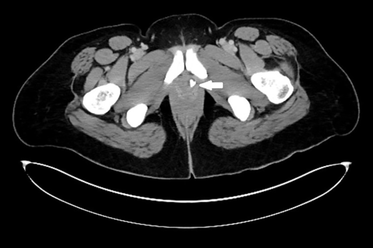
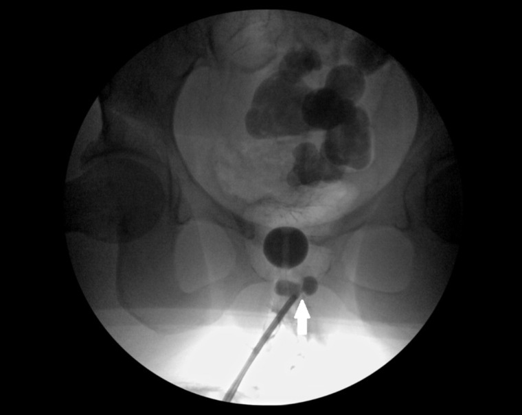
A 37-year-old African-American woman with a past medical history of hypertension, asthma, obesity, status post laparoscopic sleeve gastrectomy 4 years ago and status post laparoscopic chole-cystectomy was admitted to the emergency department on July 25, 2016 with complaints of epigastric pain, nausea, and vomiting for 7 days. The patient denied fevers, chills, and dysuria; however, she had a past history of recurrent urinary tract infections of unknown etiology. The patient was admitted to the hospital, and received symptomatic therapy with improvement of symptoms. Routine investigations were within the normal range. From a bariatric standpoint, a computed tomographic scan of the abdomen with oral and intravenous contrast was performed which showed 9 mm coarse calcifications within the left periurethral region indicative of a stone within a urethral diverticulum without any pelvic side wall adenopathy or free fluid (fig. 1). Subsequently, patient was consulted by urology services and advised for cystoscopic evaluation warranting removal of the stones and repair of the possible diverticulum. Patient was posted for the procedure as an outpatient. With general anesthesia patient was placed in lithotomy position and then prepped and draped as per the universal standard sterile surgical guidelines. A 17 F cystoscope under video monitoring was passed per urethra after female sounds were used to dilate the external meatus; digital palpation suggested stones but wasn't definitive. Urethra and bladder were inspected and diverticulum could not be visualized initially; however, upon withdrawing the scope the ureteral diverticulum was noted with fundus projecting anteriorly and a pantaloon type neck with intervening tissue which was later excised to view the wide opened neck using a pediatric cystoscope (fig. 2). Urethrogram was performed to document the exact location of the diverticulum. Graspers were used to remove the stones and the stones were sent for further laboratory evaluation and analysis. The bladder neck was occluded with a Foley balloon and a retrograde pullback urethrogram performed under fluoroscopic control the neck of the diverticulum and its connection to the urethra were delineated and excised. The scope was removed and a 20 F Foley placed. Patient was transferred to the Post Anesthesia Care Unit in a stable condition and discharged the same day to be later followed-up in the urology clinic.
Fig. 1.
CT scan of abdomen demonstrating impacted stones within urethral outpouching.
Fig. 2.
Intra-operative fluoroscopy showing the pantaloons type urethral diverticulum.
The analysis of the stones suggested a mixed variety with the first constituent being 70% ammonium urate and 30% magnesium ammonium urate (struvite) as the second constituent.
Discussion
From currently available information, risk factors for urethral diverticula include female sex, trauma, and surgical procedures of the vagina, urethra, and/or bladder. African American females are 3 times more likely to have a urethral diverticulum than their Caucasian counterparts [4].
Urethral diverticula are nearly always acquired, but their pathogenesis is not completely understood [4]. Most are formed in the posterior aspect of the lower urethra, i.e. between 3 o' clock and 9 o' clock where the Skene's glands are predominant. Thomas et al. [5] reported neo-plastic alterations in 6% of 90 pathology samples of urethral diverticulectomy. Most common histological types are adenocarcinoma and suggest a glandular origin.
Stasis of infected urine with deposition of salts and mucoid desquamation of the epithelial lining can cause formation of struvite and ammonium urate stones [6]; whereas migratory stones are usually composed of calcium oxalate and calcium phosphate stones and occur in up to 1.5–10% of urethral diverticula [7, 8].
Symptoms attributable to urethral diverticula include urinary incontinence (71.5%), recurrent urinary tract infection (57.1%), frequency (50%), urgency (35.7%), dysuria (35.7%), and dyspareunia (21.4%). Although a small proportion may remain asymptomatic [9].
Videocystometrography can confirm the presence of a diverticulum along with the calculi within but is no longer recommended [10]. MRI is the most accurate method of diagnosis. Ultrasound techniques or CT scan can also be used alternatively [9]. Urodynamic study may be obtained if the patient's symptoms include urinary incontinence.
Diagnosis of a urethral diverticulum should be considered in the advent of recurrent lower urinary tract infections. Asymptomatic diverticulum can be managed entirely by patient's choice; albeit surgical approach with extraction of stones is definitive as the passage of struvite stone is rare which might form a nidus for infection further resulting in obstructive symptoms as well as progression to malignancy. There is no consensus on the procedure of choice in the surgical community; however, transvaginal excision is commonly performed. In this case, we performed a transurethral excision of the diverticula without complications. Proper consideration should be given to avoid clinical recurrence, urethral stricture, urethrovaginal fistula and new onset urinary incontinence [8].
References
- 1.Martensson O, Duchek M. Translabial ultrasonography with pulsed colour-Doppler in the diagnosis of female urethral diverticula. Scand J Urol Nephrol. 1994;28:101–104. doi: 10.3109/00365599409180480. [DOI] [PubMed] [Google Scholar]
- 2.Greenwell TJ, Spilotros M. Urethral diverticula in women. Nat Rev Urol. 2015;12:671–680. doi: 10.1038/nrurol.2015.230. [DOI] [PubMed] [Google Scholar]
- 3.Thongprayoon C, Cheungpasitporn W, Vijayvargiya P, Anthanont P, Erickson SB. The riskof kidney stones following bariatric surgery: a systematic review and meta-analysis. Ren Fail. 2016;38:424–430. doi: 10.3109/0886022X.2015.1137186. [DOI] [PubMed] [Google Scholar]
- 4.Glowacki LS, Beecroft ML, Cook RJ, Pahl D, Churchill DN. The natural history of asymptomatic urolithiasis. J Urol. 1992;147:319–321. doi: 10.1016/s0022-5347(17)37225-7. [DOI] [PubMed] [Google Scholar]
- 5.Thomas AA, Rackley RR, Lee U, Goldman HB, Vasavada SP, Hansel DE. Urethral diverticula in 90 female patients: a study with emphasis on neoplastic alterations. J Urol. 2008;180:2463–2467. doi: 10.1016/j.juro.2008.08.040. [DOI] [PubMed] [Google Scholar]
- 6.Presman D, Rolnick D, Zumerchek J. Calculus formation within a diverticulum of the female urethra. J Urol. 1964;91:376–379. doi: 10.1016/S0022-5347(17)64133-8. [DOI] [PubMed] [Google Scholar]
- 7.Skyggebjerg KD. Female urethral diverticulum with calculus. Acta Obstet Gynecol Scand. 1986;65:797–798. doi: 10.3109/00016348609161505. [DOI] [PubMed] [Google Scholar]
- 8.Shim JS, Oh MM, Kang JI, Ahn ST, Moon DG, Lee JG. Calculi in a female urethral diverticulum. Int Neurourol J. 2011;15:55–57. doi: 10.5213/inj.2011.15.1.55. [DOI] [PMC free article] [PubMed] [Google Scholar]
- 9.Dai Y, Wang J, Shen H, Zhao RN, Li YZ. Diagnosis of female urethral diverticulum using transvaginal contrast-enhanced sonourethrography. Int Urogynecol J. 2013;24:1467–1471. doi: 10.1007/s00192-012-2036-0. [DOI] [PubMed] [Google Scholar]
- 10.Romanzi LJ, Groutz A, Blaivas JG. Urethral diverticulum in women: diverse presentations resulting in diagnostic delay and mismanagement. J Urol. 2000;164:428–433. [PubMed] [Google Scholar]


