Abstract
Carpal tunnel syndrome (CTS) is the most common entrapment neuropathy occurring in upper limbs. The etiology, however, has not been fully understood yet. Median nerve could be compressed by either increase of carpal tunnel pressure (CTP) or direct impingement when it is forced toward to carpal ligament especially in wrist flexion leading to CTS development. Thus, the increase of carpal tunnel pressure is considered an important role in CTS development. It has been identified that forces applied to the palm would affect the CTP. However, the quantitative relationship between palmar contact force and CTP is not known. The purpose of this study was to quantitatively evaluate the relationship between palmar contact force and CTP. Eight human cadaveric hands were used. The CTP was measured with a diagnostic catheter-based pressure transducer inserted into the carpal tunnel. A custom made device was used to apply forces to the palm for the desired CTP. Palmar contact forces corresponding to the determined CTP level were recorded respectively. The testing was repeated with different ranges of tension applied to the flexor digitorum superficialis tendon of the third finger. The tensions were constant at 50 g for the other flexor tendons and median nerve. The results showed that CTP increased linearly with the force applied to the palm. When CTP was 30 mmHg, mean values of the contact force to the palm was 293 g (SD: 15.2) including all tensions. These results would help to understand the effect of daily activities with hands on CTP.
Keywords: carpal tunnel syndrome, carpal tunnel pressure, palmar contact force
Introduction
Carpal tunnel syndrome (CTS) is the most common disease manifested as a peripheral nerve disorder occurring in upper limbs. Although numerous studies have been conducted to evaluate CTS, its etiology has not been fully elucidated. Many risk factors are involved in CTS initiation or progression, such as aging, gender, forceful and repetitive hand use, wrist joint deformity after trauma injury and endogenous diseases represented by metabolic disease including diabetes (English et al., 1995; Mackinnon and Novak, 1997; Masear et al., 1986; Silverstein et al., 1987; Zhou et al., 2016). Furthermore, carpal tunnel pressure (CTP) in patients with CTS is usually significantly higher than that in healthy people both at rest and during activities (Goss and Agee, 2010; Hamanaka et al., 1995; Ikeda et al., 2006; Lee et al., 2016). If CTP exceeds a certain level, usually 30 mmHg, symptoms such as discomfort, tingling and paresthesia would appear suggesting median nerve disorder (Coppieters et al., 2012; Goss and Agee, 2010; Prim et al., 2016; Seradge et al., 1995). According to Lundborg et al (Lundborg et al., 1982), sensory and motor block could occur if the compression is over 60 mmHg by 10 to 30 minutes. Also, the wrist posture during work related tasks such as typing contributes to the CTP elevation (Rempel et al., 2008). Hence, CTP is a key factor in evaluating CTS. It is also well known that repetitive operations with hands are related to the onset and prevalence of CTS (Amadio, 1985, 2001; English et al., 1995; Hadler, 1993; Latko et al., 1999; Mackinnon and Novak, 1997; Masear et al., 1986; Szabo, 1998; Zhao et al., 2007). When hands and fingers are used with forceful operation, the shape of carpal tunnel is distorted by external forces, which decreases the volume of carpal canal space and results in the increase of CTP (Keir et al., 1998b; Li et al., 2011; McGorry et al., 2014a). If these activities last for a short period or are intermittent, its effect would be negligible from the viewpoint of nerve endurance and reversibility. However, if this situation lasts for a long time or is continuously repeated, median nerve would be directly squeezed by the external force and also indirectly due to the built up of pressure within the carpal tunnel.
It has been reported that externally applied force on the palm affects CTP (Cobb et al., 1995; Gelberman et al., 1983; Lundborg et al., 1982; Szabo et al., 1983; Viikari-Juntura and Silverstein, 1999). However, the quantitative relationship between the force applied to the palm and CTP is not established. The elucidation of quantitative relationship between the magnitude of the force applied to the palm and the increase of CTP would lead to a better comprehension of the CTS pathophysiology focusing on CTP. Moreover, it would be beneficial to quantify the value of the external force applied to the palm corresponding to normal threshold of CTP. That may be also taken as an alert in daily life and at workplace to prevent or minimize the onset and progression of CTS. The purpose of this study was to investigate the quantitative relationship between the contact force applied to the palm and CTP and to evaluate the value of palmar contact force corresponding to normal threshold of CTP.
Material and Method
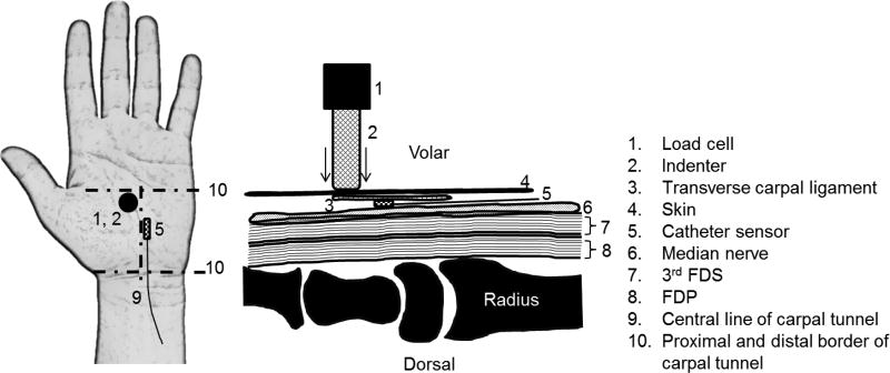
A total of 8 human cadaveric hands (4 males and 4 females, 5 right and 3 left hands, of age ranging from 20 to 80) were obtained. As an exclusion criteria, specimen with following past history was excluded ; prior wrist or hand disease, volar wrist surgery, known hand/wrist tumor or deformity, cervical radiculopathy, inflammatory arthritis, osteoarthritis in the wrist joint, flexor tendinitis, hemodialysis, peripheral nerve disease, metabolic disease, amyloidosis and major trauma to the ipsilateral wrist. Specimens were kept in a refrigerator for 24 hours to defrost before the test. In order to retain the carpal tunnel as intact as possible, minimum necessary procedure of inserting pressure sensor was done in this area. Longitudinal distance of carpal tunnel was defined as 4cm from first wrist flexion crease to distal direction. Soft tissues from the forearm area 6 cm proximal to the first wrist crease were removed and the flexor tendons and median nerve were exposed and identified. A 50 g weight was attached to individual tendons and median nerve except the third flexor digitorum superficialis (FDS) tendon. Multiple weights of 50, 200, 500, 1000 and 1500 g were applied to the third FDS to create different tendon tension conditions. Afterwards, an external fixator was installed between the distal radius epiphysis and the metacarpal diaphysis of the second finger to keep the wrist joint in a neutral supinated position. The specimen was mounted on a custom-made table and all fingers were kept in extended position with a Velcro band. A diagnostic catheter-based pressure transducer (Mikro-Cath, Millar Inc., Houston, TX) was inserted into carpal tunnel located at 2 cm proximal to first wrist crease and 0.5 cm ulnar side to the longitudinal middle line of the palm to measure real time CTP. An ultrasound system (Verasonics, Inc, Kirkland, WA 98053) was used to confirm the position of sensor before the measurement (Zhang et al., 2016; Zhang et al., 2017; Zhou et al., 2017).In order to increase CTP, a customized pressing apparatus was designed and used to press the palm, including a circular plate with 1.2 mm in diameter at the tip and was propelled by an air cylinder (Airpot, Norwalk, CT). This equipment was able to control the applied force by adjusting the amount of air flowing. A load cell (Transducer Techniques, Temecula, CA) was used to measure real time force applied. An analog-to-digital data acquisition board (DAQPad-6015, National Instruments, Austin, TX) was used to collect and integrate the signals with a sampling rate of 1000Hz. LabVIEW software (National Instruments, Austin, TX) was used as an interface to control and collect data. The pressed location on the palm was determined with reference to a previous study (Cobb et al., 1995). The center point pressed was 0.6 mm proximal to the distal line of the carpal tunnel and 0.6 mm radial to the longitudinal middle line of the palm (figure 1).
Figure 1.
Schematics of anatomy and pressing point
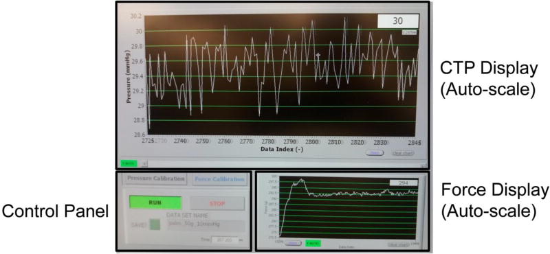
The real time measurements of CTP and palmar contact force were simultaneously monitored throughout the test (figure 2). The palmar contact force was gradually increased until the CTP reached the desired value. The pressing force was recorded when the CTP was stabilized. Then, the force was released and the CTP went back to zero. Following 2–3 minutes relaxation, the test was repeated for the next desired CTP value. The values of contact force were recorded when the desired value of CTP reached at 0, 10, 20, 30, 60, 90, 120, 150mmHg with varied tendon tensions at 50, 200, 500, 1000, and 1500 g respectively.
Figure 2.
Real time carpal tunnel pressure and palmar contact force. The upper / lower display screen shows the real time value of carpal tunnel pressure / palmar contact force to the palm respectively.
Statistical analysis
Two-way ANOVA of repeated measures were used to compare the palmar contact forces between tendon tensions as well as CTPs. Caution has been made to the assumption of sphericity. If the assumption was violated, results were corrected by using Greenhouse–Geisser estimates of sphericity. Pair-wise comparison was conducted through post-hoc Bonferroni test with α = 0.05. In addition, in order to build the quantitative relationship between palmar contact force and CTP, linear fitting was implemented to find out the equation and coefficient of determination was also calculated to determine how fitted they were.
Results
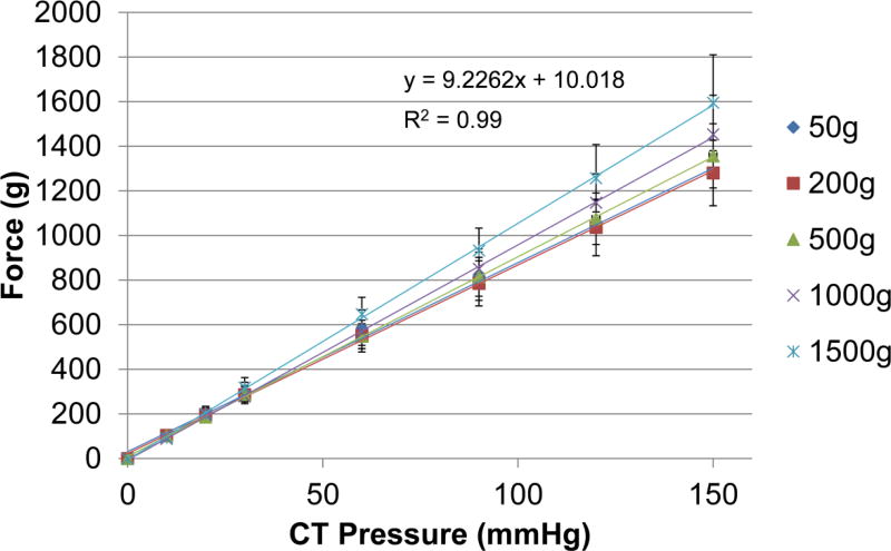
The relationship between palmar contact force and CTP under different tendon tensions is shown in Figure 3. From statistical analysis, CTP significantly increased with the increase of palmar contact force (p = .000, partial eta squared = .925). Statistically significant difference in palmar contact force was found between any two CTP conditions (all p < .05). In addition, no significant increase in palmar contact force was found when tendon tension increased (p = .312, partial eta squared = .149). Mean values of palmar contact force were 293 g (SD: 15.2) at CTP 30 mmHg, 578 g (SD: 41.1) at CTP 60 mmHg, 840 g (SD: 56.6) at CTP 90 mmHg, 1110 g (SD: 94.0) at CTP 120 mmHg and 1393 g (SD: 133.3) at CTP 150 mmHg when they were averaged in all tendon tensions. There was no interaction between CTP and tendon tension (p = .163, partial eta squared = .243). Linear regression model was applied on the results with the R2 values. The fitted equation was y = 8.4725x + 31.001 for 50 g tension, y = 8.4559x + 22.318 for 200 g, y = 8.9592x + 8.7638 for 500 g, y = 9.645x - 6.5562 for 1000 g and y = 10.598x - 5.4365 for 1500 g. The average fitted equation over all 5 tensions was calculated to be y = 9.2262x + 10.018. y represents CTP and×represents palmar contact force. All results similarly showed linear relationship between palmar contact force and CTP. All R2 values showed above 0.99 and standard error of the estimate of the average fitted line was 8.38.
Figure 3.
Relationship between palmar contact force and CTP combing graphs of all tensions. Equation of the average fitted line of all tensions, standard deviation bars and R2 value are shown.
Discussion
Our results showed that CTP linearly increased with contact force applied to the palm. We also found that tension applied to the 3rd FDS did not alter the relationship between palmar contact force and CTP. In addition it was observed that the higher tension applied, the steeper trend of the line tended. However, the increase of CTP by tensions was not significant. This indicates that CTP increase depends much more on the force applied to the palm than the longitudinal force itself from extrinsic muscle. As Lundborg et al described that externally induced compression force to the median nerve increased CTP (Lundborg et al., 1982), we could observe the same trend. Furthermore, we could obtain a proportional relationship between CTP and external force applied to the palm. This may also allude that we should be aware of the force affecting intra carpal tunnel would be greater than expected even though the palm does not feel strong, although anatomically the palm has some soft tissue layers such as skin, fat and connective tissue between the surface and transverse carpal ligament, and which function as a cushioning tissue against external forces and impacts. We believe that this result can be used as a clue to find out more details about the CTP change affected by external force. Moreover, this may lead to minimizing the risk exposed to the situation which adversely affects median nerve function and interrupts applicable works when the external force described above is applied.
Regardless of the magnitude of tendon tension, the mean values of palmar contact force were almost the same when CTP was 30 mmHg. The compressive force on the palm to reach CTP 30mmHg, which is considered as a threshold value for the onset and the aggravation of CTS, was indeed moderate (293.7 g). Although exceeding CTP 30 mmHg while working is very likely and unavoidable (Keir et al., 2007; Keir et al., 1998a; Rempel et al., 1998; Rempel and Gerr, 2008; Rempel et al., 2008; You et al., 2014), minimizing the duration of such activities should be advised. As a future work, in order to understand the relationship between hand daily activities and magnitude of the forces applied to the palm in more detail, an additional investigation would be necessary.
The normal CTP value at rest ranges from 0 to 38 mmHg, varying slightly depending on the reporters (Gelberman et al., 1981; Hamanaka et al., 1995; Luchetti et al., 1989). On the other hand, CTS patients potentially have higher pressure in the carpal tunnel as a base condition. The sustained high pressure in the carpal tunnel would cause functional damage to the median nerve (Lee et al., 2016; Luchetti et al., 1998; Murata et al., 2007; Okutsu et al., 1989; Silverstein et al., 1987; Viikari-Juntura and Silverstein, 1999). Thus, the higher the CTP, the more compressed the median nerve becomes. Our results suggest that CTS patients should take precautions when applying contact forces to the palm as this could potentially increase their high baseline CTP. Even in that sense, our results would be beneficial to prevent the disease onset and progression from the view point of preventive medicine.
Since we believe that the range of forces used in this experiment was generally not so large and was less invasive, this quantitative correlation derived from the present experiment would be also useful for in vivo studies to create a temporary CTP increased model to simulate carpal tunnel syndrome. Based on previous studies, ultrasound elastography has been successfully used to evaluate carpal tunnel pressure (Kubo et al., 2017; Wang et al., 2012). Before advancing into a stage of large clinical trial on CTS patients, these results could aid in the development and validation of noninvasive CTP measurements on healthy subjects.
There were several limitations in this study. Firstly, we verified this study using a cadaver model, however, it may not necessarily show the same result in vivo, especially in CTS patients who may have different pathophysiological conditions. Secondly, we tested the wrist only in neutral supinated position. Since it has been shown that wrist angles influence CTP (Rempel et al., 1998; Rempel et al., 2008; Werner et al., 1983; Werner and Andary, 2002), it is more natural to take into account for that. Future studies focusing on effects of various postures of the wrist would be expected. Thirdly, we only changed the tendon tension on 3rd FDS tendon, because it was the tendon closest to median nerve and the pressure sensor we inserted. Also, according to the partial eta squared values reflecting effect size, it would be stronger to include more specimens to reinforce the results in terms of tendon tensions. Finally, we only selected one small area on the palm to apply the force based on the previous publication that indicated the most sensitive contact point related to the CTP increased (Cobb et al., 1995). Usually various locations or the whole palm should be used and pressed when working with hand activities that leads to carpal tunnel elevation as evidenced in ergonomic literatures (McGorry et al., 2014b; Rempel et al., 1998; Rempel et al., 1994; Rempel et al., 2008). In a future study, a variety of areas on the palm will be pressed to reflect some practical activities associate with blue and white collared workers.
We verified the quantitative relationship between palmar contact force and carpal tunnel pressure, deriving a linear relationship between them. We also found that tendon tension did not alter this relationship. Mean value of palmar contact force corresponding to CTP 30 mmHg typically considered as a threshold pressure was 293 g (SD: 15.2). Furthermore, it would be necessary to change the area and increase the number of contact places on the palm to augment the consistency of evaluating the relationship between palmar contact force and daily hand activities.
Acknowledgments
This study was supported by a grant from National Institutes of Health/National Institute of Arthritis and Musculoskeletal and Skin Diseases (AR067421).
Footnotes
Publisher's Disclaimer: This is a PDF file of an unedited manuscript that has been accepted for publication. As a service to our customers we are providing this early version of the manuscript. The manuscript will undergo copyediting, typesetting, and review of the resulting proof before it is published in its final citable form. Please note that during the production process errors may be discovered which could affect the content, and all legal disclaimers that apply to the journal pertain.
Conflict of interest statement
There are no conflicts of interest for all authors.
References
- Amadio PC. Pyridoxine as an adjunct in the treatment of carpal tunnel syndrome. J Hand Surg Am. 1985;10:237–241. doi: 10.1016/s0363-5023(85)80112-x. [DOI] [PubMed] [Google Scholar]
- Amadio PC. Repetitive stress injury. J Bone Joint Surg Am. 2001;83-A:136–137. doi: 10.2106/00004623-200101000-00018. author reply 138–141. [DOI] [PubMed] [Google Scholar]
- Cobb TK, An KN, Cooney WP. Externally applied forces to the palm increase carpal tunnel pressure. J Hand Surg Am. 1995;20:181–185. doi: 10.1016/S0363-5023(05)80004-8. [DOI] [PubMed] [Google Scholar]
- Coppieters MW, Schmid AB, Kubler PA, Hodges PW. Description, reliability and validity of a novel method to measure carpal tunnel pressure in patients with carpal tunnel syndrome. Manual therapy. 2012;17:589–592. doi: 10.1016/j.math.2012.03.005. [DOI] [PubMed] [Google Scholar]
- English CJ, Maclaren WM, Court-Brown C, Hughes SP, Porter RW, Wallace WA, Graves RJ, Pethick AJ, Soutar CA. Relations between upper limb soft tissue disorders and repetitive movements at work. Am J Ind Med. 1995;27:75–90. doi: 10.1002/ajim.4700270108. [DOI] [PubMed] [Google Scholar]
- Gelberman RH, Hergenroeder PT, Hargens AR, Lundborg GN, Akeson WH. The carpal tunnel syndrome. A study of carpal canal pressures. J Bone Joint Surg Am. 1981;63:380–383. [PubMed] [Google Scholar]
- Gelberman RH, Szabo RM, Williamson RV, Hargens AR, Yaru NC, Minteer-Convery MA. Tissue pressure threshold for peripheral nerve viability. Clin Orthop Relat Res. 1983:285–291. [PubMed] [Google Scholar]
- Goss BC, Agee JM. Dynamics of intracarpal tunnel pressure in patients with carpal tunnel syndrome. J Hand Surg Am. 2010;35:197–206. doi: 10.1016/j.jhsa.2009.09.019. [DOI] [PubMed] [Google Scholar]
- Hadler NM. Arm pain in the work place. Bull Rheum Dis. 1993;42:6–8. [PubMed] [Google Scholar]
- Hamanaka I, Okutsu I, Shimizu K, Takatori Y, Ninomiya S. Evaluation of carpal canal pressure in carpal tunnel syndrome. J Hand Surg Am. 1995;20:848–854. doi: 10.1016/S0363-5023(05)80442-3. [DOI] [PubMed] [Google Scholar]
- Ikeda K, Osamura N, Tomita K. Segmental carpal canal pressure in patients with carpal tunnel syndrome. J Hand Surg Am. 2006;31:925–929. doi: 10.1016/j.jhsa.2006.03.004. [DOI] [PubMed] [Google Scholar]
- Keir PJ, Bach JM, Hudes M, Rempel DM. Guidelines for wrist posture based on carpal tunnel pressure thresholds. Hum Factors. 2007;49:88–99. doi: 10.1518/001872007779598127. [DOI] [PubMed] [Google Scholar]
- Keir PJ, Bach JM, Rempel DM. Effects of finger posture on carpal tunnel pressure during wrist motion. J Hand Surg Am. 1998a;23:1004–1009. doi: 10.1016/S0363-5023(98)80007-5. [DOI] [PubMed] [Google Scholar]
- Keir PJ, Bach JM, Rempel DM. Effects of finger posture on carpal tunnel pressure during wrist motion. The Journal of hand surgery. 1998b;23:1004–1009. doi: 10.1016/S0363-5023(98)80007-5. [DOI] [PubMed] [Google Scholar]
- Kubo K, Zhou B, Cheng YS, Yang TH, Qiang B, An KA, Moran SL, Amadio PC, Zhang X, Zhao C. Ultrasound Elastography for Carpal Tunnel Pressure Measurement: A Cadaveric Validation Study. Journal of Orthopaedic Research. 2017 doi: 10.1002/jor.23658. [DOI] [PMC free article] [PubMed] [Google Scholar]
- Latko WA, Armstrong TJ, Franzblau A, Ulin SS, Werner RA, Albers JW. Cross-sectional study of the relationship between repetitive work and the prevalence of upper limb musculoskeletal disorders. Am J Ind Med. 1999;36:248–259. doi: 10.1002/(sici)1097-0274(199908)36:2<248::aid-ajim4>3.0.co;2-q. [DOI] [PubMed] [Google Scholar]
- Lee HJ, Kim IS, Sung JH, Lee SW, Hong JT. Intraoperative dynamic pressure measurements in carpal tunnel syndrome: Correlations with clinical signs. Clin Neurol Neurosurg. 2016;140:33–37. doi: 10.1016/j.clineuro.2015.11.006. [DOI] [PubMed] [Google Scholar]
- Li ZM, Masters TL, Mondello TA. Area and shape changes of the carpal tunnel in response to tunnel pressure. Journal of Orthopaedic Research. 2011;29:1951–1956. doi: 10.1002/jor.21468. [DOI] [PMC free article] [PubMed] [Google Scholar]
- Luchetti R, Schoenhuber R, De Cicco G, Alfarano M, Deluca S, Landi A. Carpal-tunnel pressure. Acta Orthop Scand. 1989;60:397–399. doi: 10.3109/17453678909149305. [DOI] [PubMed] [Google Scholar]
- Luchetti R, Schoenhuber R, Nathan P. Correlation of segmental carpal tunnel pressures with changes in hand and wrist positions in patients with carpal tunnel syndrome and controls. J Hand Surg Br. 1998;23:598–602. doi: 10.1016/s0266-7681(98)80009-0. [DOI] [PubMed] [Google Scholar]
- Lundborg G, Gelberman RH, Minteer-Convery M, Lee YF, Hargens AR. Median nerve compression in the carpal tunnel--functional response to experimentally induced controlled pressure. The Journal of hand surgery. 1982;7:252–259. doi: 10.1016/s0363-5023(82)80175-5. [DOI] [PubMed] [Google Scholar]
- Mackinnon SE, Novak CB. Repetitive strain in the workplace. J Hand Surg Am. 1997;22:2–18. doi: 10.1016/S0363-5023(05)80174-1. [DOI] [PubMed] [Google Scholar]
- Masear VR, Hayes JM, Hyde AG. An industrial cause of carpal tunnel syndrome. J Hand Surg Am. 1986;11:222–227. doi: 10.1016/s0363-5023(86)80055-7. [DOI] [PubMed] [Google Scholar]
- McGorry RW, Fallentin N, Andersen JH, Keir PJ, Hansen TB, Pransky G, Lin JH. Effect of grip type, wrist motion, and resistance level on pressures within the carpal tunnel of normal wrists. Journal of Orthopaedic Research. 2014a;32:524–530. doi: 10.1002/jor.22571. [DOI] [PMC free article] [PubMed] [Google Scholar]
- McGorry RW, Fallentin N, Andersen JH, Keir PJ, Hansen TB, Pransky G, Lin JH. Effect of grip type, wrist motion, and resistance level on pressures within the carpal tunnel of normal wrists. J Orthop Res. 2014b;32:524–530. doi: 10.1002/jor.22571. [DOI] [PMC free article] [PubMed] [Google Scholar]
- Murata K, Yajima H, Maegawa N, Hattori K, Takakura Y. Investigation of segmental carpal tunnel pressure in patients with idiopathic carpal tunnel syndrome--is it necessary to release the distal aponeurotic portion of the flexor retinaculum in endoscopic carpal tunnel release surgery? Hand Surg. 2007;12:205–209. doi: 10.1142/S0218810407003559. [DOI] [PubMed] [Google Scholar]
- Okutsu I, Ninomiya S, Hamanaka I, Kuroshima N, Inanami H. Measurement of pressure in the carpal canal before and after endoscopic management of carpal tunnel syndrome. J Bone Joint Surg Am. 1989;71:679–683. [PubMed] [Google Scholar]
- Prim DA, Zhou B, Hartstone-Rose A, Uline MJ, Shazly T, Eberth JF. A mechanical argument for the differential performance of coronary artery grafts. Journal of the mechanical behavior of biomedical materials. 2016;54:93–105. doi: 10.1016/j.jmbbm.2015.09.017. [DOI] [PMC free article] [PubMed] [Google Scholar]
- Rempel D, Bach JM, Gordon L, So Y. Effects of forearm pronation/supination on carpal tunnel pressure. J Hand Surg Am. 1998;23:38–42. doi: 10.1016/S0363-5023(98)80086-5. [DOI] [PubMed] [Google Scholar]
- Rempel D, Gerr F. Intensive keyboard use and carpal tunnel syndrome: comment on the article by Atroshi et al. Arthritis Rheum. 2008;58:1882–1883. doi: 10.1002/art.23509. author reply 1883. [DOI] [PubMed] [Google Scholar]
- Rempel D, Manojlovic R, Levinsohn DG, Bloom T, Gordon L. The effect of wearing a flexible wrist splint on carpal tunnel pressure during repetitive hand activity. J Hand Surg Am. 1994;19:106–110. doi: 10.1016/0363-5023(94)90231-3. [DOI] [PubMed] [Google Scholar]
- Rempel DM, Keir PJ, Bach JM. Effect of wrist posture on carpal tunnel pressure while typing. J Orthop Res. 2008;26:1269–1273. doi: 10.1002/jor.20599. [DOI] [PMC free article] [PubMed] [Google Scholar]
- Seradge H, Jia YC, Owens W. In vivo measurement of carpal tunnel pressure in the functioning hand. J Hand Surg Am. 1995;20:855–859. doi: 10.1016/S0363-5023(05)80443-5. [DOI] [PubMed] [Google Scholar]
- Silverstein BA, Fine LJ, Armstrong TJ. Occupational factors and carpal tunnel syndrome. American journal of industrial medicine. 1987;11:343–358. doi: 10.1002/ajim.4700110310. [DOI] [PubMed] [Google Scholar]
- Szabo RM. Carpal tunnel syndrome as a repetitive motion disorder. Clin Orthop Relat Res. 1998:78–89. [PubMed] [Google Scholar]
- Szabo RM, Gelberman RH, Williamson RV, Hargens AR. Effects of increased systemic blood pressure on the tissue fluid pressure threshold of peripheral nerve. J Orthop Res. 1983;1:172–178. doi: 10.1002/jor.1100010208. [DOI] [PubMed] [Google Scholar]
- Viikari-Juntura E, Silverstein B. Role of physical load factors in carpal tunnel syndrome. Scand J Work Environ Health. 1999;25:163–185. doi: 10.5271/sjweh.423. [DOI] [PubMed] [Google Scholar]
- Wang Y, Qiang B, Zhang X, Greenleaf JF, An KN, Amadio PC, Zhao C. A non-invasive technique for estimating carpal tunnel pressure by measuring shear wave speed in tendon: a feasibility study. Journal of biomechanics. 2012;45:2927–2930. doi: 10.1016/j.jbiomech.2012.09.002. [DOI] [PubMed] [Google Scholar]
- Werner CO, Elmqvist D, Ohlin P. Pressure and nerve lesion in the carpal tunnel. Acta orthopaedica Scandinavica. 1983;54:312–316. doi: 10.3109/17453678308996576. [DOI] [PubMed] [Google Scholar]
- Werner RA, Andary M. Carpal tunnel syndrome: pathophysiology and clinical neurophysiology. Clin Neurophysiol. 2002;113:1373–1381. doi: 10.1016/s1388-2457(02)00169-4. [DOI] [PubMed] [Google Scholar]
- You D, Smith AH, Rempel D. Meta-analysis: association between wrist posture and carpal tunnel syndrome among workers. Saf Health Work. 2014;5:27–31. doi: 10.1016/j.shaw.2014.01.003. [DOI] [PMC free article] [PubMed] [Google Scholar]
- Zhang X, Meixner D, Kinnick R, Osborn T, Barthalmai B, Greenleaf J, Kalra S. Ultrasonics Symposium (IUS) IEEE International; 2016. Year Lung ultrasound surface wave elastography. [DOI] [PMC free article] [PubMed] [Google Scholar]
- Zhang X, Osborn T, Zhou B, Bartholmai B, Greenleaf JF, Kalra S. An ultrasound surface wave elastography technique for noninvasive measurement of surface lung tissue. The Journal of the Acoustical Society of America. 2017;141:3721–3721. [Google Scholar]
- Zhao C, Ettema AM, Osamura N, Berglund LJ, An KN, Amadio PC. Gliding characteristics between flexor tendons and surrounding tissues in the carpal tunnel: a biomechanical cadaver study. Journal of orthopaedic research : official publication of the Orthopaedic Research Society. 2007;25:185–190. doi: 10.1002/jor.20321. [DOI] [PubMed] [Google Scholar]
- Zhou B, Alshareef M, Prim D, Collins M, Kempner M, Hartstone-Rose A, Eberth JF, Rachev A, Shazly T. The perivascular environment along the vertebral artery governs segment-specific structural and mechanical properties. Acta Biomaterialia. 2016;45:286–295. doi: 10.1016/j.actbio.2016.09.004. [DOI] [PMC free article] [PubMed] [Google Scholar]
- Zhou B, Sit AJ, Zhang X. Noninvasive measurement of wave speed of porcine cornea in ex vivo porcine eyes for various intraocular pressures. Ultrasonics. 2017;81:86–92. doi: 10.1016/j.ultras.2017.06.008. [DOI] [PMC free article] [PubMed] [Google Scholar]


