Abstract
Influenza pandemic occurs when a new strain from other animal species overcomes the inter-species barriers and supports rapid human-to-human transmission. A critical prerequisite to this process is that hemagglutinin (HA) acquires a few key mutations to switch from avian receptors to human receptors. Previous studies suggest that H1 and H2/H3 HAs use different sets of mutations for the switch. This report shows that HA from the 1918 H1N1 pandemic virus (1918H1 HA) adopts the set of mutations used by H2/H3 HAs in receptor-preference switch when its 130-loop is made similar to those of H2/H3 HAs. Thus, the 130-loop appears to be the key determinant for the different mutations employed by pandemic H1 or H2/H3 HA. The correlation of the mutational routes and the 130-loop as unraveled in this study opens the door for efficient investigation of mutations required by other HA subtypes for inter-human airborne transmission.
Keywords: Human adaptation, inter-human airborne transmission, influenza pandemics, receptor binding, the 130-loop
Introduction
Influenza type A virus is responsible for all recorded influenza pandemics in human history. The 20th century has witnessed three major pandemics, the 1918 H1N1 “Spanish” pandemic, the 1957 H2N2 “Asian” pandemic and 1968 H3N2 “Hong Kong” pandemic. The most recent swine influenza pandemic in 2009, which resulted in about half-million human deaths globally (Dawood et al., 2012), clearly illustrated our extreme vulnerability to influenza pandemics.
Influenza pandemics occur when a new strain from other animal species acquires mutations to allow inter-species jump into humans and support efficient human-to-human airborne transmission (de Graaf and Fouchier, 2014; Donatelli et al., 2016; Imai and Kawaoka, 2012; Medina and Garcia-Sastre, 2011; Neumann and Kawaoka, 2015; Paulson and de Vries, 2013; Raman et al., 2014; Shi et al., 2014; Skehel and Wiley, 2000; Xiong et al., 2014). The minimal requirements for this jump are human-adaptation of polymerase subunit PB2 and hemagglutinin (HA) (Van Hoeven et al., 2009). Humanadapted HA exhibits preferential binding for NeuAcα(2,6)Gal-linked human receptors with concomitant loss of affinity for NeuAcα(2,3)Gal-linked avian receptors (Tumpey et al., 2007). Out of the 18 serologically different HA subtypes (H1~H16 in wild waterfowl, H17 and H18 in bats) of influenza type A viruses (Fouchier et al., 2005; Tong et al., 2012; Tong et al., 2013; WHO-MEMORANDUM, 1980), only H1, H2, and H3 HA subtypes have been established in humans. They are responsible for all seasonal influenza epidemics and recorded pandemics. These three HA subtypes utilize two distinct sets of “hallmark” mutations to adapt to the human host: Q226L and G228S for H2 and H3 HAs (Connor et al., 1994; Rogers et al., 1985; Rogers et al., 1983; Xu et al., 2010), and E190D and G225D for H1 HA (Glaser et al., 2005; Srinivasan et al., 2008; Stevens et al., 2006a; Tumpey et al., 2007). However, it was unknown why these different routes exist.
There is a rapidly growing list of other avian HA subtypes that cause severe morbidity and mortality among humans in recent years. These include the highly pathogenic H5N1 influenza since 1997 with a fatality rate of ~60%, H9N2 influenza since 1999, the first human-infecting H6N1 in June 2013, multiple severe H7N9 outbreaks in China since March 2013 with a mortality rate of ~40%, and several fatal cases of human infection by H10N8 virus since December 2013 (Centers for Disease and Prevention, 2013; Chen et al., 2014; Gao et al., 2013; Kadota, 2013; Shi et al., 2013; Wan et al., 2008; Watanabe et al., 2013; Webster et al., 2006; Wei et al., 2013; Yuan et al., 2013; Zhang et al., 2017; Zhang et al., 2015; Zhou et al., 2013). These human-infecting avian viruses pose severe threats for human pandemics; some may be only a few mutations away from a full human adaptation (Yen and Webster, 2009). Therefore, there is a pressing need for a better understanding of the mutations that may render these viruses airborne transmissibility among humans, thus contributing to influenza pandemics.
Numerous attempts have been made in the field to unravel mutations that may allow other avian HA subtypes to behave like pandemic HAs with preferential binding for human receptors. These include selecting H5N1 virus or hybrid strains for airborne transmissibility among ferrets or guinea pigs, or introducing hallmark mutations that are known to confer human adaptation on H1~H3 HAs into other HA subtypes (Chen et al., 2012; Herfst et al., 2012; Imai et al., 2012; Linster et al., 2014; Stevens et al., 2006b; Tharakaraman et al., 2013a; Tharakaraman et al., 2013b; Zhang et al., 2013b). Despite extensive studies, however, the determinants for avian-to-human receptor switch in other HA subtypes remained obscure.
In this report, through a series of mutational studies on pandemic A/South Carolina/1/1918(H1N1) (1918H1) HA followed by functional and structural characterizations, we have clearly demonstrated that the different lengths of the 130-loop in pandemic H1 HA vs. H2/H3 HAs are responsible for their distinct mutational routes in receptor-preference switch. These findings open the door for uncovering essential mutations required by other HA subtypes in human adaptation.
Results
Pandemic H1 and H2/H3 HAs have distinct 130-loops
Inspired by the observation from our earlier structural and functional study of H6 HAs that the sitting position of the Sia-1 moiety dictates the receptor-binding mode (Ni et al., 2015), we compared the complex structures of human receptor analogue LSTc (α(2,6)-linked lactoseries tetrasaccharide c) bound with HA proteins from the last four influenza pandemic viruses: 1918 H1N1 virus (1918H1 HA; PDB code: 2WRG), A/Singapore/1/1957(H2N2) virus (1957H2 HA; PDB code: 2WR7) (Liu et al., 2009), A/X-31/1968(H3N2) virus (1968H3 HA; PDB code: 2YPG) (Lin et al., 2012) and A/California/04/2009(H1N1) virus (2009H1 HA; PDB code: 3UBE) (Xu et al., 2012) (Fig.1a). Based on the view shown in Fig.1a, the receptor-binding site is formed by the 190-helix at the top, the 220-loop on the left, the 150-loop on upper right, and the 130- loop on lower right and at the base. We immediately noticed an interesting feature in the sitting positions of the Sia-1 moiety of LSTc: while it sits similarly in the receptorbinding sites of 1957H2 and 1968H3 HAs, the Sia-1 moiety sits ~0.6 Å higher and ~0.6 Å towards the 220-loop in 1918H1 HA (Fig.1a). This is also true for 2009H1 HA (Fig.1a). In addition, the 5’-acetylamino group of Sia-1 is located higher in 1957H2 HA than 1968H3 HA (Fig.1a). Most intriguingly, the differences in the Sia-1 sitting positions appeared to have a correlation with the length of the 130-loop in the region of HA1 131~134 (using 1968H3 HA numbering throughout) that constitutes the right edge of the receptor-binding site. Specifically, corresponding to the higher sitting position of the Sia-1 moiety in the receptor-binding site of 1918H1 and 2009H1 HAs than those in 1957H2 and 1968H3 HAs, and the higher sitting position of the 5’-acetylamino group of Sia-1 in 1957H2 HA than that in 1968H3 HA, the height of the 130-loops from high (closer to the 150-loop) to low (further away from the 150-loop) is in the order of 1918H1/2009H1, 1957H2 and 1968H3 HAs (Fig.1a, 1b). A structure-based sequence alignment of the 130-loop region revealed that the 130-loops in 1918H1 and 2009 H1 HAs are one-residue longer (with an additional residue of HA1 K133a) than those of 1957H2 and 1968H3 HAs (Fig.1c). However, given the same number of residues in 1957H2 and 1968H3 HAs in this loop, why does the 130-loop of 1968H3 HA adopt a substantially lower position than that of 1957H2 HA? Careful structural inspection revealed that distinct from 1957H2 HA, residues Q132 and N133 in the 130-loop of 1968H3 HA form an extensive interaction network with residues N152 and R255 located below the receptor-binding site, which may help maintain the 130-loop in 1968H3 HA at the lower position (Fig.1a).
Fig.1. Comparison of pandemic HA structures at the receptor-binding site.
a). Comparison of HA-LSTc complexes of HAs from pandemic viruses: 1918H1 (in yellow color), 2009H1 (in orange color), 1957H2 (in green color) and 1968H3 (in purple color). The Sia-1 moiety of LSTc in H1 HAs clearly sits higher and more toward the 220-loop than those in 1957H2 and 1968H3 HAs within the receptor-binding site (highlighted by two slim blue arrows). The large blue arrow highlights the different lengths of the 130-loop in these pandemic HAs. Extensive interactions of the 130-loop with residues below in 1968H3 HA may explain its lower position than that in 1957H2 HA despite the same length of the 130-loops. b). Another view of the structural comparison of HA-LSTc complexes from pandemic viruses. Same color code as in a). c). Structure-based sequence alignment of pandemic HAs in the 130-loop region. The 133a insertion (between 133 and 134 of H3 HA) found in 1918H1 and 2009H1 HAs, and the corresponding deletion in H2/H3 HAs are highlighted by a box.
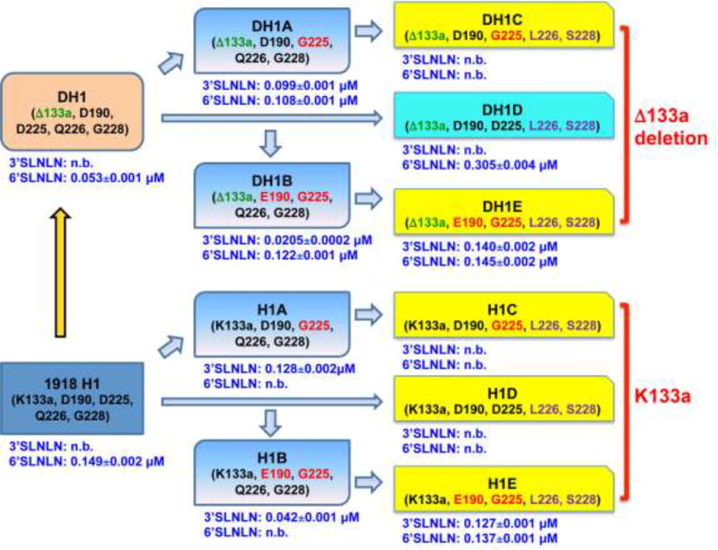
It is known that H1~H3 HA subtypes utilize two distinct sets of mutations to adapt to humans: E190D and G225D for H1 HA (Glaser et al., 2005; Srinivasan et al., 2008; Stevens et al., 2006a; Tumpey et al., 2007), and Q226L and G228S for H2 and H3 HAs (Connor et al., 1994; Rogers et al., 1985; Rogers et al., 1983; Xu et al., 2010). Thus, we asked whether the different lengths of the 130-loop are responsible for the existence of these two mutational routes, and the different sitting positions of the Sia-1 moiety in 1918/2009H1 HAs versus H2/H3 HAs? In order to directly answer this question, in what follows, we made a series of mutations on 1918H1 HA to see if a Δ133a deletion would convert it into H2/H3 HA-like. For comparison, we also made the same sets of mutations on 1918H1 HA without the Δ133a deletion (Fig.2).
Fig.2. Summary of all the 1918H1 HA mutants that were examined in this study.
Two series of mutants were made, one with Δ133a (DH1 and DH1A~E, Top row) and the other with K133a (H1 and H1A~E, bottom row). The mutations are highlighted in the following color code: Δ133a (green), E190 and G225 (red), L226 and S228 (purple). The apparent dissociation constant KD for 3’SLNLN or 6’SLNLN receptor analogues is shown for each mutant in blue. The abbreviation “n.b.” means that no binding was detected. See also Fig.S1 and Fig.S2.
Deletion of HA1 133a on the background of 1918H1 HA retained a preferential binding for human receptors
Using recombinant proteins expressed in and purified from insect cells and the label-free OCTET RED96 system (Pall ForteBio), we first determined the apparent dissociation constant (KD) for 1918H1 HA with human receptor analogue 6’SLNLN and avian receptor analogue 3’SLNLN, where LN represents lactosamine (Galβ1-4GlcNAc), 3’SLN and 6’SLN represent Neu5Acα(2,3) and Neu5Acα(2,6) linked to LN, respectively. 1918H1 HA had a strong binding (KD of 0.149 µM) for 6’SLNLN and no detectable binding for 3’SLNLN (Fig.2, Fig.S1a), agreeing with previously published studies that it has a predominant preference for human receptors (Glaser et al., 2005; Srinivasan et al., 2008; Stevens et al., 2004; Tumpey et al., 2007).
Comparing to H2/H3 HAs and seasonal human H1 HAs, 1918H1 HA has an additional residue, K133a. We made a single-residue deletion Δ133a (Mutant DH1 HA) to mimic the short 130-loop as found in H2/H3 HAs or in seasonal human H1 HAs. Mutant DH1 HA had a KD of 0.053 µM for 6’SLNLN, with no observable binding for 3’SLNLN (Fig.2, Fig.S2a). The retained preferential binding for human receptors by this mutant is in agreement with the fact that its composition (Δ133a, D190, D225, Q226 and G228) is frequently observed in HAs of human-adapted H1N1 viruses that cause seasonal influenza epidemics each year.
D225G and D190E in pandemic 1918H1 HA
1918H1 HA has the residue composition of K133a, D190, D225, Q226 and G228, while avian-origin H1 HAs have a residue composition of 133a, E190, G225, Q226 and G228. We made a single-residue substitution D225G or two-residue mutation D190E/D225G to mimic the residues found in avian H1 HAs. The mutation D225G on the 1918H1 HA background (resulting in Mutant H1A HA) enhanced the binding for 3’SLNLN to KD of 0.128 µM but meantime abolished binding to 6’SLNLN (Fig.2, Fig.S1b), thus turning it into an avian-like H1 HA. Additional mutation of D190E on H1A HA (giving rise to Mutant H1B HA) further strengthened the binding for 3’SLNLN (KD of 0.042 µM) (Fig.2, Fig.S1c). Agreeing with their preference for avian receptors, the combinations of E190/G225/Q226/G228 in H1B HA and D190/G225/Q226/G228 in H1A HA are of the highest prevalence among all known avian H1N1 isolates between 1976~2013 (94.2% and 2.6%, respectively).
D225G and D190E on the background of DH1 HA behaved differently from those on the 1918H1 HA background
The mutation D225G introduced into DH1 HA (yielding Mutant DH1A HA) resulted in dual binding, with KD of 0.099 µM for 3’SLNLN and KD of 0.108 µM for 6’SLNLN (Fig.2, Fig.S2b). Additional mutation D190E on this background (giving Mutant DH1B HA) further increased the affinity for 3’SLNLN (KD of 0.0205 µM) with an almost unchanged affinity for 6’SLNLN (KD of 0.122 µM) (Fig.2, Fig.S2c). Comparing to the mutations on the background of 1918H1 HA (H1A and H1B HAs) that bound solely to avian receptors (Fig.2, Fig.S1), the same mutations on the background of the Δ133a deletion (DH1A and DH1B HAs) clearly behaved differently (Fig.2, Fig.S2).
L226/S228 on the background of 1918H1 HA
The pair of mutations Q226L/G228S, as found in human-adapted H2/H3 HAs, was introduced into the 1918H1 HA background with different amino-acid compositions at 190/225 (1918H1 HA: D190/D225; H1A HA: D190/G225; or H1B HA: E190/G225). On the background of 1918H1 or H1A HAs, the resultant Mutant H1D HA or H1C HA, respectively, had no binding for any type of receptors (Fig.2, Fig.S1e, Fig.S1d). On the background of H1B HA, the resultant Mutant H1E HA had a reduced binding for 3’SLNLN (KD of 0.127 µM) but an enhanced binding for 6’SLNLN (KD of 0.137 µM) (Fig.2, Fig.S1f). This dual receptor binding of H1E HA is distinct from its parental Mutant H1B HA that preferentially binds to avian receptors.
Deletion of HA1 133a converts 1918H1 HA to H2/H3 HA-like
The pair of mutations Q226L/G228S on the background of DH1A HA (resulting in Mutant DH1C HA) exhibited affinity for neither type of receptors (Fig.2, Fig.S2d). On the background of DH1B HA, the Q226L/G228S pair resulted in Mutant DH1E HA with a dual receptor binding, KD of 0.140 µM for 3’SLNLN and 0.145 µM for 6’SLNLN (Fig.2, Fig.S2f). This dual binding property is reminiscent of its parental Mutant DH1B HA with which they share the same E190/G225 mutation pair. Most interestingly, the pair of mutations Q226L/G228S on Mutant DH1 HA (resulting in Mutant DH1D HA: Δ133a/D190/D225/L226/S228) had a KD of 0.305 µM for 6’SLNLN with no binding for 3’SLNLN (Fig.2, Fig.S2e). Thus, Mutant DH1D HA has acquired a quantitative preference for human receptors with concomitant loss of binding for avian receptors. This is in marked contrast to the no-binding activity exhibited by Mutant H1D HA with which the only difference is the Δ133a deletion (Fig.2, Fig.S1e). Clearly, the quantitative preference for human receptors by the DH1D HA mutant is the consequence of the Δ133a deletion. Therefore, the Δ133a deletion has apparently endorsed the conversion of 1918H1 HA to behave like H2/H3 HAs where the mutations Q226L and G228S together enabled the preferential binding for human receptors. These results confirmed our hypothesis that the length of the 130-loop is indeed responsible for the different mutational routes that 1918H1 HA or 1957H2 and 1968H3 HAs use to achieve receptor-preference switch. Out of the 6,251 unique HA sequences from human H3N2 strains isolated between 1976~2013, 82.7% have a residue composition of D190, D/N225, I/L/V226 and S228 that closely resemble that of Mutant DH1D HA.
Structural basis for the observed conversion of 1918H1 HA into H2/H3 HAs
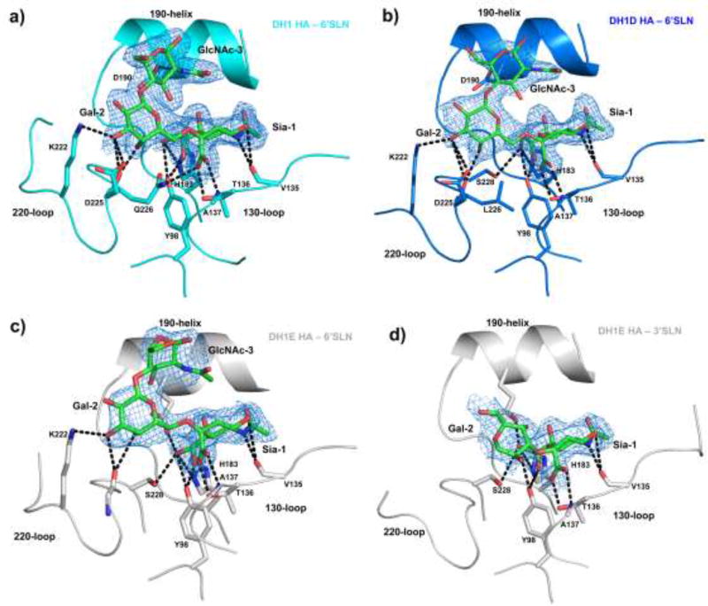
We determined the crystal structures of DH1, DH1D and DH1E HAs in complex with human receptor analogue 6’SLN (Fig.3, Table 1). DH1 HA formed a total of 13 hydrogen bonds with bound 6’SLN receptor (Fig.3a, Table 2), in consistent with its almost 10-fold stronger binding for human receptor analogue 6’SLNLN than 1918H1 HA (Fig.2). Of them, eight hydrogen bonds were found between the Sia-1 moiety and DH1 HA, at main-chain oxygen atom of V135 and nitrogen atom of A137, and at side-chain atoms of T136, Y98, H183 and Q226 (Table 2). Four hydrogen bonds were found between the Gal-2 moiety and DH1 HA at side-chain atoms of K222, and main-chain and side-chain atoms of D225. Furthermore, the GalNAc-3 moiety of 6’SLN contributed a hydrogen bond with D190 OD1 of DH1 HA (Table 2). DH1D HA had a total of 12 hydrogen bonds with the bound 6’SLN receptor analogue. Among them, eight hydrogen bonds were between the Sia-1 moiety of 6’SLN and DH1D HA (Fig.3b, Table 2). Comparing to DH1 HA, DH1D HA lost one hydrogen bond due to the mutation Q226L but gained a new hydrogen bond from the mutation G228S. Moreover, the mutation Q226L created a hydrophobic environment to favor the interaction with the hydrophobic C6 atom in the glycosidic linkage of 6’SLN. In addition, there were four hydrogen bonds between the Gal-2 moiety and DH1D HA at the side chains of K222 and D225 and the main-chain oxygen atom of D225 (Fig.3b, Table 2), as seen for DH1 HA. Compared to DH1D HA, DH1E HA had the same eight hydrogen bonds with the Sia-1 moiety, but with an additional hydrogen bond from the D190E mutation (Fig.3c, Table 2). Additionally, the Gal-2 moiety formed three hydrogen bonds with the side chain of K222 and main-chain oxygen atom of G225 (Fig.3c, Table 2).
Fig.3. Structures of 1918H1 HA mutants.
a). DH1 HA-6’SLN complex. DH1 HA structure is shown in cyan and 6’SLN is in green. b). DH1D HA-6’SLN complex. DH1D HA structure is shown in blue and 6’SLN is in green. c). DH1E HA-6’SLN complex. DH1E HA structure is shown in gray and 6’SLN is in green. d). DH1E HA-3’SLN complex. DH1E HA structure is shown in gray and 3’SLN is in green. For all panels, the composite omit 2mFo-DFc electron density map for the receptor is shown at 1σ. The hydrogen bonds detected by LigPlot+ are shown as black dashed lines.
Table 1.
Statistics of data collection and structural refinement of 1918H1 HA mutants
| DH1 HA-6’SLN | DH1D HA-6’SLN | DH1E HA-6’SLN | DH1E HA-3’SLN | |
|---|---|---|---|---|
|
| ||||
| Data collection statistics | ||||
|
| ||||
| Wavelength (Å) | 0.97872 | 0.97856 | 0.97856 | 0.97856 |
|
| ||||
| Resolution range (Å) | 48.74~2.15 (2.27~2.15) | 54.47~2.35 (2.48~2.35) | 45.69~2.45 (2.58~2.45) | 55.38~2.95 (3.11~2.95) |
|
| ||||
| Space group | P 1 21 1 | P 1 21 1 | P 1 21 1 | P 1 21 1 |
|
| ||||
| Unit cell | ||||
| a, b, c (Å) | 95.68, 81.82, 121.46 | 95.75, 80.26, 121.23 | 95.38, 81.37, 120.96 | 95.94, 81.05, 120.95 |
| α, β, γ (°) | 90, 90.79, 90 | 90, 91.35, 90 | 90, 95.45, 90 | 90, 91.13, 90 |
|
| ||||
| Total reflections | 436,178 (63,284) | 337,887 (48,689) | 302,929 (44,056) | 142,031 (20,833) |
|
| ||||
| Unique reflections | 102,164 (14,841) | 76,785 (11,130) | 68,049 (9,868) | 39,403 (5,731) |
|
| ||||
| Multiplicity | 4.3 (4.3) | 4.4 (4.4) | 4.5 (4.5) | 3.6 (3.6) |
|
| ||||
| Completeness (%) | 100.0 (100.0) | 100.0 (99.9) | 100.0 (100.0) | 100.0 (100.0) |
|
| ||||
| Mean I/sigma(I) | 9.4 (2.6) | 9.1 (2.6) | 10.3 (2.5) | 6.9 (2.0) |
|
| ||||
| Wilson B-factor (Å2) | 25.0 | 31.3 | 36.5 | 50.6 |
|
| ||||
| Rmerge | 0.139 (0.515) | 0.138 (0.510) | 0.142 (0.573) | 0.212 (0.679) |
|
| ||||
| Refinement statistics | ||||
|
| ||||
| Rwork | 0.182 (0.220) | 0.199 (0.226) | 0.199 (0.244) | 0.213 (0.285) |
|
| ||||
| Rfree | 0.208 (0.263) | 0.236 (0.262) | 0.228 (0.270) | 0.250 (0.340) |
|
| ||||
| Number of non-hydrogen atoms | 13,012 | 12,566 | 12,382 | 11,594 |
|
| ||||
| Macromolecules | 11,403 | 11,402 | 11,393 | 11,393 |
|
| ||||
| Ligands | 260 | 246 | 216 | 201 |
|
| ||||
| Water | 1,349 | 918 | 773 | — |
|
| ||||
| Protein residues | 1,472 | 1,472 | 1,470 | 1,469 |
|
| ||||
| RMS | ||||
|
| ||||
| Bonds (Å) | 0.006 | 0.005 | 0.007 | 0.005 |
|
| ||||
| Angles (°) | 1.20 | 1.15 | 1.24 | 1.17 |
|
| ||||
| Ramachandran | ||||
|
| ||||
| Favored (%) | 98.13 | 97.99 | 98.54 | 98.41 |
|
| ||||
| Allowed (%) | 1.87 | 1.94 | 1.39 | 1.59 |
|
| ||||
| Outliers (%) | 0.00 | 0.07 | 0.07 | 0.00 |
|
| ||||
| Clashscore | 2.81 | 3.52 | 3.04 | 2.56 |
|
| ||||
| Average B-factor (Å2) | 33.00 | 36.79 | 35.85 | 38.61 |
|
| ||||
| Macromolecules | 32.21 | 36.73 | 35.65 | 38.46 |
| Ligands | 42.65 | 46.31 | 47.87 | 46.73 |
|
| ||||
| Water | 37.89 | 35.02 | 35.41 | — |
|
| ||||
| PDB accession code | 5VMC | 5VMF | 5VMG | 5VMJ |
Statistics for the highest-resolution shell are shown in parentheses.
Table 2.
Hydrogen bonds between HA and bound receptor analogues
| Ligand | DH1 HA – 6’SLN | DH1D HA – 6’SLN | DH1E HA -6’SLN | DH1E HA-3’SLN | ||||
|---|---|---|---|---|---|---|---|---|
| Interaction pairs |
Distance (Å) |
Interaction pairs |
Distance (Å) |
Interaction pairs |
Distance (Å) |
Interaction pairs |
Distance (Å) |
|
| Sia-1 | O1A······T136 OG1 | 2.58 | O1A······T136 OG1 | 2.63 | O1A······T136 OG1 | 2.68 | O1A······T136 OG1 | 2.75 |
| O1A······Q22 6 NE2 | 3.00 | |||||||
| O1B······A137 N | 2.66 | O1B······A13 7 N | 2.62 | O1B······A13 7 N | 2.82 | O1B······A13 7 N | 2.88 | |
| O4······V135 O | 3.51 | O4······V135 O | 3.69 | O4······V135 O | 3.63 | O4······V135 O | 3.58 | |
| N5······V135 O | 2.99 | N5······V135 O | 2.83 | N5······V135 O | 2.92 | N5······V135 O | 2.82 | |
| O8······Y98 OH | 2.76 | O8······Y98 OH | 2.68 | O8······Y98 OH | 2.71 | O8······Y98 OH | 3.06 | |
| O9······Y98 OH | 2.97 | O9······Y98 OH | 2.61 | O9······Y98 OH | 2.86 | O9······Y98 OH | 3.24 | |
| O9······H183 NE2 | 3.14 | O9······H183 NE2 | 2.85 | O9······H183 NE2 | 3.05 | O9······H183 NE2 | 3.11 | |
| O9······E190 OE1 | 2.48 | O9······E190 OE1 | 2.38 | |||||
| O9······S228 OG | 3.11 | O9······S228 OG | 2.85 | O9······S228 OG | 3.12 | |||
| Gal-2 | O3······K222 NZ | 3.00 | O3······K222 NZ | 2.79 | O3······K222 NZ | 3.16 | ||
| O3······D225 OD2 | 2.81 | O3······D225 OD2 | 3.65 | |||||
| O3······D225 O | 3.28 | O3······D225 O | 3.70 | O3······G225 O | 3.07 | |||
| O4······D225 O | 2.56 | O4······D225 O | 2.75 | O4······G225 O | 3.34 | |||
| GalNAc-3 | N2······D190 OD1 | 2.97 | ||||||
Hydrogen atoms were calculated by LigPlot+.
We also determined the crystal structure of DH1E HA with avian receptor analogue 3’SLN. In agreement with DH1E HA’s dual-receptor binding, the DH1E HA – 3’SLN complex maintained the same set of hydrogen bonds with the Sia-1 moiety (Fig.3d, Table 2) as observed in DH1E HA – 6’SLN. However, different from the DH1E HA – 6’SLN complex, no hydrogen bonds were found between DH1E HA and the Gal-2 moiety of 3’SLN.
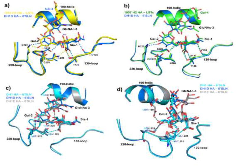
The structure of DH1D HA with 6’SLN was compared to 1918H1 HA in complex with human receptor analogue LSTc (PDB code: 2WRG) in Fig.4a and with 1957H2 HA in complex with LSTc (PDB code: 2WR7) (Liu et al., 2009) in Fig.4b. Clearly the short 130-loop resulted from the Δ133a deletion in DH1D HA made it to closely resemble 1957H2 HA, but was distinct from 1918H1 HA, in terms of the conformation of the bound receptor (Fig.4a, 4b). Specifically, the Sia-1 moiety of the bound 6’SLN in DH1D HA was lower and towards the 130-loop at the right by ~0.6 Å than that of LSTc in 1918H1 HA (Fig.4a), to be at a similar sitting position to that of LSTc in 1957H2 HA (Fig.4b). In addition, the structures of three 1918H1 HA deletion mutants (DH1, DH1D and DH1E HAs) in complex with 6’SLN were compared in Fig.4c, 4d. Clearly the Sia-1 moiety of 6’SLN interacted to all three HA mutants in a similar manner, despite the different residues at 190, 225, 226 and 228 (DH1: D190/D225/Q226/G228; DH1D: D190/D225/L226/S228; DH1: E190/G225/L226/S228). These structural comparisons further confirmed that the length of the 130-loop is a major determinant for the sitting position of the Sia-1 moiety within the receptor-binding site of HA (Fig.4a~d).
Fig.4. Comparison of HA structures.
a). Comparison of 1918H1 HA-LSTc complex (in yellow color) with DH1D HA-6’SLN (in blue color). The Sia-1 moiety of LSTc in 1918H1 HA clearly sits higher and more toward the 220-loop than that of 6’SLN in DH1D HA. b). Comparison of 1957H2 HA-LSTc complex (in green color) with DH1D HA-6’SLN (in blue color). The Sia-1 moiety of LSTc in 1957H2 HA clearly binds similarly to that of 6’SLN in DH1D HA. c, d). Structural comparison of three 1918H1 HA deletion mutants (DH1 HA in cyan, DH1D HA in blue and DH1E HA in gray) in binding to human receptor analogue 6’SLN in two different views. In all panels, hydrogen bonds between the bound receptor and HA are shown as dashed lines with the same color as each corresponding HA.
Furthermore, we found that from the Gal-2 moiety, the receptor analogue exited the receptor-binding site of DH1E HA using a path that was distinct from those of DH1 and DH1D HAs (Fig.4d). This difference was likely due to the contributions from residues 190 and 225 that were common in DH1 and DH1D HAs (D190 and D225), but different with DH1E HA (E190 and G225) (Fig.4d). The different exiting paths of human receptors from the receptor-binding site of the three mutant 1918H1 HAs (Fig.4d) resembled those observed among 1918H1 and 2009H1 (with D190 and D225), 1957H2 and 1968H3 HAs (with E190 and G225) (Fig.1b), highlighting the differential interactions contributed by D190/D225 vs. E190/G225 within the receptor-binding site.
Discussion
It is known that human-adapted H1 HAs harbor the mutations E190D and G225D, while human-adapted H2/H3 HAs carry the mutations Q226L and G228S (Connor et al., 1994; Glaser et al., 2005; Rogers et al., 1985; Rogers et al., 1983; Shi et al., 2014; Srinivasan et al., 2008; Stevens et al., 2006a; Tumpey et al., 2007; Xu et al., 2010). However, given the conserved nature of the receptor-binding site among different subtypes of influenza type A virus HA, there were longstanding puzzles in the field regarding why these different mutational routes were used by H1~H3 HAs and why the introduction of some of these “hallmark” mutations into other subtypes such as H5 and H7 HAs failed to confer human-like phenotypes.
Comparison of the crystal structures of pandemic H1~H3 HAs unraveled that 1957H2 and 1968H3 HAs have a short 130-loop, while the 130-loop of 1918H1 and 2009H1 HAs is one-residue longer (with an additional residue, K133a) (Fig.1a~c). This observation led to our extensive structural and functional investigation reported here, demonstrating that a single-residue deletion of K133a (Δ133a) converted 1918H1 HA into H2/H3 HA-like where the mutations Q226L and G228S bestowed a preferential binding for human receptors. In marked contrast, without the Δ133a mutation, the 1918H1 HA mutant harboring Q226L and G228S (Mutant H1D HA) had no detectable binding for any type of receptors (Fig.2, Fig.S1e). In addition, while the mutations D225G and D190E/D225G converted 1918H1 HA into an avian HA, they rendered DH1 HA almost equal binding affinities for human and avian receptors. Furthermore, structural comparison of human receptor analogues in complex with 1918H1 HA, 1957H2 HA, and DH1D HA (Fig.4a, 4b) as well as DH1, DH1D, DH1E HAs (Fig.4c) unraveled that the length of the 130-loop dictates the sitting position of bound receptors. Altogether, these receptor-binding and structural data support that the 130-loop is the key structural determinant that defines the different sitting positions of bound receptors within the receptor-binding site of pandemic H1 or H2/H3 HAs, thus leading to different mutational routes in receptor-preference switch.
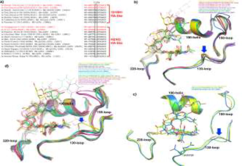
We next aligned all known HA structures in complex with human receptor analogue LSTc (each with at least three sugar moieties of LSTc visible in the structure) to allow a fair comparison (Fig.5). Structure-based sequence alignment revealed that these HAs have either a long 130-loop with an HA1 133a insertion as pandemic 1918H1 and 2009H1 HAs (1918H1 HA-like), or a short 130-loop without the insertion as H2/H3 HAs (H2/H3 HA-like) (Fig.5a). As the pandemic H1 HAs, avian and swine H1 HAs all have a long 130-loop, in agreement with the avian and swine origins of the 1918 and 2009 pandemic H1N1 viruses, respectively. In addition, HA proteins from H6 viruses and earlier H5N1 isolates (Ni et al., 2015; Zhang et al., 2013a) have a 1918H1 HA-like long 130-loop. Interestingly, the Sia-1 moieties of LSTc bound to this subgroup of HAs all are at a similar sitting position as that in 1918H1 HA, but different from that of 1957H2 HA (Fig.5b). The only exception is A/Taiwan/2/2013(H6N1) HA (PDB code: 5BR6) where the presence of HA1 N137 likely caused a right-shift of Sia-1 toward the 130-loop (Ni et al., 2015) (Fig.5c). On the other hand, HAs of seasonal human H1, all H2 and H3, H4, recent H5, H7, and H9 have a short 130-loop (Fig.5a). Strikingly, the Sia-1 moieties of LSTc bound to this subgroup of HAs all are at a similar sitting position as those in 1957H2/1968H3 HAs, but different from that of 1918H1 HA (Fig.5d). Therefore, these data clearly support the crucial role of the 130-loop in dictating the modes of receptor binding with these HAs (H1~H7, and H9), while at times residues within the receptor binding site, such as HA1 N137 in A/Taiwan/2/2013(H6N1) HA, can modulate such interactions.
Fig.5. Comparison of HA structures and sequences in the 130-loop.
a). Structure-based sequence alignment of HAs in all known HA-LSTc structures (with at least three sugar moieties visible) in the 130-loop region. 1918H1 and 2009H1 and H2/H3 HAs are highlighted in red. The HAs are classified into two subgroups, 1918H1 HA-like or H2/H3 HA-like, according to the length of the 130-loop. The 133a insertion (between 133 and 134 of H3 HA) found in 1918H1 HA-like subgroup and the corresponding deletion in H2/H3 HA-like subgroup are highlighted. b). Structural comparison of 1918H1 HA-like with 1957H2 HA. Due to the large number of structures and for easy visual comparison, the receptors in 1918H1 HA and 1957H2 HA are shown as sticks as references while all others as wires. c). Structural comparison of A/Taiwan/2/2013(H6N1) HA with 1918H1 and 1957H2 HAs. The residues at HA1 137 are shown. d). Structural comparison of H2/H3 HA-like with 1918H1 HA. Similarly, for easy visual comparison, the receptors in 1918H1 HA and 1957H2 HA are shown as sticks as references while all others as wires.
Indeed, a brief survey of published data in the literature strongly support the role of the 130-loop in receptor-preference switch, and exemplify how this novel knowledge may aid in investigation of mutations required by a given HA in human adaptation. For example, A/Indonesia/5/2005(H5N1) HA has a 1918H1 HA-like long 130-loop. The introduction of mutations Q226L/G228S, as used by H2/H3 HAs in human adaptation, together with two other mutations (H110Y/T160A), did not lead to a preferential binding for human receptors (with KD of 372 and 554 µM for human and avian receptor analogues, respectively) (Herfst et al., 2012; Zhang et al., 2013a). Similarly, for A/Vietnam/1194/2004(H5N1) HA with a long 130-loop, the mutant containing Q226L together with others (N158D/N224K/T318I) only had a 3-fold stronger binding for human receptors over avian receptors (Imai et al., 2012; Xiong et al., 2013). Moreover, for HA from A/chicken/Vietnam /NCVD-093/08(H5N1) (ckViet08) with a 1918H1 HA-like long 130-loop, mutations such as T160A/Q226L/G228S and T160A/N224K/Q226L only led to dual binding or no binding at all (Tharakaraman et al., 2013b; Zhu et al., 2015). However, when a deletion mutation ΔL133a (which rendered ckViet08 HA to be H2/H3 HA-like) was combined with N158D/N224K/Q226L (note, N158D and T160A have the same effect by removing a potential glycosylation site on the 150-loop), the resulting mutant started to bind much more strongly to human receptors than avian receptors (Tharakaraman et al., 2013b). On the other hand, while A/duck/Egypt/10185SS/2010(H5N1) HA with a natural Δ133a deletion and D158/A160/K224 preferentially recognized avian receptors, the introduction of Q226L bestowed an exclusive binding for human receptors (Tharakaraman et al., 2013b; Zhu et al., 2015). Moreover, recent studies on H7N9 HA revealed that, while the naturally occurring Q226L was not sufficient to allow preferential binding for human receptors, three additional mutations (V186G, K193T and G228S) conferred a complete switch for human receptors (de Vries et al., 2017). The last example came from H4 HA (Song et al., 2017). With an H2/H3 HA-like 130-loop, Song et al. convincingly demonstrated that the pair of mutations Q226L/G228S bestowed the avian-to-human receptor switch (Song et al., 2017).
Previous studies have suggested a role of 133a insertion or deletion, together with other features in the receptor-binding site, in affecting the receptor-binding properties of HAs (Koerner et al., 2012; Tharakaraman et al., 2013a; Tharakaraman et al., 2013b; Zhu et al., 2015). Here in this study, through a series of in-depth functional and structural characterization of 1918H1 HA and its various mutants, we demonstrated that the 130-loop is a key determinant for HA in receptor-preference switch. The results of our study help address the longstanding question in the field of why distinct mutational routes are used by H1 HA vs. H2/H3 HAs. However, these intriguing findings by no means imply that D190/D225 or L226/S228 are the “magic bullet”, which would endorse a complete preference for human receptors for 1918H1 HA-like or H2/H3 HA-like 130-loop, respectively, independent of the residue composition within the receptor-binding site for a given HA. On the contrary, it is our hope that the findings from this study will provide an invaluable tool to accelerate evaluation of contributions from other mutations in rendering some subtypes of avian HAs to be human-like. In a real application, the length of the 130-loop and more importantly the sitting position of the Sia-1 moiety of the receptor can help first establish whether a given HA would likely behave as 1918H1 HA or H2/H3 HAs. This will set the stage to much more effectively explore the contributions of other residues at or surrounding the receptor-binding site, including the features proposed by Sasisekharan and colleagues (Tharakaraman et al., 2013a; Tharakaraman et al., 2013b). HAs of seasonal human H1 viruses, despite a short 130-loop, are known to employ the E190D/G225D route. Future studies are required to investigate into this interesting phenomenon. Ultimately, a deeper understanding of the molecular mechanisms of receptor-preference switch by different subtypes of influenza A virus HA will help timely detection of newly emerging influenza strains of immediate pandemic threats.
Materials and Methods
Gene construct, expression, and purification of 1918H1 HA and its mutants
The gene encoding influenza A/South Carolina/1/1918 (H1N1) HA (1918H1 HA) was assembled from short DNA oligos (Stemmer et al., 1995) synthesized at Integrated DNA Technologies and cloned into a modified pFastBac1 vector (Invitrogen), where HA was preceded by a gp64 signal peptide and followed by a TEV cleavage site, a T4 fibritin foldon and a His6-tag as previously described (Ni et al., 2013; Ni et al., 2014). Various mutations in the receptor-binding site of 1918H1 HA were introduced by site-directed mutagenesis and verified by sequencing the full length of 1918H1 HA gene. The recombinant baculoviruses harboring 1918H1 HA or its various mutants were generated using the Bac-to-Bac system (Invitrogen). HA proteins were normally expressed in High Five cells at MOI~5.0 for 72 hours, but a shorter time of expression (48 hours) was used if an HA mutant was found to be unstable. The supernatant was harvested and dialyzed against Dialysis Buffer containing 10 mM Tris•HCl, pH 7.5, 50 mM NaCl. After dialysis, HA in the supernatant was captured by incubating with Ni-NTA agarose resin. After extensive wash, HA was treated by trypsin digestion that cleaved at the HA1–HA2 cleavage site and at the C-terminal TEV site. This trypsin digestion released HA proteins from the Ni-NTA agarose resin, and the released HA proteins were further purified sequentially by ion exchange (Source Q) and size exclusion (Superdex 200 10/300 GL) chromatography (GE Healthcare). The fractions containing HA trimers from size exclusion chromatography were pooled, concentrated and protein concentration was determined by Bradford assay and confirmed by SDS-PAGE.
Determination of apparent dissociation constants between HA and receptor analogues by Bio-Layer Interferometry
The biotinylated receptor analogues (3’SLNLN and 6’SLNLN) (Consortium of Functional Glycomics, cfg_rRequest_2413) were immobilized on streptavidin biosensor in 1×Kinetics Buffer (PBS with 0.1% BSA, 0.02% Tween-20, and 0.05% sodium azide, Pall ForteBio). To measure the binding affinity, association curves were recorded with HA protein diluted to a series of concentrations in 1× Kinetics Buffer followed by dissociation in 1×Kinetics Buffer only using OCTET RED96 (Pall ForteBio). The Kon (association rate) and Koff (dissociation rate) were obtained by fitting the curves using a 1:1 binding model. The apparent dissociation constant (KD) was calculated as KD = Koff/Kon.
Crystallization, data collection and structural refinement and analysis
Purified HA proteins (DH1, DH1D and DH1E HAs) were concentrated to about 10 mg/mL and exchanged to crystallization buffer containing 10 mM Hepes, pH 7.5. Initial crystallization screening was carried out using various commercial crystallization kits. Needle-cluster crystals were obtained in the condition of 0.10 M NaF and 14% PEG 3350. Concentrations of NaF and PEG3500 were optimized for each of these HA mutants to improve the size and quality of the crystals. Crystals were harvested in the crystal growth condition containing 15~20% glycerol. The receptor analogues at 5 mM (3’SLN and 6’SLN) were soaked into crystals for about 10 min. A longer incubation time or the use of longer receptor analogues (LSTa and LSTc) cracked the crystals and was thus avoided. X-ray diffraction data were collected at LS-CAT Sector 21, Advanced Photon Sources. The data were processed using programs in CCP4 (CCP4, 1994). The structures were solved by molecular replacement in PHENIX (Adams et al., 2010) using the 1918H1 HA structure as search model (PDB accession code: 2WRG) (Liu et al., 2009). The structural models underwent multiple rounds of refinement by PHENIX (Adams et al., 2010) and manual building using COOT (Emsley and Cowtan, 2004) until convergence. Hydrogen bonds were calculated by LigPlot+ with allowable bond length of 2.2~4.0 Å (Laskowski and Swindells, 2011). Structural alignment shown in Fig.1 and Fig.5 was done by superimposing the 190-helix (HA1 188~195), 130-loop (HA1 134~138) and 220-loop (HA1 220~229) in 1968H3 HA and the corresponding regions in all other HAs. All structural figures were made using PyMOL (DeLano, 2002).
Supplementary Material
Highlights of our work.
The 130-loop is a key determinant for mutations used by pandemic H1 or H2/H3 HAs.
The 130-loop dictates the modes of receptor binding in HA subtypes.
Receptor binding modes can help unravel HA mutations required in adapting to humans.
Acknowledgments
We gratefully thank Consortium of Functional Glycomics for the biotinylated receptor analogues used in this study (cfg_rRequest_2413). Q.W. thanks support from the National Institutes of Health (R01-AI067839, R01-GM116280) and the Welch Foundation (Q-1826). This research used resources of the Advanced Photon Source, a U.S. Department of Energy (DOE) Office of Science User Facility operated for the DOE Office of Science by Argonne National Laboratory under Contract No. DE-AC02-06CH11357. Use of the LS-CAT Sector 21 was supported by the Michigan Economic 20 Development Corporation and the Michigan Technology Tri-Corridor (Grant 085P1000817).
Footnotes
Publisher's Disclaimer: This is a PDF file of an unedited manuscript that has been accepted for publication. As a service to our customers we are providing this early version of the manuscript. The manuscript will undergo copyediting, typesetting, and review of the resulting proof before it is published in its final citable form. Please note that during the production process errors may be discovered which could affect the content, and all legal disclaimers that apply to the journal pertain.
Conflict of Interest
The authors declare no conflict of interest.
Author Contributions
Q.W. conceived and supervised the study and drafted the manuscript; F.N. and E.K. conducted the study. All authors reviewed and edited the manuscript and agreed on the final manuscript.
References
- Adams PD, Afonine PV, Bunkoczi G, Chen VB, Davis IW, Echols N, Headd JJ, Hung LW, Kapral GJ, Grosse-Kunstleve RW, et al. PHENIX: a comprehensive Python-based system for macromolecular structure solution. Acta Crystallogr D Biol Crystallogr. 2010;66:213–221. doi: 10.1107/S0907444909052925. [DOI] [PMC free article] [PubMed] [Google Scholar]
- CCP4. The CCP4 suite: Programs for protein crystallography. Acta Cryst. 1994;D50:760–763. doi: 10.1107/S0907444994003112. [DOI] [PubMed] [Google Scholar]
- Centers for Disease, C., and Prevention. Emergence of avian influenza A(H7N9) virus causing severe human illness - China, February–April 2013. MMWR Morbidity and mortality weekly report. 2013;62:366–371. [PMC free article] [PubMed] [Google Scholar]
- Chen H, Yuan H, Gao R, Zhang J, Wang D, Xiong Y, Fan G, Yang F, Li X, Zhou J, et al. Clinical and epidemiological characteristics of a fatal case of avian influenza A H10N8 virus infection: a descriptive study. Lancet. 2014;383:714–721. doi: 10.1016/S0140-6736(14)60111-2. [DOI] [PubMed] [Google Scholar]
- Chen LM, Blixt O, Stevens J, Lipatov AS, Davis CT, Collins BE, Cox NJ, Paulson JC, Donis RO. In vitro evolution of H5N1 avian influenza virus toward human-type receptor specificity. Virology. 2012;422:105–113. doi: 10.1016/j.virol.2011.10.006. [DOI] [PMC free article] [PubMed] [Google Scholar]
- Connor RJ, Kawaoka Y, Webster RG, Paulson JC. Receptor specificity in human, avian, and equine H2 and H3 influenza virus isolates. Virology. 1994;205:17–23. doi: 10.1006/viro.1994.1615. [DOI] [PubMed] [Google Scholar]
- Dawood FS, Iuliano AD, Reed C, Meltzer MI, Shay DK, Cheng PY, Bandaranayake D, Breiman RF, Brooks WA, Buchy P, et al. Estimated global mortality associated with the first 12 months of 2009 pandemic influenza A H1N1 virus circulation: a modelling study. The Lancet Infectious diseases. 2012;12:687–695. doi: 10.1016/S1473-3099(12)70121-4. [DOI] [PubMed] [Google Scholar]
- de Graaf M, Fouchier RA. Role of receptor binding specificity in influenza A virus transmission and pathogenesis. EMBO J. 2014;33:823–841. doi: 10.1002/embj.201387442. [DOI] [PMC free article] [PubMed] [Google Scholar]
- de Vries RP, Peng W, Grant OC, Thompson AJ, Zhu X, Bouwman KM, de la Pena ATT, van Breemen MJ, Ambepitiya Wickramasinghe IN, de Haan CAM, et al. Three mutations switch H7N9 influenza to human-type receptor specificity. PLoS Pathog. 2017;13:e1006390. doi: 10.1371/journal.ppat.1006390. [DOI] [PMC free article] [PubMed] [Google Scholar]
- DeLano WL. The PyMOL molecular graphics systems. Cambridge, MA: Schrodinger, LLC; 2002. [Google Scholar]
- Donatelli I, Castrucci MR, De Marco MA, Delogu M, Webster RG. Human-Animal Interface: The Case for Influenza Interspecies Transmission. Adv Exp Med Biol. 2016 doi: 10.1007/5584_2016_136. [DOI] [PubMed] [Google Scholar]
- Emsley P, Cowtan K. Coot: model-building tools for molecular graphics. Acta Crystallogr D Biol Crystallogr. 2004;60:2126–2132. doi: 10.1107/S0907444904019158. [DOI] [PubMed] [Google Scholar]
- Fouchier RA, Munster V, Wallensten A, Bestebroer TM, Herfst S, Smith D, Rimmelzwaan GF, Olsen B, Osterhaus AD. Characterization of a novel influenza A virus hemagglutinin subtype (H16) obtained from black-headed gulls. J Virol. 2005;79:2814–2822. doi: 10.1128/JVI.79.5.2814-2822.2005. [DOI] [PMC free article] [PubMed] [Google Scholar]
- Gao R, Cao B, Hu Y, Feng Z, Wang D, Hu W, Chen J, Jie Z, Qiu H, Xu K, et al. Human infection with a novel avian-origin influenza A (H7N9) virus. N Engl J Med. 2013;368:1888–1897. doi: 10.1056/NEJMoa1304459. [DOI] [PubMed] [Google Scholar]
- Glaser L, Stevens J, Zamarin D, Wilson IA, Garcia-Sastre A, Tumpey TM, Basler CF, Taubenberger JK, Palese P. A single amino acid substitution in 1918 influenza virus hemagglutinin changes receptor binding specificity. J Virol. 2005;79:11533–11536. doi: 10.1128/JVI.79.17.11533-11536.2005. [DOI] [PMC free article] [PubMed] [Google Scholar]
- Herfst S, Schrauwen EJ, Linster M, Chutinimitkul S, de Wit E, Munster VJ, Sorrell EM, Bestebroer TM, Burke DF, Smith DJ, et al. Airborne transmission of influenza A/H5N1 virus between ferrets. Science. 2012;336:1534–1541. doi: 10.1126/science.1213362. [DOI] [PMC free article] [PubMed] [Google Scholar]
- Imai M, Kawaoka Y. The role of receptor binding specificity in interspecies transmission of influenza viruses. Curr Opin Virol. 2012;2:160–167. doi: 10.1016/j.coviro.2012.03.003. [DOI] [PMC free article] [PubMed] [Google Scholar]
- Imai M, Watanabe T, Hatta M, Das SC, Ozawa M, Shinya K, Zhong G, Hanson A, Katsura H, Watanabe S, et al. Experimental adaptation of an influenza H5 HA confers respiratory droplet transmission to a reassortant H5 HA/H1N1 virus in ferrets. Nature. 2012;486:420–428. doi: 10.1038/nature10831. [DOI] [PMC free article] [PubMed] [Google Scholar]
- Kadota J. Emergence of a new strain of the avian influenza A (H7N9) virus. Respiratory investigation. 2013;51:49. doi: 10.1016/j.resinv.2013.05.001. [DOI] [PubMed] [Google Scholar]
- Koerner I, Matrosovich MN, Haller O, Staeheli P, Kochs G. Altered receptor specificity and fusion activity of the haemagglutinin contribute to high virulence of a mouse-adapted influenza A virus. J Gen Virol. 2012;93:970–979. doi: 10.1099/vir.0.035782-0. [DOI] [PubMed] [Google Scholar]
- Laskowski RA, Swindells MB. LigPlot+: multiple ligand-protein interaction diagrams for drug discovery. J Chem Inf Model. 2011;51:2778–2786. doi: 10.1021/ci200227u. [DOI] [PubMed] [Google Scholar]
- Lin YP, Xiong X, Wharton SA, Martin SR, Coombs PJ, Vachieri SG, Christodoulou E, Walker PA, Liu J, Skehel JJ, et al. Evolution of the receptor binding properties of the influenza A(H3N2) hemagglutinin. Proc Natl Acad Sci U S A. 2012;109:21474–21479. doi: 10.1073/pnas.1218841110. [DOI] [PMC free article] [PubMed] [Google Scholar]
- Linster M, van Boheemen S, de Graaf M, Schrauwen EJ, Lexmond P, Manz B, Bestebroer TM, Baumann J, van Riel D, Rimmelzwaan GF, et al. Identification, characterization, and natural selection of mutations driving airborne transmission of A/H5N1 virus. Cell. 2014;157:329–339. doi: 10.1016/j.cell.2014.02.040. [DOI] [PMC free article] [PubMed] [Google Scholar]
- Liu J, Stevens DJ, Haire LF, Walker PA, Coombs PJ, Russell RJ, Gamblin SJ, Skehel JJ. Structures of receptor complexes formed by hemagglutinins from the Asian Influenza pandemic of 1957. Proc Natl Acad Sci U S A. 2009;106:17175–17180. doi: 10.1073/pnas.0906849106. [DOI] [PMC free article] [PubMed] [Google Scholar]
- Medina RA, Garcia-Sastre A. Influenza A viruses: new research developments. Nature reviews Microbiology. 2011;9:590–603. doi: 10.1038/nrmicro2613. [DOI] [PMC free article] [PubMed] [Google Scholar]
- Neumann G, Kawaoka Y. Transmission of influenza A viruses. Virology. 2015;479–480:234–246. doi: 10.1016/j.virol.2015.03.009. [DOI] [PMC free article] [PubMed] [Google Scholar]
- Ni F, Kondrashkina E, Wang Q. Structural basis for the divergent evolution of influenza B virus hemagglutinin. Virology. 2013;446:112–122. doi: 10.1016/j.virol.2013.07.035. [DOI] [PMC free article] [PubMed] [Google Scholar]
- Ni F, Kondrashkina E, Wang Q. Structural and Functional Studies of Influenza Virus A/H6 Hemagglutinin. PLoS One. 2015;10:e0134576. doi: 10.1371/journal.pone.0134576. [DOI] [PMC free article] [PubMed] [Google Scholar]
- Ni F, Nnadi Mbawuike I, Kondrashkina E, Wang Q. The roles of hemagglutinin Phe-95 in receptor binding and pathogenicity of influenza B virus. Virology. 2014;450–451:71–83. doi: 10.1016/j.virol.2013.11.038. [DOI] [PMC free article] [PubMed] [Google Scholar]
- Paulson JC, de Vries RP. H5N1 receptor specificity as a factor in pandemic risk. Virus Res. 2013;178:99–113. doi: 10.1016/j.virusres.2013.02.015. [DOI] [PMC free article] [PubMed] [Google Scholar]
- Raman R, Tharakaraman K, Shriver Z, Jayaraman A, Sasisekharan V, Sasisekharan R. Glycan receptor specificity as a useful tool for characterization and surveillance of influenza A virus. Trends Microbiol. 2014;22:632–641. doi: 10.1016/j.tim.2014.07.002. [DOI] [PMC free article] [PubMed] [Google Scholar]
- Rogers GN, Daniels RS, Skehel JJ, Wiley DC, Wang XF, Higa HH, Paulson JC. Host-mediated selection of influenza virus receptor variants. Sialic acid-alpha 2,6Gal-specific clones of A/duck/Ukraine/1/63 revert to sialic acid-alpha 2,3Gal-specific wild type in ovo. J Biol Chem. 1985;260:7362–7367. [PubMed] [Google Scholar]
- Rogers GN, Paulson JC, Daniels RS, Skehel JJ, Wilson IA, Wiley DC. Single amino acid substitutions in influenza haemagglutinin change receptor binding specificity. Nature. 1983;304:76–78. doi: 10.1038/304076a0. [DOI] [PubMed] [Google Scholar]
- Shi W, Shi Y, Wu Y, Liu D, Gao GF. Origin and molecular characterization of the human-infecting H6N1 influenza virus in Taiwan. Protein Cell. 2013;4:846–853. doi: 10.1007/s13238-013-3083-0. [DOI] [PMC free article] [PubMed] [Google Scholar]
- Shi Y, Wu Y, Zhang W, Qi J, Gao GF. Enabling the 'host jump': structural determinants of receptor-binding specificity in influenza A viruses. Nature reviews Microbiology. 2014;12:822–831. doi: 10.1038/nrmicro3362. [DOI] [PubMed] [Google Scholar]
- Skehel JJ, Wiley DC. Receptor binding and membrane fusion in virus entry: the influenza hemagglutinin. Annu Rev Biochem. 2000;69:531–569. doi: 10.1146/annurev.biochem.69.1.531. [DOI] [PubMed] [Google Scholar]
- Song H, Qi J, Xiao H, Bi Y, Zhang W, Xu Y, Wang F, Shi Y, Gao GF. Avian-to-Human Receptor-Binding Adaptation by Influenza A Virus Hemagglutinin H4. Cell Rep. 2017;20:1201–1214. doi: 10.1016/j.celrep.2017.07.028. [DOI] [PubMed] [Google Scholar]
- Srinivasan A, Viswanathan K, Raman R, Chandrasekaran A, Raguram S, Tumpey TM, Sasisekharan V, Sasisekharan R. Quantitative biochemical rationale for differences in transmissibility of 1918 pandemic influenza A viruses. Proc Natl Acad Sci U S A. 2008;105:2800–2805. doi: 10.1073/pnas.0711963105. [DOI] [PMC free article] [PubMed] [Google Scholar]
- Stemmer WP, Crameri A, Ha KD, Brennan TM, Heyneker HL. Single-step assembly of a gene and entire plasmid from large numbers of oligodeoxyribonucleotides. Gene. 1995;164:49–53. doi: 10.1016/0378-1119(95)00511-4. [DOI] [PubMed] [Google Scholar]
- Stevens J, Blixt O, Glaser L, Taubenberger JK, Palese P, Paulson JC, Wilson IA. Glycan microarray analysis of the hemagglutinins from modern and pandemic influenza viruses reveals different receptor specificities. J Mol Biol. 2006a;355:1143–1155. doi: 10.1016/j.jmb.2005.11.002. [DOI] [PubMed] [Google Scholar]
- Stevens J, Blixt O, Tumpey TM, Taubenberger JK, Paulson JC, Wilson IA. Structure and receptor specificity of the hemagglutinin from an H5N1 influenza virus. Science. 2006b;312:404–410. doi: 10.1126/science.1124513. [DOI] [PubMed] [Google Scholar]
- Stevens J, Corper AL, Basler CF, Taubenberger JK, Palese P, Wilson IA. Structure of the uncleaved human H1 hemagglutinin from the extinct 1918 influenza virus. Science. 2004;303:1866–1870. doi: 10.1126/science.1093373. [DOI] [PubMed] [Google Scholar]
- Tharakaraman K, Jayaraman A, Raman R, Viswanathan K, Stebbins NW, Johnson D, Shriver Z, Sasisekharan V, Sasisekharan R. Glycan receptor binding of the influenza A virus H7N9 hemagglutinin. Cell. 2013a;153:1486–1493. doi: 10.1016/j.cell.2013.05.034. [DOI] [PMC free article] [PubMed] [Google Scholar]
- Tharakaraman K, Raman R, Viswanathan K, Stebbins NW, Jayaraman A, Krishnan A, Sasisekharan V, Sasisekharan R. Structural determinants for naturally evolving H5N1 hemagglutinin to switch its receptor specificity. Cell. 2013b;153:1475–1485. doi: 10.1016/j.cell.2013.05.035. [DOI] [PMC free article] [PubMed] [Google Scholar]
- Tong S, Li Y, Rivailler P, Conrardy C, Castillo DA, Chen LM, Recuenco S, Ellison JA, Davis CT, York IA, et al. A distinct lineage of influenza A virus from bats. Proc Natl Acad Sci U S A. 2012;109:4269–4274. doi: 10.1073/pnas.1116200109. [DOI] [PMC free article] [PubMed] [Google Scholar]
- Tong S, Zhu X, Li Y, Shi M, Zhang J, Bourgeois M, Yang H, Chen X, Recuenco S, Gomez J, et al. New world bats harbor diverse influenza A viruses. PLoS Pathog. 2013;9:e1003657. doi: 10.1371/journal.ppat.1003657. [DOI] [PMC free article] [PubMed] [Google Scholar]
- Tumpey TM, Maines TR, Van Hoeven N, Glaser L, Solorzano A, Pappas C, Cox NJ, Swayne DE, Palese P, Katz JM, et al. A two-amino acid change in the hemagglutinin of the 1918 influenza virus abolishes transmission. Science. 2007;315:655–659. doi: 10.1126/science.1136212. [DOI] [PubMed] [Google Scholar]
- Van Hoeven N, Pappas C, Belser JA, Maines TR, Zeng H, Garcia-Sastre A, Sasisekharan R, Katz JM, Tumpey TM. Human HA and polymerase subunit PB2 proteins confer transmission of an avian influenza virus through the air. Proc Natl Acad Sci U S A. 2009;106:3366–3371. doi: 10.1073/pnas.0813172106. [DOI] [PMC free article] [PubMed] [Google Scholar]
- Wan H, Sorrell EM, Song H, Hossain MJ, Ramirez-Nieto G, Monne I, Stevens J, Cattoli G, Capua I, Chen LM, et al. Replication and transmission of H9N2 influenza viruses in ferrets: evaluation of pandemic potential. PLoS One. 2008;3:e2923. doi: 10.1371/journal.pone.0002923. [DOI] [PMC free article] [PubMed] [Google Scholar]
- Watanabe T, Kiso M, Fukuyama S, Nakajima N, Imai M, Yamada S, Murakami S, Yamayoshi S, Iwatsuki-Horimoto K, Sakoda Y, et al. Characterization of H7N9 influenza A viruses isolated from humans. Nature. 2013;501:551–555. doi: 10.1038/nature12392. [DOI] [PMC free article] [PubMed] [Google Scholar]
- Webster RG, Peiris M, Chen H, Guan Y. H5N1 outbreaks and enzootic influenza. Emerg Infect Dis. 2006;12:3–8. doi: 10.3201/eid1201.051024. [DOI] [PMC free article] [PubMed] [Google Scholar]
- Wei SH, Yang JR, Wu HS, Chang MC, Lin JS, Lin CY, Liu YL, Lo YC, Yang CH, Chuang JH, et al. Human infection with avian influenza A H6N1 virus: an epidemiological analysis. The Lancet Respiratory medicine. 2013;1:771–778. doi: 10.1016/S2213-2600(13)70221-2. [DOI] [PMC free article] [PubMed] [Google Scholar]
- WHO-MEMORANDUM. A revision of the system of nomenclature for influenza viruses. Bull World Health Organ. 1980;58:585–591. [PMC free article] [PubMed] [Google Scholar]
- Xiong X, Coombs PJ, Martin SR, Liu J, Xiao H, McCauley JW, Locher K, Walker PA, Collins PJ, Kawaoka Y, et al. Receptor binding by a ferret-transmissible H5 avian influenza virus. Nature. 2013;497:392–396. doi: 10.1038/nature12144. [DOI] [PubMed] [Google Scholar]
- Xiong X, McCauley JW, Steinhauer DA. Receptor binding properties of the influenza virus hemagglutinin as a determinant of host range. Curr Top Microbiol Immunol. 2014;385:63–91. doi: 10.1007/82_2014_423. [DOI] [PubMed] [Google Scholar]
- Xu R, McBride R, Nycholat CM, Paulson JC, Wilson IA. Structural characterization of the hemagglutinin receptor specificity from the 2009 H1N1 influenza pandemic. J Virol. 2012;86:982–990. doi: 10.1128/JVI.06322-11. [DOI] [PMC free article] [PubMed] [Google Scholar]
- Xu R, McBride R, Paulson JC, Basler CF, Wilson IA. Structure, receptor binding, and antigenicity of influenza virus hemagglutinins from the 1957 H2N2 pandemic. J Virol. 2010;84:1715–1721. doi: 10.1128/JVI.02162-09. [DOI] [PMC free article] [PubMed] [Google Scholar]
- Yen HL, Webster RG. Pandemic influenza as a current threat. Curr Top Microbiol Immunol. 2009;333:3–24. doi: 10.1007/978-3-540-92165-3_1. [DOI] [PubMed] [Google Scholar]
- Yuan J, Zhang L, Kan X, Jiang L, Yang J, Guo Z, Ren Q. Origin and molecular characteristics of a novel 2013 avian influenza A(H6N1) virus causing human infection in Taiwan. Clin Infect Dis. 2013;57:1367–1368. doi: 10.1093/cid/cit479. [DOI] [PubMed] [Google Scholar]
- Zhang F, Bi Y, Wang J, Wong G, Shi W, Hu F, Yang Y, Yang L, Deng X, Jiang S, et al. Human infections with recently-emerging highly pathogenic H7N9 avian influenza virus in China. J Infect. 2017 doi: 10.1016/j.jinf.2017.04.001. [DOI] [PubMed] [Google Scholar]
- Zhang H, de Vries RP, Tzarum N, Zhu X, Yu W, McBride R, Paulson JC, Wilson IA. A human-infecting H10N8 influenza virus retains a strong preference for avian-type receptors. Cell Host Microbe. 2015;17:377–384. doi: 10.1016/j.chom.2015.02.006. [DOI] [PMC free article] [PubMed] [Google Scholar]
- Zhang W, Shi Y, Lu X, Shu Y, Qi J, Gao GF. An airborne transmissible avian influenza H5 hemagglutinin seen at the atomic level. Science. 2013a;340:1463–1467. doi: 10.1126/science.1236787. [DOI] [PubMed] [Google Scholar]
- Zhang Y, Zhang Q, Kong H, Jiang Y, Gao Y, Deng G, Shi J, Tian G, Liu L, Liu J, et al. H5N1 hybrid viruses bearing 2009/H1N1 virus genes transmit in guinea pigs by respiratory droplet. Science. 2013b;340:1459–1463. doi: 10.1126/science.1229455. [DOI] [PubMed] [Google Scholar]
- Zhou J, Wang D, Gao R, Zhao B, Song J, Qi X, Zhang Y, Shi Y, Yang L, Zhu W, et al. Biological features of novel avian influenza A (H7N9) virus. Nature. 2013;499:500–503. doi: 10.1038/nature12379. [DOI] [PubMed] [Google Scholar]
- Zhu X, Viswanathan K, Raman R, Yu W, Sasisekharan R, Wilson IA. Structural Basis for a Switch in Receptor Binding Specificity of Two H5N1 Hemagglutinin Mutants. Cell Rep. 2015;13:1683–1691. doi: 10.1016/j.celrep.2015.10.027. [DOI] [PMC free article] [PubMed] [Google Scholar]
Associated Data
This section collects any data citations, data availability statements, or supplementary materials included in this article.


