Highlights
-
•
This article represents a unique case of accessory/double nail of middle finger of hand.
-
•
The key purpose is to highlight this case as there is no such data in existing literature.
-
•
This surgical pathology can cause pain, discomfort and cosmetic concern.
-
•
Being a rare entity, this condition can cause diagnostic confusion.
-
•
Treatment is surgical excision.
Keywords: Double nail, Accessory nail, Nail unit resection, Histology
Abstract
Introduction
A rudimentary, accessory or double nail of the middle finger of hand is extremely rare. Although rudimentary, accessory or double nail of the toes only described four times before, in the literature. Most cases are incidentally detected and only few patients seek help because they have discomfort and pain or cosmetic reasons. Some have a positive family history, but most patients cannot give any information concerning heredity or any previous trauma.
Presentation
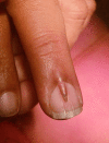
A 28 year old male presented with an abnormal growth of the nail of middle finger of left hand,which was hard and pointed. It was causing pain and discomfort to patient when touched. This growth was made of keratinized material and inseparable from nail bed, which showed a longitudinal depression corresponding to a slight protuberance of the cuticle of primary nail.
Discussion
Being an extremely rare entity, this situation can lead to difficulty in diagnosis, so its symptoms and pattern of occurrence should be thoroughly noted. Surgical excision is the treatment of choice for patient’s symptomatic relief and cosmetic concern. Nail unit resection procedure can easily be performed under local anesthesia.
Conclusion
Accessory nail of finger should be removed surgically due to pain and discomfort or cosmetic reasons and histopathology should be done afterwards.
1. Introduction
Accessory or double nails are sometimes congenital or even acquired condition. In some patients they cause symptoms while in others they are asymptomatic depending also upon the type of work or profession of the patient. Symptomatology also depends upon the type of foot-wear or hand-wear used.
In a cosmetically conscious world, it has become increasingly important for patients to get these removed or excised, even if no symptoms of pain and discomfort are present. There is no data available so far to prove that they usually get infected or any malignant changes if not treated for long time, although there might be some concerns regarding hygiene.
2. Presentation of case
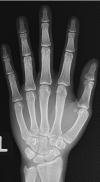
This particular patient, a 28 year old male presented at out-door department with abnormal growth of the nail of middle finger of left hand which was hard and pointed. It was causing pain and discomfort to the patient on manual work and patient also showed cosmetic concerns. There was no previous history of any trauma or infection or any surgery to this finger. There was no such family history. Patient was otherwise healthy with no other systemic or metabolic disease. Digital X-ray of left hand showed no unusual findings.
Patient planned to take an opinion from the specialist surgeon as this growth was causing him symptoms and psychological distress and his general practitioner was unable to make a definitive diagnosis. On examination, this growth was made of keratinized material and inseparable from nail bed which showed a longitudinal depression corresponding to a slight protuberance of the cuticle of primary nail. Provisionally diagnosed as a case of accessory nail and it was planned to get it removed surgically under local anesthesia.
Nail unit resection was carried out after exposing the nail bed and surgical wound closed with non absorbable fine suture material i.e. with prolene 4/0 after securing hemostasis. After its removal under local anesthesia, the specimen was sent for histopathology which confirmed it as nail tissue. Patient was examined after a week and stiches removed. No other complications reported by patient and his psychological feedback was satisfactory.
After identifying this case as a unique clinical entity, a group of surgeons with proper consent and permission of the patient, decided to put this as a case report study. All relevant information of patient and thorough literature review was done, all photographic evidence was obtained. Histological evidence achieved which confirmed this condition.
3. Discussion
A rudimentary, accessory or double nail of the middle finger of hand is extremely rare. Although rudimentary accessory or double nail of the toes only described four times before. Most cases are accidentally detected and only few patients seek help, because they have discomfort or pain. Some have a positive family history, but most patients cannot give any information concerning heredity. Clinically, the nail of this patient’s middle finger of left hand was abnormally wide and split or showed a longitudinal depression corresponding to a slight protuberance of the cuticle.
A hereditary dysplasia of the fifth toenail identical to our observations was first published in 1969 [1]. Surprisingly, descriptions of double nails are rare [[2], [3]], despite its relatively common occurrence. However, double little toenails were observed in dermatological practices in Germany, Norway, the Netherlands, Switzerland, Belgium, Portugal, and Thailand, as well as in immigrants from a variety of other countries including Africa (Benin). Thus, this is certainly not a special racial or ethnic feature.
Although rarely stated, the condition appears to be autosomal dominant and occurring in families, both in males and in females. As most patients did not even mention their double little toenails, it can be assumed that they are not embarrassed by them. Symptoms do not necessarily depend on the width of the nail but rather on the type of manual work or severity of accompanying anomalies in cases of toe accessory nails.
Treatment, if requested, is easy, by segmental excision of the accessory nail including the entire double nail with the matrix, proximal fold and hyponychium. The defect usually closed primarily with triple sutures after mobilization of the lateral aspect skin of the distal phalanx. Triple sutures allow 3-0 suture material to be used as much of the tension is exerted and distributed while tying a single knot.
Pathomechanisms and genetics are interesting aspects to be considered. Fully developed supernumerary digits develop nails. It is known that a bone core has to be present at the distal phalanx for the development of a nail anlage. Atelephalangia is associated with lack of nail formation or nail hypoplasia [[4], [5]], although total or complete anonychia also occurs without bone changes [6]. On the other hand, nail epithelium also has a special role in bone formation and digit regeneration. Nail cells were indeed found to influence underlying mesenchymal cells to regenerate digit bone [7].
In conclusion, a double or accessory nail of middle finger of hand is a rarely reported condition. It is autosomal dominant with variable expression [[8], [9], [10]]. Symptoms such as pain and discomfort are present. Histological examination of double nail shows short but otherwise normal nail or less well developed nail structure.
The treatment of choice is segmental excision of the entire accessory nail unit from nail bed, with mobilization of the lateral skin and primary suture.
4. Conclusion
Accessory nail of finger is an extremely rare pathology, but if encountered, it could be removed surgically due to symptoms or cosmetic reasons and histopathology should be done afterwards.
Conflict of interest
None.
Funding
None.
Ethical approval
Exempted from ethical approval by the ethical committee of our institution.
Consent
Confirmed.
Author contribution
Afzal Randhawa: ideated the study and drafted the article.
Shahid Hussain: revising it critically for important intellectual content.
Maliha Bilal: acquisition of data and intellectual content.
Naila Shabbir: substantial contribution to conception and design.
Adnan Qadir: acquisition of data and drafted the article.
Nawab Ali: final approval of the version to be submitted.
Guarantor
Javed Iqbal.
Footnotes
Supplementary data associated with this article can be found, in the online version, at https://doi.org/10.1016/j.ijscr.2018.02.006.
Appendix A. Supplementary data
The following are Supplementary data to this article:


References
- 1.Hundeiker M. Hereditäre Nageldysplasie der 5. Zehe. Hautarzt. 1969;20:281–282. (PubMed) [PubMed] [Google Scholar]
- 2.Haneke E. Therapie von Nagelfehlbildungen. In: Landthaler M., Hohenleutner U., editors. vol. 12. Blackwell Wissenschafts-Verlag; Berlin/Wien: 1997. pp. 180–187. (Fortschritte der operativen Dermatologie). [Google Scholar]
- 3.Chi C.C., Wang S.H. Inherited accessory nail of the fifth toe cured by surgical matricectomy. Dermatol. Surg. 2004;30:1177–1179. doi: 10.1111/j.1524-4725.2004.30351.x. (PubMed) [DOI] [PubMed] [Google Scholar]
- 4.Oldridge M., Temple I.K., Santos H.G., Gibbons R.J., Mustafa Z., Chapman K.E., Loughlin J., Wilkie A.O.M. Brachydactyly type B: linkage to chromosome 9q22 and evidence for genetic heterogeneity. Am. J. Hum. Genet. 1999;64:578–585. doi: 10.1086/302255. (PMC free article [PubMed]) [DOI] [PMC free article] [PubMed] [Google Scholar]
- 5.Schwabe G.C., Tinschert S., Buschow C., Meinecke P., Wolff G., Gillessen-Kaesbach G., Oldridge M., Wilkie A.O.M., Kömec R., Mundlos S. Distinct mutations in the receptor tyrosine kinase gene ROR2 cause, brachydactyly type B. Am. J. Hum. Genet. 2000;67:822–831. doi: 10.1086/303084. (PMC free article [PubMed]) [DOI] [PMC free article] [PubMed] [Google Scholar]
- 6.Balta I., Kalkan G. A case report on autosomal recessive total congenital anonychia. Pediatr. Dermatol. 2013;30:e268–e269. doi: 10.1111/pde.12143. (PubMed) [DOI] [PubMed] [Google Scholar]
- 7.Takeo M., Chou W.C., Sun Q., Lee W., Rabbani P., Loomis C., Taketo M.M., Ito M. Wnt activation in nail epithelium couples nail growth to digit regeneration. Nature. 2013;499:228–232. doi: 10.1038/nature12214. (PMC free article [PubMed]) [DOI] [PMC free article] [PubMed] [Google Scholar]
- 8.Castilla E.E., da Graca Dutra M., Lugarinho da Fonseca R., Paz J.E. Hand and foot postaxial polydactyly: two different traits. Am. J. Med. Genet. 1997;73:48–54. doi: 10.1002/(sici)1096-8628(19971128)73:1<48::aid-ajmg10>3.0.co;2-r. (PubMed) [DOI] [PubMed] [Google Scholar]
- 9.Iso R. Congenital nail defects of the index finger and reconstructive surgery. Orthop. Surg. (Tokyo) 1969;20:1383–1384. (PubMed) [PubMed] [Google Scholar]
- 10.Haneke E., Kienlein-Kletschka B. Congenital onychodysplasia: iso-Kikuchi syndrome. Hautarzt. 1984;35:468–471. (PubMed) [PubMed] [Google Scholar]


