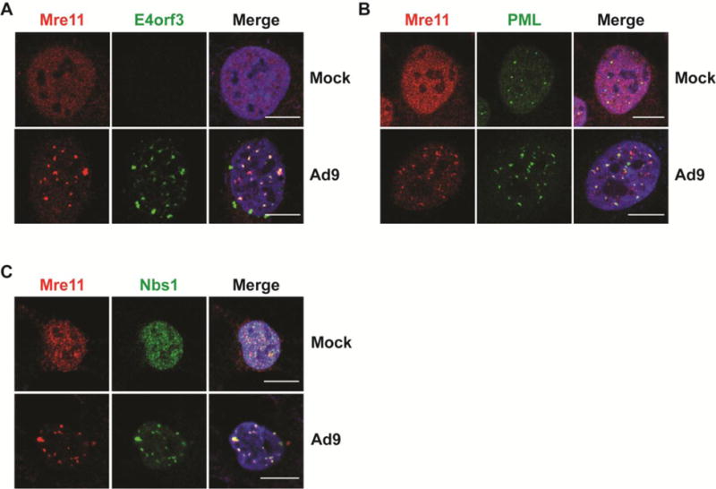
Figure 6. MRN colocalizes with E4orf3 and PML during Ad9 infection.

(A) Representative immunofluorescence results from Ad9-infected U2OS cells (24 hpi) showing Mre11 (red) and Ad9-E4orf3 (A), PML (B), or Nbs1 (C) in green. Merged images include DAPI in blue. Scale bar = 10 μm.
