Abstract
Diagnosis and treatment of ectopic cesarean scar pregnancy has become a challenge for contemporary obstetrics. With an increase in the number of pregnancies concluded with a cesarean section and with the development of transvaginal ultrasonography, the frequency of cesarean scar pregnancy diagnoses has increased as well. The aim of the study is to evaluate various diagnostic methods (ultrasonography in particular) and analyze effective treatment methods for cesarean scar pregnancy. An ultrasound scan, Doppler examination and magnetic resonance imaging are all useful in early detection of asymptomatic cesarean scar pregnancy, thus enabling effective treatment and preservation of fertility. Dilatation and curettage is not recommended as it carries significant risk of bleeding and very high risk of hysterectomy and fertility loss. Systemic methotrexate treatment should not be applied on the routine basis due to its low efficacy, high risk of fertility loss and adverse effects. Local methotrexate therapy (under ultrasound or hysteroscopy guidance) should be considered a perfect management method as it offers fertility preservation in asymptomatic pregnant patients without concomitant hemodynamic disorders. Synchronous usage of several treatment methods is an effective way to manage cesarean scar pregnancy. The combination of local methotrexate with simultaneous aspiration of gestational tissues under ultrasound or hysteroscopy guidance seems optimal. Subsequently, the remaining gestational tissues can be removed hysteroscopically in combination with vascular coagulation at the implantation site. In more advanced cases, local methotrexate treatment should be considered followed by laparoscopic or laparotomic wedge resection with subsequent surgical correction of the cesarean section scar.
Keywords: cesarean scar pregnancy, ectopic pregnancy, ultrasonography, transvaginal transducer, methotrexate
Introduction
Diagnosis and treatment of cesarean scar pregnancy (CSP) has become a challenge for contemporary obstetrics. With a significant increase in the percentage of pregnancies concluded with a cesarean section and with the development of transvaginal (TV) ultrasonography (US), the frequency of CSP diagnoses has increased as well. Cesarean scar pregnancy is a type of ectopic pregnancy where the fertilized egg is implanted in the muscle or fibrous tissue of the scar after a previous cesarean section(1).
Aim
The aim of the study is to evaluate various diagnostic tools (US in particular) and to analyze methods of effective CSP treatment.
Epidemiology
The overall prevalence of ectopic pregnancy is approximately 2% relative to all labors. In most cases (about 97%), an ectopic pregnancy is located in a fallopian tube(2). The frequency of cesarean scar pregnancy is reported to be 1:1,800 to 1:2,226 (0.05–0.04%) of all pregnancies. In women after a cesarean section, the frequency of CSP is approximately 0.15%, which constitutes 6.1% of all ectopic pregnancies in patients after at least one cesarean operation(3). According to Rotas et al., over a half of CSP cases (52%) were found in patients after one cesarean section only; the mean gestational age was 7.5 ± 2.5 weeks and the prevailing sign was vaginal bleeding, with no pain(4). It has also been proven that fertilization in vitro and embryo transfer (IVF-ET) in women with a history of cesarean section increases the risk of CSP(5). The first case of CSP was reported in 1978 by Larsen and Solomon. It was a 6-week pregnancy after uterine curettage due to a suspicion of inevitable abortion with persisting heavy bleeding and intense abdominal pain. Laparotomy revealed the presence of gestational tissues in the recess of a cesarean section scar(6). In practice, early detection of this phenomenon has become possible as ultrasound scans and transvaginal probes have been introduced to obstetric practice and imaging quality has improved(7,8). Earlier, CSP in the first trimester was treated as spontaneous abortion, while in the second and third trimesters it led to hemorrhage or uterine rupture. Up to the year 2002, only 19 CSP cases had been reported in English literature(9). With the dramatic increase in the frequency of cesarean sections and perfection of ultrasound imaging quality, the number of CSP diagnoses has increased significantly.
Diagnosis
Cesarean scar pregnancy can be a potential threat to the pregnant patient’s life since it can lead to uterine rupture, hemorrhage, coagulation disorders and even death of the pregnant patient if a correct diagnosis and sufficiently aggressive treatment are not established and implemented early enough. This threat is confirmed by two cases of CSP diagnosed in week 6 and 16 of gestation in the Department of Gynecology of the Regional Polyclinical Hospital in Płock, Poland. Table 1 presents the course, treatment and prognosis in these two cases.
Tab. 1.
Time of CSP diagnosis and its impact on treatment effects
| Gestational age | 6 weeks | 16 weeks |
|---|---|---|
| Hospitalization time | 3 days | 10 days |
| Transfusion of packed red blood cells | 0 mL | 1,200 mL |
| Transfusion of fresh frozen plasma | 0 mL | 600 mL |
| Antibiotic therapy | 3 days | 10 days |
| Fertility loss (uterine amputation) | no | yes |
| Direct risk of death | no | yes |
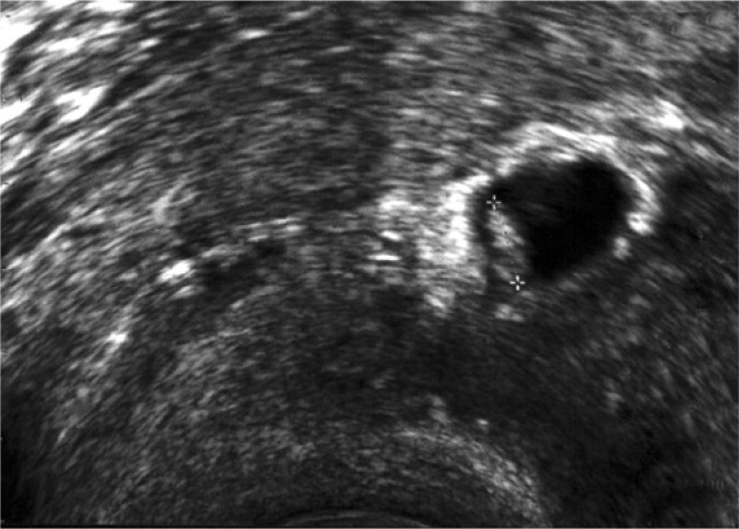
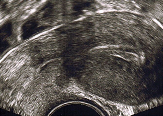
By contrast with the detection of ectopic pregnancy in a cesarean section scar in the 16th week of gestation, early diagnosis of this pathology (at approximately week 6 of gestation) (Fig. 1) enabled non-surgical treatment, resulted in short-term hospitalization and offered fertility preservation (Fig. 2, Tab. 1).
Fig. 1.
TV US. Longitudinal section of the uterus. Cesarean scar pregnancy (week 6 day 1) (authors’ own material)
Fig. 2.
TV US. Longitudinal section of the uterus and cervix 5 months after effective cesarean scar pregnancy treatment (authors’ own material)
A diagnosis of cesarean scar pregnancy based on symptoms and pelvic examination alone is difficult as CSP is asymptomatic in its initial phases. Later, signs of this type of pregnancy are frequently non-specific. Vaginal bleeding and abdominal pain are also often present in other obstetric conditions. Thanks to the progress of transvaginal US imaging, establishing a proper diagnosis has become much easier(8,10). The first detection of a 7-week cesarean scar pregnancy on transvaginal US occurred in 1990(11). Studies indicate that CSP can be detected between day 33 and 94 of pregnancy(12). Even at present, despite advanced technology, misdiagnoses are common. According to Zhang et al., early CSP is frequently misdiagnosed as normal intrauterine pregnancy, missed abortion, inevitable abortion, gestational trophoblastic disease or cervical pregnancy(13). As pregnancy develops, the diagnosis of CSP on US becomes more difficult. In early pregnancy, US enables correct CSP diagnosis and implementation of minimally invasive effective treatment. However, in advanced pregnancy (above week 12), US (usually transabdominal) produces images that are difficult to interpret, and final diagnosis is possible only during surgery.
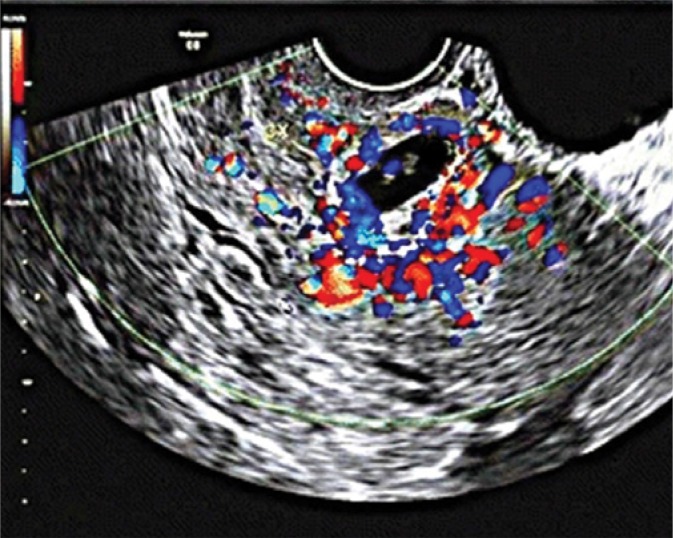
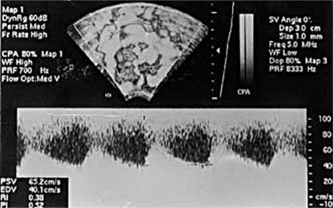
Publications suggest that early pregnancy US in patients after a cesarean section should be conducted with 5–12 MHz probes as they are optimal for CSP detection. Doppler examination is significant for establishing a correct diagnosis because it shows qualitative and quantitative vascularization around the cesarean section scar (Fig. 3). In CSP, color Doppler imaging shows functional placental vascularization caused by increased blood flow with peak systolic velocity (PSV) greater than 20 cm/s and pulsatililty index (PI) lower than 1 (Fig. 4)(5,14). In the case of systemic methotrexate (MTX) therapy, serial color Doppler scans are useful for CSP monitoring and seem to correlate well with serum β subunit of human chorionic gonadotropin (β-hCG). In many cases, turbulent high-velocity and low-impedance flow remains relatively unchanged until β-hCG returns to normal values. Patients with these features of flow should be informed about the risk of uterine rupture and internal hemorrhage due to high-velocity flow even if β-hCG decreases gradually during observation. Moreover, high peak systolic velocity should be a clear warning sign not to perform dilatation and curettage (D&C) for CSP termination due to a risk of heavy bleeding from such an area(14,15).
Fig. 3.
TV US with color Doppler imaging. Longitudinal section of the uterus with CSP (week 6 day 3) with visible fetal heart rate; CSP protruding towards the urinary bladder with strong peripheral color Doppler signal(5)
Fig. 4.
Color Doppler US. Turbulent flow surrounding CSP, with high peak systolic velocity (65.2 m/s) and low resistance index (0.52)((14)
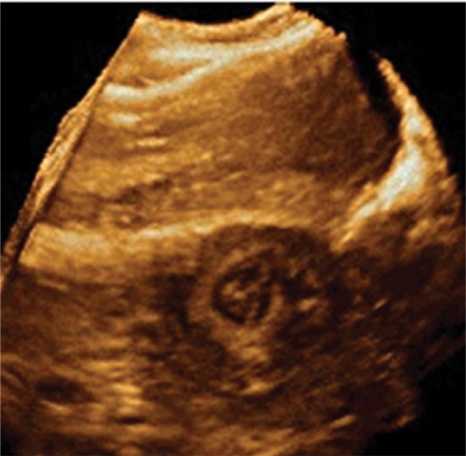
When performing an ultrasound examination, one should visualize the uterus in the sagittal section, take a close look at the cervix and uterine body as well as at the uterine cavity and cervical canal. Typical for CSP are the following: no gestational sac within the uterine cavity and cervical canal, visualization of the gestational sac and/or placenta in the cesarean section scar, very thin muscle layer between the gestational sac and the urinary bladder wall (from 1–3 mm to 4.6 mm) and intensive vascularization around the scar(16,17). Other signs suggesting CSP include so-called negative organ sliding sign, i.e. the lack of gestational sac movement upon gentle pressure with a probe in the vagina. These criteria exclude other diagnoses, such as cervico-isthmic pregnancy, cervical pregnancy or inevitable spontaneous abortion(18). CSP diagnosis and patient selection for proper therapeutic management also makes use of three-dimensional (3D) ultrasonography. Although this imaging technique cannot replace the two-dimensional one, it can bring genuine benefits in selected cases, e.g. precise spatial location of the gestational sac and assessment of its relationships with the urinary bladder wall and other structures of the lower pelvis (Fig. 5).
Fig. 5.
3D US. Gestational sac protruding towards the urinary bladder(18)
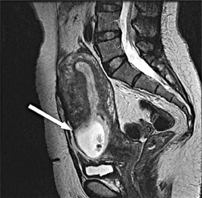
Moreover, magnetic resonance imaging (MRI) may also prove useful in CSP diagnosis. It enables accurate measurement of the distance between the urinary bladder, myometrium and gestational sac, and offers good visualization of the uterine cavity and cervical canal (Fig. 6)(19).
Fig. 6.
Sagittal T2-weighted MRI. CSP (week 8 day 3) in the lower segment of the anterior uterine wall in a cesarean section scar (arrow). Thin myometrium between the gestational sac and urinary bladder(19)
Vial et al. believe that there are two types of CSP. In the first one, the fertilized egg becomes implanted in the scar and further develops in the direction of the cervical isthmus and uterine cavity. This gives a chance for live birth, but the risk of massive bleeding from the implantation site is very high. The other type of CSP is associated with deep implantation in a damaged cesarean scar and development of pregnancy that leads to uterine rupture and hemorrhage in the first trimester of gestation(16).
Therapeutic management
Cesarean section predisposes to cesarean scar pregnancy and to placenta percreta(20). Until recently, these two abnormalities were treated as two completely separate entities. The latest data, however, indicate that they are not two separate pathologies, but consequences of a single abnormality(21). If an expectant attitude is assumed, CSP will probably transform into a pregnancy with placenta percreta in the scar and in the lower segment. This situation may lead to hysterectomy due to hemorrhage in almost every case. Early diagnosis and treatment offer much better prognosis(17,22). When making a decision about the management in CSP, the following must be considered: size of pregnancy, presence or absence of uterine continuity, β-hCG level, wish to remain fertile and patient’s hemodynamic state(18).
One of the classical and the first methods used in these cases is uterine curettage with secondary intrauterine insertion of a Foley catheter(23). Fang et al., in their publication from 2017, indicate 95% efficacy of this management in CSP between day 31 and 67(24). The Foley catheter, as an effective method of hemostasis, is also used in combination with MTX, both systemic and local, with a subsequent puncture and suction of the CSP under US guidance(25). Kanat-Pektas et al.(26) conducted a cross-sectional analysis of CSP cases from 1978–2014. CSP was most often treated with systemic MTX, uterine artery embolization (UAE), dilatation and curettage (D&C), hysterotomy and hysteroscopy. These methods were used in 33.9%, 21.9%, 14.1%, 10.6% and 6.7% of CSP cases, respectively. Combined treatment methods were applied more rarely: TA US (transabdominal ultrasound) or TV US + local MTX treatment in 6.6% of cases, TV US + gestational sac aspiration in 3.7% of cases, TV US or TA US + local injection of vasopressin or potassium chloride in 1% of cases and bilateral hypogastric artery ligation in 0.1% of cases. Hysterectomy was necessary in 1.5% of patients. In many cases, hysterectomy is the secondary necessary treatment, as an “effect” of unsuccessful first-line therapy. The analysis of Kanat-Pektas et al. suggests that the frequency of hysterectomy was 3.6%, 1.1%, 0.0%, 7.3% and 1.7% of CSP cases treated with systemic MTX, UAE, hysteroscopy, D&C and hysterotomy, respectively. Dilatation and curettage is linked with the highest risk of secondary hysterectomy. The risk if half as high with systemic MTX. However, there was not one case of hysterectomy after hysteroscopy. The following procedures were conducted during hysteroscopy (ordered according to their frequency):
hysteroscopic removal of gestational tissue;
hysteroscopic hysterotomy;
hysteroscopic local MTX injection;
hysteroscopic local ethanol injection;
hysteroscopic aspiration of gestational sac after local MTX injection.
The efficacy of each of these methods as first-line treatment differs significantly and reaches: 92.1% for hysterotomy, 61.6% for D&C, 39.1% for hysteroscopy, 18.3% for UAE and 8.7% for systemic MTX therapy.
The most effective hysterotomy (in laparoscopy or laparotomy) is mainly used in more advanced CSP cases as well as in uterine rupture and massive hemorrhage. Wedge resection and surgical management of the implantation site enable fertility preservation(26). Precise location of CSP in laparoscopy may pose a problem due to the urinary bladder that covers it. In these cases, the procedure was postponed by 1 or 2 weeks for CSP to grow to over 3 cm, thus allowing its accurate identification during surgery(19).
In most cases, better therapeutic effects can be obtained by combined and multi-step treatment. For instance, after application of systemic MTX, the most commonly used and the most effective are: D&C, UAE, hysteroscopy and additional intragenstational MTX administration under TV US guidance(26).
Initially, systemic MTX was considered first-line treatment in CSP. However, despite initial optimistic reports about its efficacy, a cross-sectional analysis from 1978–2014 showed its relatively low efficacy, at the level of only 8.7%. Moreover, certain reports indicated the complication rate at the level of 62.1%(27). Complications of this treatment include: nausea, stomatitis, vaginal spotting and bleeding, pneumonia and alopecia. It is worth emphasizing, however, that chances for another successful pregnancy after this treatment belong to the highest compared to other methods. Nevertheless, the literature analysis shows that systemic MTX administration should not be recommended as first-line treatment of CSP. Apart from other causes listed above, this results from the long time one must wait for the drug to work (average 4–16 weeks)(28). The mean time of complete gestational tissue regression after systemic MTX monotherapy is, according to Seow, 2 months to even 1 year despite earlier return of β-hCG levels to normal(14). Sometimes, this therapy is assessed as ineffective, which may be linked with an increased risk of more and more serious complications for the patient. However, Rotos, in his analysis from 2006, argues that simultaneous systemic and local MTX administration for CSP with β-hCG levels above 10,000 mLU/mL may be a very effective method that does not require further interventions(4). Systemic MTX administration seems to be the most effective for CSP with β-hCG below 5,000 IU/mL, and should be limited to two doses, 1 mg/kg body weight each, to avoid adverse effects(14).
In the case of using uterine artery embolization as first-line treatment, further intervention is needed in over 80% of cases and encompasses the following (ordered from the most to least effective): D&C, TV US + local MTX administration, hysteroscopy and systemic MTX administration. It has been found that the ability to maintain future pregnancy after UAE was minimal due to considerable flow restriction in the uterine arteries. UAE is therefore recommended as first-line treatment only in cases with heavy bleeding or suspicion of high-grade abnormality in the uterine vascularity.
Hysteroscopy enables direct visualization of the gestational sac and performance of vascular coagulation at the site of implantation, which might prevent heavy bleeding. The first report of this form of CSP treatment was published by Wang et al. in 2005(28). This procedure is minimally invasive, characterized by low blood loss, short in duration and gives a chance for fertility preservation. However, approximately 61% of hysteroscopies do require further intervention in the form of systemic mifepristone (RU-486), systemic methotrexate or D&C. In a retrospective analysis of 2,037 CSP cases, Birch Petersen et al. distinguished as many as 14 therapy models(29):
expectant management;
systemic MTX treatment;
gestational sac needle aspiration + MTX;
D&C;
hysteroscopy;
transvaginal resection of CSP;
UAE + D&C without MTX;
UAE + D&C + hysteroscopy;
UAE + D&C + MTX;
local and systemic MTX treatment;
laparoscopy;
local MTX treatment;
repeated high-intensity focused ultrasound (HIFU) ablation;
repeated HIFU ablation + hysteroscopic gestational suction.
Considering efficacy and safety, five CSP treatment methods are recommended depending on their availability, intensity of symptoms and surgical skill: transvaginal resection, laparoscopy, UAE + D&C + hysteroscopy, UAE + D&C and hysteroscopy(29). It must be mentioned that UAE is not widely available in primary and secondary care hospitals, and requires the presence of a trained interventional radiologist, which significantly restricts its usage in daily practice. An interesting new-generation procedure helpful in CSP management is a surgical method that utilizes high-intensity focused ultrasound (HIFU). To date, it has been used only for the treatment of prostate pathologies, but proved 100% successful in the analyzed cases of CSP.
During CSP treatment, it is significant and necessary to monitor the level of β-hCG as it is an indicator of treatment efficacy. This primarily concerns cases in which MTX, UAE or HIFU are applied.
Conclusion
Transvaginal US imaging is helpful in detection of asymptomatic ectopic pregnancy implanted in the cesarean section scar. Early identification of this form of pregnancy warrants effective treatment with no negative effects on fertility. Particularly useful is Doppler imaging and, in the most difficult cases, MRI. Ultrasound imaging, mainly transvaginal and rarely transabdominal, is a significant diagnostic means utilized not only to diagnose but also to treat CSP as part of a combined approach. Dilatation and curettage with subsequent intrauterine Foley catheter insertion may be recommended, but only due to its availability, simplicity and relatively high efficacy. However, bearing in mind significant risk of hemorrhage and high risk of secondary hysterectomy and fertility loss, this form of treatment should only be used in selected cases of early diagnosed CSP. Systemic methotrexate treatment should not be applied on the routine basis due to relatively low efficacy, high risk of hysterectomy and fertility loss, and the risk of various adverse effects. On the other hand, local methotrexate therapy (under ultrasound or hysteroscopy guidance) should be considered a perfect management method as it offers fertility preservation in asymptomatic pregnant patients without concomitant hemodynamic disorders. The most effective CSP treatment is simultaneous application of 2–3 techniques. The combination of local MTX with simultaneous gestational sac aspiration under ultrasound or hysteroscopy guidance seems optimal and minimally invasive. In the second stage, the remaining gestational tissues can be removed hysteroscopically in combination with vascular coagulation of the implantation site.
In more advanced cases (CSP exceeding 3 cm), local methotrexate administration should be considered, followed by laparoscopic or laparotomic CSP wedge resection with subsequent surgical correction of the cesarean section scar.
Promising results in CSP treatment have been obtained with an innovative HIFU technique that utilizes high-intensity focused ultrasound.
Conflict of interest
Authors do not report any financial or personal connections with other persons or organizations, which might negatively affect the contents of this publication and/or claim authorship rights to this publication.
References
- 1.Le A, Shan L, Xiao T, Zhuo R, Xiong H, Wang Z. Transvaginal surgical treatment of cesarean scar ectopic pregnancy. Arch Gynecol Obstet. 2013;287:791–796. doi: 10.1007/s00404-012-2617-7. [DOI] [PubMed] [Google Scholar]
- 2.Skrzypczak J. Ciąża o przebiegu nieprawidłowym. In: Bręborowicz G, editor. Położnictwo i ginekologia. Vol. 1. Warszawa: PZWL; 2015. pp. 92–100. [Google Scholar]
- 3.Ash A, Smith A, Maxwell D. Caesarean scar pregnancy. BJOG. 2007;114:253–263. doi: 10.1111/j.1471-0528.2006.01237.x. [DOI] [PubMed] [Google Scholar]
- 4.Rotas MA, Haberman S, Levgur M. Cesarean scar ectopic pregnancies: etiology, diagnosis, and management. Obstet Gynecol. 2006;107:1373–1381. doi: 10.1097/01.AOG.0000218690.24494.ce. [DOI] [PubMed] [Google Scholar]
- 5.Ouyang Y, Li X, Yi Y, Gong F, Lin G, Lu G. First-trimester diagnosis and management of Cesarean scar pregnancies after in vitro fertilization-embryo transfer: a retrospective clinical analysis of 12 cases. Reprod Biol Endocrinol. 2015;13:126. doi: 10.1186/s12958-015-0120-2. [DOI] [PMC free article] [PubMed] [Google Scholar]
- 6.Larsen JV, Solomon MH. Pregnancy in a uterine scar sacculus – an unusual cause of postabortal haemorrhage. A case report. S Afr Med J. 1978;53:142–143. [PubMed] [Google Scholar]
- 7.Moschos E, Sreenarasimhaiah S, Twickler DM. First‐trimester diagnosis of cesarean scar ectopic pregnancy. J Clin Ultrasound. 2008;36:504–511. doi: 10.1002/jcu.20471. [DOI] [PubMed] [Google Scholar]
- 8.Wlaźlak E, Surkont G, Shek KL, Dietz HP. Can we predict urinary stress incontinence by using demographic, clinical, imaging and urodynamic data? Eur J Obstet Gynecol Reprod Biol. 2015;193:114–117. doi: 10.1016/j.ejogrb.2015.07.012. [DOI] [PubMed] [Google Scholar]
- 9.Fylstra DL. Ectopic pregnancy within a cesarean scar: A review. Obstet Gynecol Surv. 2002;57:537–543. doi: 10.1097/00006254-200208000-00024. [DOI] [PubMed] [Google Scholar]
- 10.Surkont G, Wlaźlak E, Bitner A, Tyliński W, Stetkiewicz T, Suzin J. Analysis of sonographic features of endometrial diseases in postmenopausal asymptomatic women. Przegl Menopauz. 2006;2:96–101. [Google Scholar]
- 11.Rempen A, Albert P. [Diagnosis and therapy of early pregnancy implanted in the scar of cesarean section] Z Geburtshilfe Perinatol. 1990;194:46–48. [PubMed] [Google Scholar]
- 12.Kwaśniewska A, Stupak A, Krzyżanowski A, Pietura R, Kotarski J. Cesarean scar pregnancy: uterine artery embolization combined with a hysterectomy at 13 weeks’ gestation – acase report and review of the literature. Ginekol Pol. 2014;85:961–967. doi: 10.17772/gp/1890. [DOI] [PubMed] [Google Scholar]
- 13.Zhang Y, Gu Y, Wang JM, Li Y. Analysis of cases with cesarean scar pregnancy. J Obstet Gynaecol Res. 2013;39:195–202. doi: 10.1111/j.1447-0756.2012.01892.x. [DOI] [PubMed] [Google Scholar]
- 14.Seow KM, Huang LW, Lin YH, Lin MY, Tsai YL, Hwang JL. Cesarean scar pregnancy: issues in management. Ultrasound Obstet Gynecol. 2004;23:247–253. doi: 10.1002/uog.974. [DOI] [PubMed] [Google Scholar]
- 15.Montgomery Irvine L, Padwick ML. Serial serum HCG measurements in a patient with an ectopic pregnancy: A case for caution. Hum Reprod. 2000;15:1646–1647. doi: 10.1093/humrep/15.7.1646. [DOI] [PubMed] [Google Scholar]
- 16.Vial Y, Petignat P, Hohlfeld P. Pregnancy in a cesarean scar. Ultrasound Obstet Gynecol. 2000;16:592–593. doi: 10.1046/j.1469-0705.2000.00300-2.x. [DOI] [PubMed] [Google Scholar]
- 17.Timor-Tritsch IE, Monteagudo A, Santos R, Tsymbal T, Pineda G, Arslan AA. The diagnosis, treatment, and follow-up of cesarean scar pregnancy. Am J Obstet Gynecol. 2012;207:44.e1–44.e13. doi: 10.1016/j.ajog.2012.04.018. [DOI] [PubMed] [Google Scholar]
- 18.El Guindi W, Alalfy M, Abasy A, Ellithy A, Nabil A, Abdalfatah O, et al. A report of four cases of caesarean scar pregnancy in a period of 24 months. J Med Diagn Meth. 2013;2:2. [Google Scholar]
- 19.Chiang AJ, La V, Chou CP, Wang PH, Yu KJ. Ectopic pregnancy in a cesarean section scar. Fertil Steril. 2011;95:2388–2389. doi: 10.1016/j.fertnstert.2011.03.104. [DOI] [PubMed] [Google Scholar]
- 20.Osser OV, Jokubkiene L, Valentin L. High prevalence of defects in Cesarean section scars at transvaginal ultrasound examination. Ultrasound Obstet Gynecol. 2009;34:90–97. doi: 10.1002/uog.6395. [DOI] [PubMed] [Google Scholar]
- 21.Woźniak S, Czuczwar P, Pyra K, Szkodziak P, Woźniakowska E, Milart P, et al. Ciąża w bliźnie po cięciu cesarskim – znaczenie wczesnego rozpoznania. Analiza Przypadków w Ginekologii i Położnictwie. 2013;1:6–21. [Google Scholar]
- 22.Sinha P, Mishra MJ. Caesarean scar pregnancy: A precursor of placenta percreta/accreta. J Obstet Gynaecol. 2012;32:621–623. doi: 10.3109/01443615.2012.698665. [DOI] [PubMed] [Google Scholar]
- 23.Jurkovic D, Hillaby K, Woelfer B, Lawrence A, Salim R, Elson CJ. First-trimester diagnosis and management of pregnancies implanted into the lower uterine segment Cesarean section scar. Ultrasound Obstet Gynecol. 2003;21:220–227. doi: 10.1002/uog.56. [DOI] [PubMed] [Google Scholar]
- 24.Fang Q, Sun L, Tang Y, Qian C, Yao X. Quantitative risk assessment to guide the treatment of cesarean scar pregnancy. Int J Gynaecol Obstet. 2017;139:78–83. doi: 10.1002/ijgo.12240. [DOI] [PubMed] [Google Scholar]
- 25.Timor‐Tritsch IE, Cali G, Monteagudo A, Khatib N, Berg RE, Forlani F, et al. Foley balloon catheter to prevent or manage bleeding during treatment for cervical and Cesarean scar pregnancy. Ultrasound Obstet Gynecol. 2015;46:118–123. doi: 10.1002/uog.14708. [DOI] [PubMed] [Google Scholar]
- 26.Kanat-Pektas M, Bodur S, Dundar O, Bakır VL. Systematic review: What is the best first- line approach for cesarean section ectopic pregnancy? Taiwan J Obstet Gynecol. 2016;55:263–269. doi: 10.1016/j.tjog.2015.03.009. [DOI] [PubMed] [Google Scholar]
- 27.Timor-Tritsch IE, Monteagudo A. Unforeseen consequences of the increasing rate of cesarean deliveries: early placenta accreta and cesarean scar pregnancy. A review. Am J Obstet Gynecol. 2012;207:14–29. doi: 10.1016/j.ajog.2012.03.007. [DOI] [PubMed] [Google Scholar]
- 28.Wang CJ, Yuen LT, Chao AS, Lee CL, Yen CF, Soong YK. Caesarean scar pregnancy successfully treated by operative hysteroscopy and suction curettage. BJOG. 2005;112:839–840. doi: 10.1111/j.1471-0528.2005.00532.x. [DOI] [PubMed] [Google Scholar]
- 29.Birch Petersen K, Hoffmann E, Rifbjerg Larsen C, Svarre Nielsen H. Cesarean scar pregnancy: a systematic review of treatment studies. Fertil Steril. 2016;105:958–967. doi: 10.1016/j.fertnstert.2015.12.130. [DOI] [PubMed] [Google Scholar]


