Abstract
M-mode echocardiography has been playing an important role in the diagnosis of fetal tachyarrhythmia. We recently encountered a neonatal case of atrial flutter with 2:1 atrioventricular conduction. However, M-mode erroneously indicated 1:1 atrioventricular movement. While the movement of the atrial wall far from the atrioventricular valve was much faster than that of the ventricular wall, the atrial wall adjacent to the atrioventricular valve fully synchronized to that of the ventricular wall. Thus, to avoid this novel pitfall, it would be important to add an additional assessment focusing on the movement of the atrial wall far from the ventricle.
Keywords: Fetal echocardiography, arrhythmia, myocardial wall motion, diagnostic imaging tools
Introduction
Accurate diagnosis of fetal tachyarrhythmia is important for appropriate management and prediction of prognosis. Currently, the accurate diagnosis of this condition relies on fetal echocardiography because fetal electrocardiography and magnetocardiography are not yet commonly used in clinical practice.1 Particularly, M-mode echocardiography, which can simultaneously display the atrial and ventricular wall motions, and thus the P and QRS relationship on electrocardiography, has been playing an important role in the diagnosis of fetal arrhythmia.2 The preferred M-mode beam direction is in the 4-chamber view, simultaneously through the atrial free wall and the opposite ventricular free wall close to the atrioventricular junction because these atrial and ventricular portions show the most pronounced lateral excursion during the cardiac cycle.1 We recently encountered a case showing a novel pitfall of this approach.
Case Report
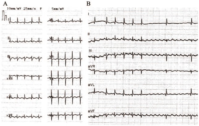
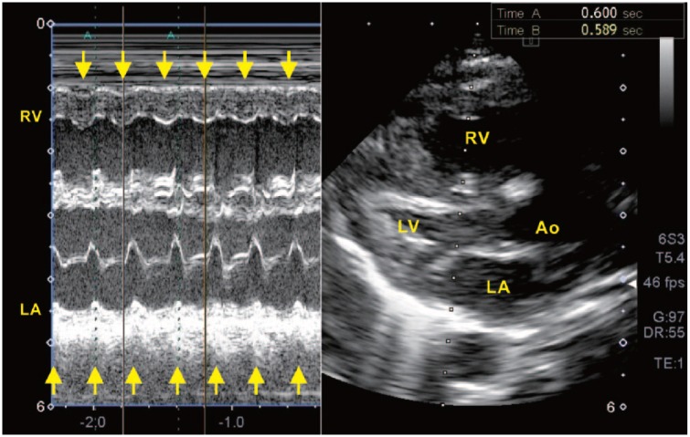
A full-term male infant was delivered through an emergent cesarean section at 39 weeks and 6 days, owing to newly found tachycardia (about 200 bpm) at routine fetal screening. His birth weight was 3399 g, and his Apgar score was 8-8. After delivery, his condition remained stable except for persistent tachycardia, mild tachypnea, and transient need for small amounts of oxygen. His 12-lead electrocardiography revealed narrow QRS tachycardia (Figure 1A) and indicated atrial flutter (AF)3 with 2:1 atrioventricular conduction. To verify the diagnosis and the relationship between atrial and ventricular contractions, we performed M-mode echocardiography. Surprisingly, M-mode, which simultaneously displays right ventricular and left atrial motions (Figure 2), clearly indicated 1:1 atrioventricular conduction. Given these inconsistent results, we performed rapid injection of adenosine triphosphate to make a transient atrioventricular block for an accurate diagnosis. Electrocardiography after adenosine triphosphate infusion revealed AF with 2:1 atrioventricular conduction (Figure 1B) and that the M-mode diagnosis of 1:1 atrioventricular conduction was incorrect.
Figure 1.
(A) Twelve-lead electrocardiography before treatment showing regular narrow QRS tachycardia with a heart rate of 203 bpm. Lead II does not have an isoelectric baseline and indicates atrial flutter with 2:1 atrioventricular conduction. (B) Electrocardiogram (II, III, aVF-lead) after adenosine triphosphate infusion revealing atrial flutter with an F rate of about 400 bpm (2:1 atrioventricular conduction).
Figure 2.
M-mode echocardiography simultaneously displays the movements of the RV and LA close to the atrioventricular junction. Arrows indicate right ventricular and left atrial contractions. M-mode indicates 1:1 atrioventricular conduction. Ao, aorta; LA, left atrium; LV, left ventricle; RV, right ventricle.
Discussion
The atrioventricular M-mode obtained by preferred beam direction1 incorrectly suggested 1:1 atrioventricular conduction in the case of AF with 2:1 atrioventricular conduction. Why did M-mode erroneously indicate 1:1 atrioventricular conduction? A retrospective analysis of movie files clarified the phenomenon (Movie Clips S1, normal speed; S2, half speed). The movement of the atrial wall far from the atrioventricular valve (opposite side of the ventricular apex) was much faster than that of the ventricular wall. In contrast, the movement of the atrial wall close to atrioventricular valve was fully synchronized to ventricular wall movement. This “atrioventricular constraint” might be the cause of the pseudo 1:1 atrioventricular conduction in this case of AF with 2:1 atrioventricular conduction. Because this phenomenon that not all parts of the atrium contract equally might not be limited to neonates and could also occur in fetuses, our observation indicates that fetal M-mode echocardiography of the atrial wall close to the atrioventricular valve may lead to incorrect diagnosis of tachyarrhythmia in some cases. This is a novel pitfall of M-mode in the diagnosis of fetal arrhythmia.
To avoid this pitfall due to “atrioventricular constraint” during the fetal period in which an electrocardiogram cannot be obtained, we believe that it is important to acquire an additional M-mode that solely focuses on the top of the atria that is far from the ventricle, for measurement of the true atrial rate. Furthermore, slow replay (eg, at half speed) would highly facilitate the visual assessment of the relationship between atrial and ventricular contractions. As a limitation of this case report, our observation was obtained only after birth. No fetal echocardiography was performed, as such we have no information about fetal heart rhythm except for the tachycardia of 200 bpm. In fetal echocardiography, simultaneous Doppler measurement of the superior vena cava and aortic blood flow would provide further insight into the relationship between atrial and ventricular contraction.4 Although the M-mode (Figure 2) clearly captured both ventricular and atrial movements, this was not obtained with the standard 4-chamber view used in fetal evaluation of arrhythmias. Thus, it needs to be further confirmed by future studies whether the pseudo 1:1 atrioventricular conduction exists and affects the diagnosis of fetal AF.
Acknowledgments
The authors would like to thank the medical staff of Saitama Medical Center, Saitama Medical University who were involved in the treatment of this patient.
Footnotes
Funding:The author(s) received no financial support for the research, authorship, and/or publication of this article.
Declaration of conflicting interests:The author(s) declared no potential conflicts of interest with respect to the research, authorship, and/or publication of this article.
Author Contributions: AK wrote the first draft of the manuscript. AK, AO, SM, and AI contributed to data collection and revision of the manuscript. SM, YI, HI, MT, and HS contributed to further interpretation and analysis of the data. SM made critical revisions and final approval. All authors reviewed and approved the final manuscript.
Ethical approval: Ethical committee of Saitama Medical Center, Saitama Medical University does not require the ethical review for a case report.
Informed consent: The parents of this infant gave written consent for the publication of this report.
ORCID iD: Satoshi Masutani  https://orcid.org/0000-0002-1551-9089.
References
- 1. Weber R, Stambach D, Jaeggi E. Diagnosis and management of common fetal arrhythmias. J Saudi Heart Assoc. 2011;23:61–66. [DOI] [PMC free article] [PubMed] [Google Scholar]
- 2. Maeno Y, Hirose A, Kanbe T, Hori D. Fetal arrhythmia: prenatal diagnosis and perinatal management. J Obstet Gynaecol Res. 2009;35:623–629. [DOI] [PubMed] [Google Scholar]
- 3. Lisowski LA, Verheijen PM, Benatar AA, et al. Atrial flutter in the perinatal age group: diagnosis, management and outcome. J Am Coll Cardiol. 2000;35:771–777. [DOI] [PubMed] [Google Scholar]
- 4. Fouron JC, Fournier A, Proulx F, et al. Management of fetal tachyarrhythmia based on superior vena cava/aorta Doppler flow recordings. Heart. 2003;89:1211–1216. [DOI] [PMC free article] [PubMed] [Google Scholar]


