Abstract
Lyme disease is a serious vector-borne infection that is caused by the Borrelia burgdorferi sensu lato family of spirochetes, which are transmitted to humans through the bite of infected Ixodes ticks. The primary etiological agent in North America is Borrelia burgdorferi sensu stricto. As geographic risk regions expand, it is prudent to support robust surveillance programs that can measure tick infection rates, and communicate findings to clinicians, veterinarians, and the general public. The molecular technique of nested polymerase chain reaction (nPCR) has long been used for this purpose, and it remains a central, inexpensive, and robust approach in the detection of Borrelia in both ticks and wildlife.
This article demonstrates the application of nPCR to tick DNA extracts to identify infected specimens. Two independent B. burgdorferi targets, genes encoding Flagellin B (FlaB) and Outer surface protein A (OspA), have been used extensively with this technique. The protocol involves tick collection, DNA extraction, and then an initial round of PCR to detect each of the two Borrelia-specific loci. Subsequent polymerase chain reaction (PCR) uses the product of the first reaction as a new template to generate smaller, internal amplification fragments. The nested approach improves upon both the specificity and sensitivity of conventional PCR. A tick is considered positive for the pathogen when inner amplicons from both Borrelia genes can be detected by agarose gel electrophoresis.
Keywords: Molecular Biology, Issue 132, Lyme disease, Borrelia burgdorferi, Ixodidae, tick, surveillance, DNA extraction, nested PCR, agarose gel electrophoresis, OspA, FlaB
Introduction
Lyme disease (LD) is the most prevalent vector-borne infection in the northern hemisphere, and its incidence continues to increase1. This debilitating illness is caused by spirocheteal pathogens of the Lyme borreliosis (LB) complex (commonly referred to as Borrelia burgdorferi sensu lato, or s.l.), which historically comprises the predominant North American pathogen, B. burgdorferi sensu stricto (s.s), in addition to B. afzelii and B. garinii, which are widespread throughout Europe and Asia, and are species of emerging clinical relevance2,3. These bacteria are transmitted to humans through the bite of infected Ixodes ticks2. Although the main North American vectors are I. scapularis and I. pacificus, multiple species within this genus have been found to harbor and transmit the bacterium4. In humans, B. burgdorferi can cause multisystem symptoms affecting the skin, joints, heart, nervous system, endocrine glands, gastrointestinal tract, and internal organs5,6,7,8,9,10. The Center for Disease Control and Prevention currently estimates over 300,000 new human cases annually in the United States11,12. While prognosis is often favorable when the illness is diagnosed and treated in the early stages, studies have suggested that anywhere between 10% and 60% of patients who received the recommended antibiotic regimen continued to experience symptoms after therapy cessation, in a phenomenon termed Post Treatment Lyme Disease Syndrome (PTLDS)13,14,15. Moreover, delays in clinical intervention can arise as a result of lack of awareness of a tick bite, non-specific presentation of the initial illness, and the low sensitivity of traditional serology-based diagnostics when used at the onset of infection. Failure to treat swiftly and appropriately enables symptom progression that can manifest in increasingly debilitating complications16,17. Lyme disease prevention is therefore a cornerstone of risk management. Strategies to combat this growing threat include robust surveillance measures to indicate the pathogen prevalence in ticks, and identify geographic regions of concern.
This article demonstrates the utility of nested polymerase chain reaction (nPCR) as a molecular screening tool by which to identify infected ticks. To increase specificity, two Borrelia genes are used for parallel amplification. Flagellin B (FlaB) encodes a major filament protein of the flagellum18, and the gene is located on the single linear chromosome, while the lipoprotein product of Outer surface protein A (OspA) mediates tick midgut colonization, and is plasmid-encoded19,20. The workflow consists of tick collection, DNA extraction, and then an initial round of PCR to detect Borrelia-specific loci. Subsequent PCR uses the product of the first reaction as a new template to generate smaller, internal amplification fragments. A tick is considered positive for Borrelia burgdorferi when inner amplicons from both Borrelia genes can be detected by agarose gel electrophoresis.
Both the nPCR technique, and the FlaB and OspA gene targets, have been widely used for ecological surveillance and clinical detection of the Lyme spirochete since the early 1990s21,22,23,24,25. Prior to the development of molecular protocols, ticks were assessed by subjecting gut contents to anti-Borrelia immunofluorescence (IF) staining, microbial culture, or a combination thereof26. These approaches suffer from inherent limitations, including the slow growth and fastidious nature of Borrelia27, antibody performance issues, and the requirement of live ticks for IF processing21. PCR was subsequently adopted to provide fast, sensitive, and specific identification of infected vectors. It offered marked improvements over the traditional methodology, including culture-independent direct application to diverse specimen types, such as dead and archived ticks that would otherwise be unsuitable for testing21,22. Nested experimental designs further improve upon the specificity and sensitivity of classical PCR by employing two distinct sets of gene-specific primers in two consecutive rounds of amplification25,28.
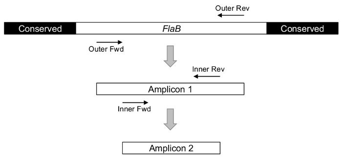
Experimental success is critically dependent on strategic gene selection and amplicon design. To accurately estimate the presence of Borrelia burgdorferi, primers should detect the relevant pathogens without cross-reacting with relapsing fever spirochetes also in the Borrelia genus. In the case of FlaB, this specificity is achieved by targeting a variable internal region of the gene, rather than the relatively conserved flanking sequences that are shared by diverse bacteria (Figure 1)24,29,30.
Figure 1: The concept of nPCR, as illustrated using Borrelia FlaB gene as a target. The 5' and 3' termini of the gene are common to organisms beyond Borrelia burgdorferi s.l., rendering these regions unsuitable for the specific assessment of Lyme-causing pathogens. Using the less-conserved interior sequences as primer targets, two rounds of PCR are executed to detect a final, internal amplicon. Please click here to view a larger version of this figure.
In a test of their discriminatory capacity, interior FlaB primers were evaluated with over 80 different Borrelia isolates, and found to identify only those associated with Lyme disease24. The lower detection limit of nPCR has been documented at between six24 and ten31 bacteria from pure culture, although the sensitivity can be further improved by Southern blotting the amplicon, and hybridizing a radiolabelled or chemiluminescent probe. Coupling the techniques reduces the detection threshold to a single spirochete31. By direct comparison, conventional, unprobed single-round PCR was found to report the presence of a minimum of 104 spirochetes31. It should be noted, however, that assay sensitivity will be lower when working with complex environmental and clinical samples, due to the overwhelming presence of unrelated DNA and potential inhibitory substances. These challenges can be largely circumvented by the use of nPCR.
Despite advances in molecular technologies in the past decades, nPCR remains a staple technique in modern surveillance efforts. If care is taken in the design and execution of this protocol, it is a powerful, adaptable, and relatively straightforward approach to capture the presence of pathogens in tick vectors.
Protocol
1. DNA Isolation from Ticks
- Acquire ticks via field collection, or passive surveillance from veterinarians and members of the public. Ticks should be killed by freezing, placed in a sealed bag, and mailed at room temperature.
- Using a submission form, gather information on the date and geographic location of the encounter, tick attachment status, host species, and recent travel history of the host.
- Since nested PCR is inherently vulnerable to contamination, ensure that the laboratory workspace is set up to minimize cross-exposure of samples. This involves executing different elements of the protocol in separate, dedicated spaces well isolated from each other that are thoroughly cleaned and sterilized, and ensuring that all instruments are free of contaminants.
- Upon receiving the specimen, photograph the tick and determine species, developmental stage, sex, and engorgement status by comparison to an identification key32 .
- Under aseptic conditions, bisect the tick using a sterile razor blade or scalpel, and place the two tick fragments into separate microcentrifuge tubes.
- To extract total DNA, use any isolation procedure that yields PCR-compatible template; this protocol demonstrates a simple chelation-based approach.
- Begin by adding an appropriate volume (often between 50 - 200 µL) of a lytic chelation reagent to the tick fragment. The specific amount will depend on the specimen size and engorgement status; guidelines should be provided by the manufacturer. Homogenize using a microtube pestle.
- Incubate samples in a water bath at 60 °C for 45 min, and vortex briefly.
- Centrifuge samples for 4 min at 16,276 x g (13,300 rpm) in a desktop microcentrifuge.
- Transfer supernatant to a fresh, labeled microtube containing 50 µL isopropanol, mix by inversion, and re-centrifuge as in (1.2.3).
- Decant the supernatant, and rinse the DNA pellet with 50 µL of 70% ethanol.
- Remove excess ethanol with a pipette, and allow the pellet to air-dry for 15 min at room temperature.
- To resuspend DNA, add 50 µL of 1 mM Tris pH 7.0, and incubate samples in a water bath for 1 h at 60 °C. The DNA can now be stored at -20 °C for future molecular analysis.
2. Nested PCR Detection of Borrelia OspA and FlaB.
NOTE: An overview of general PCR principles and practices is provided by Lorenz, 201233.
Synthesize or obtain Borrelia gene-specific outer and inner oligonucleotide primers. See Table 1 for primer sets, their respective amplicon sizes, and melting temperatures.
| Primer Name | Gene Target | Sequence (5’-3’) | Amplicon Size | Annealing Temperature |
| FlaB Out Fw | FlaB | gcatcactttcagggtctca | 503 bp | 55°C |
| FlaB Out Rv | FlaB | tggggaacttgattagcctg | ||
| FlaB In Fw | FlaB | ctttaagagttcatgttggag | 447 bp | 58°C |
| FlaB In Rv | FlaB | tcattgccattgcagattgt | ||
| OspA Out Fw | OspA | cttgaagttttcaaagaagat | 487 bp | 55°C |
| OspA Out Rv | OspA | caactgctgacccctctaat | ||
| OspA In Fw | OspA | acaagagcagacggaaccag | 350 bp | 58°C |
| OspA In Rv | OspA | ttggtgccatttgagtcgta |
Table 1: Inner and outer primers for nPCR of Borrelia burgdorferiFlaB and OspA. In practice, the FlaB primers detect B. burgdorferi s.s. and other closely related Borrelia genospecies, while OspA primers capture only B. burgdorferi s.s. Amplification from both loci would indicate B. burgdorferi. s.s.
- Pre-sterilize a PCR cabinet with UV light and 70% ethanol. NOTE: To minimize potential sample contamination, this workspace should be distinct from the location of tick dissection, DNA extraction, and gel electrophoresis.
- For the initial PCR run, use the outer primers in conjunction with the template DNA recovered in step 1.0 above, and set up the reaction mixture as described in Table 2. In parallel, perform positive control runs consisting of verified Borrelia DNA, and negative control runs including no-template reactions to detect reagent and aerosol contamination. Add DNA at the end to minimize potential contamination of reagents. Ensure each tube is closed at all times when reagents are not being added and close tubes immediately after DNA addition and before other tubes are opened.
- Program a thermal cycler as follows: 95 °C for 5 min; 40 cycles of 95 °C for 15 s, annealing temperature for 30 s, 72 °C for 45 s; 72 °C for 5 min; and hold at 4 °C.
- Perform the second round of PCR similar to the first reaction, except use inner primers with 2 µL of the first PCR product (produced in 2.2.2). NOTE: Reaction volumes are again provided in Table 2. Care must be taken to avoid cross-contamination of amplicons by ensuring that template DNA is added last and that only tubes corresponding to one sample are open at a time.
Component PCR # 1 Volumes PCR # 2 Volumes Taq Polymerase Master Mix 2X 12.5 µL 12.5 µL Nuclease-free Water 8.5 µL 8.5 µL 10 µM Forward and Reverse primers 1.0 µL Outer Primers 1.0 µL Inner Primers DNA Template 2.0 µL, from sample extraction (1.2.7) 2.0 µL, from round 1 of PCR (2.2.2) - Program the thermal cycler as before, but adjust the annealing temperature to accommodate the inner primers (refer to Table 1).
3. Agarose Gel Electrophoresis and Imaging
NOTE: For basic instructions on separating DNA by electrophoresis, see Lee et al., 201234.
Prepare a 1.2% agarose gel using 20X SB buffer (0.2 M NaOH, 0.8 M Boric Acid pH 8), diluted to 1X, and add approximately 5 µL of a DNA stain, before pouring, if pre-staining of the gel is desired.
- Load 10 µL of the product from the second (inner) PCR reaction from each experimental and control sample, alongside a 100 bp ladder (5 µL).
- Electrophorese for 1 h at 107 V (5V/cm).
View and document the gel using a transilluminator and associated camera and software.
Representative Results
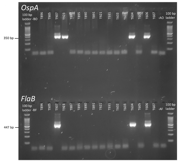
nPCR is an elegant approach to increase the specificity and sensitivity of pathogen detection, particularly when complex environmental samples are under investigation. As depicted in Figure 1, two rounds of PCR targeting a strategic region of the Borrelia FlaB locus (and OspA, not pictured) can report the presence of the Borrelia burgdorferi bacteria through the generation of short inner amplicons. When resolved by gel electrophoresis, the FlaB and OspA reaction products from each tick can be visually scored and compared against controls. In Figure 2, unique specimen identifiers are provided along the top of each gel, and no-template and aerosol controls are represented at the far left and right, respectively, adjacent to the ladders.


Robust amplicon bands are clearly visible in select experimental samples, and they are easily distinguished from surplus primers and residual DNA (Figure 2). Based on the experimental design principles set forth, a tick is considered positive for Borrelia when parallel negative controls demonstrate no amplification, and inner amplicons are produced from both FlaB and OspA primers. In this case, specimens T907, T604, and T606 met surveillance criteria for the Lyme pathogen (Figure 2). By contrast, T923 was only positive for OspA, an outcome for which there are multiple possible explanations: A) The tick of origin was negative for Borrelia, but the outer or inner OspA PCR preparation was spuriously contaminated with template DNA (note that systemic contamination of reagents was ruled out via negative controls), B) the tick carried Lyme Borrelia, but low template amounts or experimental error prevented amplification of FlaB, or C) an organism was present that contained the conserved OspA sequence, but lacked Flagellin or had insufficient identity in the targeted region of FlaB to anneal to the primers. Indeed, OspA sequence similarities have been noted in a variety of organisms, including plants and animals28. The converse situation, amplification of the more conserved Flagellin gene, but not the OspA gene, could indicate a related Borrelia species. In practice, this protocol yields more OspA single-positive results than do the FlaB primers, suggesting that scenario 'A' is the least likely explanation. Without further experimental analysis of the contentious specimen, however, it is not possible to determine the source of the single-positive result. Increasing the number of technical replicates performed on equivocal samples may help to reconcile their true status, since any contaminants that were spuriously introduced to previous reactions would not be present in the archived DNA. However, if subsequent reactions with these primers fail to provide conclusive results, other Borrelia loci could be investigated by PCR. Amplicons generated from these reactions could also be sequenced and compared among isolates to help assign identity, and estimate the degree of strain divergence. Sapi and colleagues provide an example of this workflow using human clinical samples35.
All factors considered, the outlined protocol and interpretation criteria have been estimated to yield false positive and false negative rates of 0.17% and 0.0063%, respectively.
Figure 2: Detection ofBorrelia burgdorferi in individual ticks by nPCR of FlaB and OspA. Codes assigned to each tick are represented across the top of the gels, and identities align vertically to report the detection of OspA and FlaB in each specimen. Lanes entitled B at the left of the image represent no-template controls, while A (right hand lanes) are aerosol controls to capture contamination of the laboratory environment. Please click here to view a larger version of this figure.
Discussion
Over decades of use, PCR-based techniques have consistently proven their value in the detection of Borrelia from arthropod and mammalian specimens, whether Borrelia come from environmental or clinical origins. PCR offers many improvements over pre-existing approaches to surveillance. Notably, it is not reliant on the development and performance of antibody reagents, and instead can be easily adapted to detect new targets of interest simply by modifying primer sequences. PCR also accommodates the evaluation of multiple loci in parallel, either independently or via a multiplex reaction. It can be applied to diverse specimen inputs, including fresh and archived ticks22, animal or human bodily fluids, and resected tissue25. PCR also yields amplicons that can be further processed, for example by restriction enzyme digestion, probe hybridization, or sequencing, to provide increased insight into microbial identity21.
As a clinical tool, PCR is conceptually preferable to the standard serological diagnostics, as it provides a direct indication of bacterial presence, rather than relying on host immune response as a secondary measure of infection. However, Lyme borreliosis is associated with a relatively modest microbial burden (<50 organisms/mL of urine or plasma) and transient spirochetemia, which can give rise to false negative results25. The sensitivity of this technique in the clinic varies considerably depending on the tissue type, stage of infection, and sample condition, falling between 12.5% and 62% in existing studies of blood, cerebrospinal fluid, and biopsied tissue36. Molecular sensitivity limitations are not a concern if PCR is undertaken subsequent to bacterial culture, however recovery of viable spirochetes from clinical specimens has proven similarly challenging. Only recently have protocols been optimized for higher yield35. Meanwhile, the gut of an infected adult tick can harbor on average anywhere from 2,000 to over 50,000 Borrelia37,38,39, which is solidly within the detection range of nPCR. Thus, the protocol is well suited to tick surveillance efforts.
Despite its many advantages, nPCR does pose certain challenges, and the capacity of this technique to accurately report tick infection therefore depends on strategic selection of genes and amplicons, meticulous experimental workflows that recover quality DNA from specimens while minimizing cross-exposure of samples, and the use of appropriate controls that can report contaminants in the laboratory environment and reagents. Prior to any experimentation, the scope and intentions of the investigation should be clearly defined so that appropriate genetic loci, and regions therein, can be selected. If the objective is to provide an unbiased screen for the Lyme-causing Borrelia burgdorferi s.l. complex, primers should be created to detect all of the associated strains with similar affinity and amplification efficiency, without capturing unrelated organisms25. New primers can be designed and assessed in silico using software tools such as Primer-BLAST40, although they should also be validated experimentally against standard reference isolates before being applied to uncharacterized samples. The goal of the DNA extraction procedure is to recover intact microbial template from a mixed environmental sample (tick homogenate). An optional step before proceeding with PCR is to evaluate the integrity of the extracted DNA. Additionally, parallel reactions could be set up to target a housekeeping gene in the tick. The latter method can also indicate the presence of inhibitors in the reaction mixture, providing increased confidence that negative Borrelia reactions are due to the absence of the organism, and not to the presence of an inhibitory contaminant.
The increased sensitivity of the nested PCR approach comes at the expense of potential contamination that generates false-positive results. Exogenous template could be introduced to a sample during tick dissection and DNA recovery, or in the process of setting up the outer and inner amplification reactions. It is therefore especially important to follow best practice protocols for PCR to avoid template or amplicon cross-contamination. These include the use of separate work stations with independent airflows, containment in PCR and biological safety cabinets where appropriate, thorough chemical and physical cleaning and sterilization of surfaces and reagents, and cautious handling of DNA41. No-template controls are also vital in identifying contamination of stock reagents. A particular vulnerability of nPCR is the amplicon handling that occurs when transferring the products of the first reaction into the second PCR vessel. Since the target DNA has already undergone exponential enrichment in positive samples, this step is especially prone to cross-contamination, and a single-tube nPCR protocol has been developed to circumvent this limitation. In this approach, both sets of primers for a given gene are added together to a reaction tube that remains sealed for both rounds of PCR31. The outer and inner primer pairs must be thermodynamically distinct, such that the outer couple amplifies template at a high annealing temperature that is prohibitive for the inner primers. In the second round, the annealing temperature is lowered to accommodate the internal pair. Not only does this bypass a potentially confounding step, it also permits the use of additional anti-contamination measures31,42. However, this modification may sacrifice some assay sensitivity. Regardless of the approach, increasing the number of technical replicates performed independently on a sample will help to identify spurious contamination.
Molecular techniques have continued to evolve since the introduction of nPCR, and these newer approaches offer select advantages over the original designs, albeit at increased financial expense. As the name suggests, quantitative PCR (qPCR or real time (RT)-PCR) allows for the enumeration of pathogens in a sample, while conventional techniques are qualitative in nature43. The sensitivity of qPCR is reportedly similar to that of nPCR38, although it can fluctuate depending on primer characteristics28. A variation of qPCR that uses molecular beacons (MB) in place of conventional TaqMan probes has also shown promise in the detection of Borrelia in clinical specimens44,45,46. Due to their unique secondary structure, molecular beacons reportedly produce lower background fluorescence and higher legitimate signals, thereby providing superior sensitivity47. Pre-clinical evaluations suggest a potential detection threshold of between one and ten spirochetes44,45. Moreover, the technique has been successfully applied in multiplex reactions to simultaneously detect a mammalian host gene44 and other tick-borne pathogens45, which is not as readily achievable with nPCR. Other design modifications include droplet digital PCR (ddPCR) technology, which can quantify DNA without the use of a standard curve39,48. The lower detection limit of this technique for Borrelia is likewise around ten spirochetes/sample39. Compared to nPCR, these approaches also have the advantage of reduced potential for contamination, as they only require a single amplification protocol.
If the objective of surveillance is instead to profile the varied bacterial contents of a tick, 16S rRNA metagenomic screening is an attractive option49. Although this approach is costlier, requires specialized DNA sequencers not found in all molecular biology laboratories, and necessitates more sophisticated bioinformatics-based interpretation, it can capture a broad spectrum of microbes to more accurately represent the pathogen load of the vector.
While qPCR and its derivatives are methods of choice for quantitative applications, and metagenomics screens provide broad inventories of the tick microbiome, targeted surveillance efforts are often concerned with binary reporting of the presence or absence of one or a few pathogens in a vector. Under such circumstances, these newer, more elaborate approaches may introduce unnecessary complexity and financial burden into the process. For these reasons, nPCR has withstood the test of time as a pivotal technique in tick testing.
Disclosures
The authors have nothing to disclose.
Acknowledgments
The authors wish to thank Kami Harris for primer design, Ashley McKibbon and Jessica Thomas-Ebbett for filming, and John Blakely, Alexandra Foley-Eby, Chris Langley, Julie Lewis, Kelsey McIntyre, Ashley McKibbon, Chris Zinck, and Rosie the dog for appearing in the video. A. M. K. was supported by the Canadian Lyme Disease Research Foundation, and project funding was provided by the Natural Sciences and Engineering Research Council of Canada (NSERC). M. K. B. W. received salary funding from the New Brunswick Innovation Foundation (NBIF).
References
- Schotthoefer AM, Frost HM. Ecology and epidemiology of Lyme borreliosis. Clin. Lab. Med. 2015;35(4):723–743. doi: 10.1016/j.cll.2015.08.003. [DOI] [PubMed] [Google Scholar]
- Franke J, Hildebrandt A, Dorn W. Exploring gaps in our knowledge on Lyme borreliosis spirochaetes - Updates on complex heterogeneity, ecology, and pathogenicity. Ticks Tick Borne Dis. 2013;4(1-2):11–25. doi: 10.1016/j.ttbdis.2012.06.007. [DOI] [PubMed] [Google Scholar]
- Rudenko N, Golovchenko M, Vancova M, Clark K, Grubhoffer L, Oliver JH., Jr Isolation of live Borrelia burgdorferi sensu lato spirochaetes from patients with undefined disorders and symptoms not typical for Lyme borreliosis. Clin. Microbiol. Infect. 2016;22(3):267.e9–267.e15. doi: 10.1016/j.cmi.2015.11.009. [DOI] [PubMed] [Google Scholar]
- Scott JD, Clark KL, Anderson JF, Foley JE, Young MR, Durden LA. Lyme Disease Bacterium, Borrelia burgdorferi Sensu Lato, Detected in Multiple Tick Species at Species at Kenora, Ontario, Canada. J Bacteriol Parasitol. 2017;08(01):1–10. [Google Scholar]
- Sperling J, Middelveen M, Klein D, Sperling F. Evolving perspectives on lyme borreliosis in Canada. Open Neurol J. 2012;6:94–103. doi: 10.2174/1874205X01206010094. [DOI] [PMC free article] [PubMed] [Google Scholar]
- Pachner AR. Early disseminated Lyme disease: Lyme meningitis. Am. J. Med. 1995;98(Supplement 1):30S–43S. doi: 10.1016/s0002-9343(99)80042-4. [DOI] [PubMed] [Google Scholar]
- Mikkilä HO, Seppälä IJ, Viljanen MK, Peltomaa MP, Karma A. The expanding clinical spectrum of ocular lyme borreliosis. Ophthalmology. 2000;107(3):581–587. doi: 10.1016/s0161-6420(99)00128-1. [DOI] [PubMed] [Google Scholar]
- Mc Causland FR, Niedermaier S, Bijol V, Rennke HG, Choi ME, Forman JP. Lyme disease-associated glomerulonephritis. Nephrol. Dial. Transplant. 2011;26(9):3054–3056. doi: 10.1093/ndt/gfr335. [DOI] [PubMed] [Google Scholar]
- Tunev SS, Hastey CJ, Hodzic E, Feng S, Barthold SW, Baumgarth N. Lymphoadenopathy during Lyme Borreliosis Is Caused by Spirochete Migration-Induced Specific B Cell Activation. PLoS Pathog. 2011;7(5):e1002066–e1002014. doi: 10.1371/journal.ppat.1002066. [DOI] [PMC free article] [PubMed] [Google Scholar]
- Kostić T, et al. Manifestations of Lyme carditis. Int. J. Cardiol. 2017;232:24–32. doi: 10.1016/j.ijcard.2016.12.169. [DOI] [PubMed] [Google Scholar]
- Hinckley AF, et al. Lyme Disease Testing by Large Commercial Laboratories in the United States. Clin. Infect. Dis. 2014;59(5):676–681. doi: 10.1093/cid/ciu397. [DOI] [PMC free article] [PubMed] [Google Scholar]
- Nelson CA, et al. Incidence of Clinician-Diagnosed Lyme Disease, United States 2005-2010. Emerg. Infect. Dis. 2015;21(9):1625–1631. doi: 10.3201/eid2109.150417. [DOI] [PMC free article] [PubMed] [Google Scholar]
- Aucott JN, Rebman AW, Crowder LA, Kortte KB. Post-treatment Lyme disease syndrome symptomatology and the impact on life functioning: is there something here? Qual Life Res. 2012;22(1):75–84. doi: 10.1007/s11136-012-0126-6. [DOI] [PMC free article] [PubMed] [Google Scholar]
- Aucott JN, Crowder LA, Kortte KB. Development of a foundation for a case definition of post-treatment Lyme disease syndrome. Int. J. Infect. Dis. 2013;17(6):e443–e449. doi: 10.1016/j.ijid.2013.01.008. [DOI] [PubMed] [Google Scholar]
- Adrion ER, Aucott J, Lemke KW, Weiner JP. Health care costs, utilization and patterns of care following Lyme disease. PLoS ONE. 2015. [DOI] [PMC free article] [PubMed]
- Cameron DJ. Consequences of treatment delay in Lyme disease. Journal of Evaluation in Clinical Practice. 2007;13(3):470–472. doi: 10.1111/j.1365-2753.2006.00734.x. [DOI] [PubMed] [Google Scholar]
- Johnson L, Aylward A, Stricker RB. Healthcare access and burden of care for patients with Lyme disease: a large United States survey. Health policy. 2011;102(1):64–71. doi: 10.1016/j.healthpol.2011.05.007. [DOI] [PubMed] [Google Scholar]
- Motaleb MA, et al. Borrelia burgdorferi periplasmic flagella have both skeletal and motility functions. Proc. Natl. Acad. Sci. U.S.A. 2000;97(20):10899–10904. doi: 10.1073/pnas.200221797. [DOI] [PMC free article] [PubMed] [Google Scholar]
- Rosa PA, Tilly K, Stewart PE. The burgeoning molecular genetics of the Lyme disease spirochaete. Nat. Rev. Microbiol. 2005;3(2):129–143. doi: 10.1038/nrmicro1086. [DOI] [PubMed] [Google Scholar]
- Kenedy MR, Lenhart TR, Akins DR. The role of Borrelia burgdorferi outer surface proteins. FEMS Immunol. Med. Microbiol. 2012;66(1):1–19. doi: 10.1111/j.1574-695X.2012.00980.x. [DOI] [PMC free article] [PubMed] [Google Scholar]
- Persing DH, Telford SR, Spielman A, Barthold SW. Detection of Borrelia burgdorferi infection in Ixodes dammini ticks with the polymerase chain reaction. J. Clin. Microbiol. 1990;28(3):566–572. doi: 10.1128/jcm.28.3.566-572.1990. [DOI] [PMC free article] [PubMed] [Google Scholar]
- Persing DH, et al. Detection of Borrelia burgdorferi DNA in museum specimens of Ixodes dammini ticks. Science. 1990;249(4975):1420–1423. doi: 10.1126/science.2402635. [DOI] [PubMed] [Google Scholar]
- Wise DJ, Weaver TL. Detection of the Lyme disease bacterium, Borrelia burgdorferi, by using the polymerase chain reaction and a nonradioisotopic gene probe. J. Clin. Microbiol. 1991;29(7):1523–1526. doi: 10.1128/jcm.29.7.1523-1526.1991. [DOI] [PMC free article] [PubMed] [Google Scholar]
- Johnson BJ, Happ CM, Mayer LW, Piesman J. Detection of Borrelia burgdorferi in ticks by species-specific amplification of the flagellin gene. Am. J. Trop. Med. Hyg. 1992;47(6):730–741. doi: 10.4269/ajtmh.1992.47.730. [DOI] [PubMed] [Google Scholar]
- Schmidt BL. PCR in laboratory diagnosis of human Borrelia burgdorferi infections. Clin. Microbiol. Rev. 1997;10(1):185–201. doi: 10.1128/cmr.10.1.185. [DOI] [PMC free article] [PubMed] [Google Scholar]
- Ogden NH, et al. Ixodes scapularis ticks collected by passive surveillance in Canada: analysis of geographic distribution and infection with Lyme borreliosis agent Borrelia burgdorferi. J. Med. Entomol. 2006;43(3):600–609. doi: 10.1603/0022-2585(2006)43[600:ISTCBP]2.0.CO;2. [DOI] [PubMed] [Google Scholar]
- Doern GV. Detection of selected fastidious bacteria. Clin. Infect. Dis. 2000;30(1):166–173. doi: 10.1086/313586. [DOI] [PubMed] [Google Scholar]
- Nolte O. Nucleic Acid Amplification Based Diagnostic of Lyme (Neuro-)borreliosis - Lost in the Jungle of Methods, Targets, and Assays? Open Neurol J. 2012;6(1):129–139. doi: 10.2174/1874205X01206010129. [DOI] [PMC free article] [PubMed] [Google Scholar]
- Wallich R, Moter SE, Simon MM, Ebnet K, Heiberger A, Kramer MD. The Borrelia burgdorferi flagellum-associated 41-kilodalton antigen (flagellin): molecular cloning, expression, and amplification of the gene. Infect. Immun. 1990;58(6):1711–1719. doi: 10.1128/iai.58.6.1711-1719.1990. [DOI] [PMC free article] [PubMed] [Google Scholar]
- Picken RN. Polymerase chain reaction primers and probes derived from flagellin gene sequences for specific detection of the agents of Lyme disease and North American relapsing fever. J. Clin. Microbiol. 1992;30(1):99–114. doi: 10.1128/jcm.30.1.99-114.1992. [DOI] [PMC free article] [PubMed] [Google Scholar]
- Picken MM, Picken RN, Han D, Cheng Y, Strle F. Single-tube nested polymerase chain reaction assay based on flagellin gene sequences for detection ofBorrelia burgdorferi sensu lato. Eur. J. Clin. Microbiol. Infect. Dis. 1996;15(6):489–498. doi: 10.1007/BF01691317. [DOI] [PubMed] [Google Scholar]
- Keirans JE, Litwak TR. Pictorial key to the adults of hard ticks, family Ixodidae (Ixodida: Ixodoidea), east of the Mississippi River. J. Med. Entomol. 1989;26(5):435–448. doi: 10.1093/jmedent/26.5.435. [DOI] [PubMed] [Google Scholar]
- Lorenz TC. Polymerase Chain Reaction: Basic Protocol Plus Troubleshooting and Optimization Strategies. J Vis Exp. 2012. p. e3998. [DOI] [PMC free article] [PubMed]
- Lee PY, Costumbrado J, Hsu CY, Kim YH. Agarose Gel Electrophoresis for the Separation of DNA Fragments. J Vis Exp. 2012. p. e3923. [DOI] [PMC free article] [PubMed]
- Sapi E, Pabbati N, Datar A, Davies EM, Rattelle A, Kuo BA. Improved culture conditions for the growth and detection of Borrelia from human serum. Int J Med Sci. 2013;10(4):362–376. doi: 10.7150/ijms.5698. [DOI] [PMC free article] [PubMed] [Google Scholar]
- Waddell LA, Greig J, Mascarenhas M, Harding S, Lindsay R, Ogden N. The Accuracy of Diagnostic Tests for Lyme Disease in Humans, A Systematic Review and Meta-Analysis of North American Research. PLoS ONE. 2016;11(12):e0168613–e0168623. doi: 10.1371/journal.pone.0168613. [DOI] [PMC free article] [PubMed] [Google Scholar]
- Brunet LR, Spielman A, Telford SR., III Density of Lyme disease spirochetes within deer ticks collected from zoonotic sites. Am. J. Trop. Med. Hyg. 1995;53(3):300–302. doi: 10.4269/ajtmh.1995.53.300. [DOI] [PubMed] [Google Scholar]
- Wang G, et al. Real-time PCR for simultaneous detection and quantification of Borrelia burgdorferi in field-collected Ixodes scapularis ticks from the Northeastern United States. Appl. Environ. Microbiol. 2003;69(8):4561–4565. doi: 10.1128/AEM.69.8.4561-4565.2003. [DOI] [PMC free article] [PubMed] [Google Scholar]
- King JL, Smith AD, Mitchell EA, Allen MS. Validation of droplet digital PCR for the detection and absolute quantification of Borrelia DNA in Ixodes scapularis ticks. Parasitology. 2017;144(4):359–367. doi: 10.1017/S0031182016001864. [DOI] [PubMed] [Google Scholar]
- Ye J, Coulouris G, Zaretskaya I, Cutcutache I, Rozen S, Madden TL. Primer-BLAST: a tool to design target-specific primers for polymerase chain reaction. BMC Bioinformatics. 2012;13(1):134. doi: 10.1186/1471-2105-13-134. [DOI] [PMC free article] [PubMed] [Google Scholar]
- Kwok S, Higuchi R. Avoiding false positives with PCR. Nature. 1989;339(6221):237–238. doi: 10.1038/339237a0. [DOI] [PubMed] [Google Scholar]
- Rys PN, Persing DH. Preventing false positives: quantitative evaluation of three protocols for inactivation of polymerase chain reaction amplification products. J. Clin. Microbiol. 1993;31(9):2356–2360. doi: 10.1128/jcm.31.9.2356-2360.1993. [DOI] [PMC free article] [PubMed] [Google Scholar]
- Pahl A, Kühlbrandt U, Brune K, Röllinghoff M, Gessner A. Quantitative detection of Borrelia burgdorferi by real-time PCR. J. Clin. Microbiol. 1999;37(6):1958–1963. doi: 10.1128/jcm.37.6.1958-1963.1999. [DOI] [PMC free article] [PubMed] [Google Scholar]
- Saidac DS, Marras SAE, Parveen N. Detection and quantification of Lyme spirochetes using sensitive and specific molecular beacon probes. BMC Microbiol. 2009;9(1):43. doi: 10.1186/1471-2180-9-43. [DOI] [PMC free article] [PubMed] [Google Scholar]
- Chan K, Marras SAE, Parveen N. Sensitive multiplex PCR assay to differentiate Lyme spirochetes and emerging pathogens Anaplasma phagocytophilum and Babesia microti. BMC Microbiol. 2013;13(1):295. doi: 10.1186/1471-2180-13-295. [DOI] [PMC free article] [PubMed] [Google Scholar]
- Schlachter S, Chan K, Marras SAE, Parveen N. Detection and Differentiation of Lyme Spirochetes and Other Tick-Borne Pathogens from Blood Using Real-Time PCR with Molecular Beacons. Methods Mol. Biol. 2017;1616:155–170. doi: 10.1007/978-1-4939-7037-7_10. [DOI] [PubMed] [Google Scholar]
- Wang L, Blasic JR, Jr, Holden MJ, Pires R. Sensitivity comparison of real-time PCR probe designs on a model DNA plasmid. Anal. Biochem. 2005;344(2):257–265. doi: 10.1016/j.ab.2005.06.038. [DOI] [PubMed] [Google Scholar]
- Sze MA, Abbasi M, Hogg JC, Sin DD. A Comparison between Droplet Digital and Quantitative PCR in the Analysis of Bacterial 16S Load in Lung Tissue Samples from Control and COPD GOLD 2. PLos ONE. 2014;9(10):e110351–e110356. doi: 10.1371/journal.pone.0110351. [DOI] [PMC free article] [PubMed] [Google Scholar]
- Sperling JL, et al. Comparison of bacterial 16S rRNA variable regions for microbiome surveys of ticks. Ticks Tick Borne Dis. 2017. pp. 1–9. [DOI] [PubMed]


