Abstract
Human fetal brain neural stem cells are a unique non-genetically modified model system to study the impact of various stimuli on human developmental neurobiology. Rather than use an animal model or genetically modified induced pluripotent cells, human neural stem cells provide an effective in vitro system to examine the effects of treatments, screen drugs, or examine individual differences. Here, we provide the detailed protocols for methods used to expand human fetal brain neural stem cells in culture with serum-free media, to differentiate them into various neuronal subtypes and astrocytes via different priming procedures, and to freeze and recover these cells. Furthermore, we describe a procedure of using human fetal brain neural stem cells to study Zika virus infection.
Keywords: Developmental Biology, Issue 132, Neural stem cell, differentiation, proliferation, neuron, Zika virus, disease modeling
Introduction
Zika virus (ZIKV) is a flavivirus transmitted either sexually, or by the mosquito vectors Aedes aegypti and Aedes albopictus mosquitoes. ZIKV was recently identified as a severe public health threat due to its ease of transmission and affiliated neurological symptoms1. One of the most concerning neurological effects is the development of microcephaly in fetuses born to pregnant infected mothers2,3. Microcephaly is a neurodevelopmental disorder where the head is smaller than the typical size during fetal development and at birth, with the circumference less than 2 standard deviations below the mean4. The smaller head circumference is commonly accompanied by a variety of comorbidities such as developmental delays, seizures, vision and hearing loss, and feeding difficulty.
Recent studies have used animal models or induced pluripotent stem cells to study the effect of ZIKV infection on neurodevelopment5,6,7,8. While these studies have contributed to our knowledge of ZIKV, the use of different species or genetically modified cells may be time consuming and/or add additional variables that may confound the effect of ZIKV on developing neural cells5,6,7,8. However, the difficulty with hNSC culture, particularly the non-adherent neurosphere culture described in this protocol, is that the culture is very sensitive to the methods used to conduct the culture9. Any change in medium components, or even the physical handling of the culture vessel, is enough to elicit a reaction from the cells9. To address these issues, we developed an in vitro human fetal brain-derived neural stem cell (hNSCs) culture to mechanistically interrogate the effect of ZIKV on fetal neural stem cells. Using our method, hNSCs were maintained for over 80 passages without apparent phenotypic changes10. Furthermore, chromosomal alterations such as trisomy were either none or minimal11. This hNSC culture grows as a non-adherent neurosphere culture. One advantage of neurospheres is that the spheres create a unique environmental niche in culture that is more reflective of in vivo niches compared to two-dimensional cultures9. Another advantage of this protocol is that multiple cell types can be derived from the hNSC culture, enabling an investigator to observe the impact of a given variable on hNSC survival and differentiation. This protocol is applicable to individuals looking to answer mechanistic questions regarding central nervous system development or dysfunction. The following protocol describes how to expand a hNSC culture to infect with ZIKV, and subsequently differentiate the hNSCs to observe the impact of infection on the differentiation process. It also includes methods to store hNSCs for long-term usage, and to differentiate hNSCs into various types of neurons that allow further investigation of ZIKV-induced deficits contributing to brain malformation11. We believe this protocol is also of interest to investigators seeking to understand the impact of any environmental stimulus such as infection or toxins on neural stem cell survival and differentiation.
Protocol
Human neural stem cells were originally derived from discarded human fetal cortexes in the first trimester12. All protocol procedures adhere to the University of Texas Medical Branch ethics guidelines concerning the use of human tissue samples, and the cell lines were approved by the Institutional Biosafety Committee.
1. Stock medium preparation and stem cell recovery
- Prepare culture medium stock (DFHGPS) by combining the reagents in steps 1.1.1.-1.1.5.
- Add 210 mL of Dulbecco's Modified Eagle Medium with high glucose and L-glutamine (D). Store it at 4 ˚C.
- Add 70 mL of Ham's F12 Nutrient mix with L-glutamine supplement (F). Store it at 4 ˚C.
- Add 4.2 mL of 15 mM HEPES Buffer (H) and store it at room temperature.
- Add 4.2 mL of 10% D-glucose Solution (G) and store it at room temperature.
- Add 2.88 mL of penicillin/streptomycin (PS) solution.. Aliquot the stock solution into 3 mL aliquots and store the aliquots at -20 °C until needed. NOTE: The final concentration of penicillin in the medium will be 100 units/mL, and the concentration of streptomycin will be 100 µg/mL. This medium will be referred to as DFHGPS for the remainder of the protocol and should be stored at 4 °C for use within 1-2 weeks. If needed, DFHGPS may be scaled up proportionally.
The day before cells are recovered, coat a T75 flask with 5 mL of conditioned medium, obtained from previous cultures of the hNSC cell line, and leave in a 37 °C incubator with 8.5% carbon dioxide (CO2) overnight. NOTE: If currently culturing hNSCs, save the medium that cells have been growing in during a medium change. This is conditioned medium as it has been "conditioned" by the cells. Conditioned medium can be saved in a screw cap tube and stored at 4 °C for approximately 7 days. Contact the corresponding author to receive hNSCs. Conditioned medium is preferred but not absolutely required, particularly when first starting the culture of hNSCs.
The day of recovery, warm 20 mL of DFHGPS in a 50 mL screw cap tube in a 37 °C water bath for at least 10 min and keep it in the water bath until ready to use.
In a separate 15 mL screw cap tube, warm 10 mL of DFHGPS in a 37 °C water bath for at least 10 min and keep it in the water bath until ready to use in step 1.13.
Retrieve a container of ice and all medium components (Table 1). Thaw all medium components on the ice and leave them on the ice while preparing for the medium change.
Obtain 5 mL of conditioned medium stock (stored at 4 °C) and keep it in a 37 °C water bath until ready to use in step 1.14. See note after step 1.2 if there is no conditioned medium or current human fetal neural stem cell culture.
Retrieve the 50 mL screw cap tube from step 1.3 and place it in the biosafety cabinet (BSC). Transfer 10 mL of DFHGPS into a new 15 mL screw cap tube, and then place the 50 mL tube containing the remaining 10 mL of DFHGPS medium back in the 37 °C water bath.
Obtain a cryo-vial of hNSCs stored in liquid nitrogen. See note after step 1.2 if there is no current human fetal neural stem cell culture. NOTE: Human neural stem cells were originally derived from discarded human fetal brains12 and maintained in culture without genetic modifications10. 5 × 10⁶ cells should be thawed and plated on a T75 flask. Each cryo-vial should contain 5 × 10⁶ cells; therefore, one vial per flask is needed.
Thaw a vial of cells in 37 °C water bath by inverting the vial every 10 s for approximately 1 min or until ice is melted and the contents of the vial are completely liquid. Do not submerge the whole vial under water to avoid potential contamination.
In the BSC, use a P1000 micro-pipette to aspirate the thawed cell solution and add it drop-wise to the 15 mL tube from step 1.7, containing 10 mL of DFHGPS. To add the thawed cell solution drop-wise, slowly press down on the plunger so that only a drop or two is released at once. Meanwhile, swirl the 15 mL tube to allow a quick mixture.
Centrifuge the cell suspension at 200 x g for 5 min at room temperature. Discard the supernatant, making sure to retain the cell pellet.
During step 1.11, retrieve the 50 mL tube containing the remaining 10 mL from step 1.7. After the centrifugation, resuspend the cell pellet in the 10 mL DFHGPS and centrifuge again at 200 x g for 5 min at room temperature.
During the final spin, prepare the new growth medium by following Table 1 to add growth factors to the 10 mL of DFHGPS medium from step 1.4 (Table 1) in BSC. Leave the new medium containing growth factors in the BSC until ready to use.
Retrieve the T75 flask from step 1.2 and discard the conditioned medium coating the flask by aspiration. Then add the 5 mL of conditioned media from step 1.6. to the flask using a serological pipette. Be careful not to scratch the bottom of the flask.
Retrieve the tube of cells from the centrifuge and discard the supernatant by aspirating it, leaving the cell pellet intact.
Resuspend cell pellet in 10 mL of new media from step 1.13 by using a 5 mL serological pipette and pipetting up and down several times. Add the suspension to the coated T75 flask from step 1.14. Use the remaining 5 mL to rinse the tube that contained the cell pellet, and then add those 5 mL to the flask as well.
Rock the T75 flask back and forth 3 times to ensure the cells are evenly distributed across the flask. Make sure the cap of the flask is loose to allow air flow. Under a light microscope observe the cells, make notes, and take images if possible. NOTE: The cells should look translucent and spherical. Make a note if there are cells that look dark, opaque, or have asymmetrical boarders as this can indicate cell death. Also watch for any fungal or bacterial contamination, such as the media becoming yellow in color.
Place the flask of cells in a 37 °C incubator with 8.5% CO2. NOTE: Use 8.5% instead of 5.0% CO2 to maintain pH of this serum-free culture media in the range of 7.1-7.4.
2. hNSC Medium Change
NOTE: Change medium every 3-4 days .
Turn on the BSC and clean thoroughly with 70% ethanol. Observe cells under a light microscope. NOTE: Make notes on the appearance of the cells and sphere sizes. Three days after recovery or passage, cells will cluster together and begin to form non-adherent neurospheres. Caution: If cells adhere to the bottom of the flask, the culture will need to be discarded as cell adherence will increase premature differentiation and cells will be unable to maintain a stem state. If there are too many cells in the flask, the cells may form large spheres (greater than 2 mm in diameter) in which case, cells in the center of the sphere will begin to differentiate due to lack of access to growth factors in the medium. If spheres larger than 1 mm in diameter are observed, cells can be passaged (steps 3.1-3.22).
Follow steps 1.4 and 1.5, and prepare 10 mL of fresh medium (see step 1.13).
Tilt flask to the side, allowing spheres to sink to the bottom of the flask.
Remove 5 mL of conditioned media from the top of the pooled medium using a 5 mL serological pipette, taking care not to aspirate any neurospheres. Place the 5 mL of conditioned media into a clean 15 mL tube.
Remove an additional 5 mL of conditioned media from the flask, again carefully avoiding the spheres, and place the 5 mL into the same 15mL tube.
Add 10 mL of new media from step 2.2 to the remaining 5 mL of conditioned media and cells in the flask. Set the flask down flat and observe cells under a light microscope.
If it appears cells have been aspirated during collection of conditioned medium, spin the conditioned medium at 100 x g for 5 min at room temperature. Re-suspend any cells that accumulate at the bottom in 1 mL of conditioned medium, and then add them to the culture flask.
Store the 10 mL of unused conditioned media from steps 2.5/2.6 at 4 ˚C in a 15 mL screw cap tube.
Repeat steps 1.17/1.18.
3. hNSC Passage
NOTE: hNSCs should be passage every 9-11 days if cells grow normally, as population doubling time is approximately 3 days13.
If expanding the NSCs into a new flask, make sure all new flasks are coated as in step 1.2.
Clean the BSC as in step 2.1, and conduct steps 1.4 and 1.5.
Prepare medium as in step 1.13. Each T75 flask requires 10 mL of medium. For multiple T75 flasks, multiply 10 mL of media by number of flasks.
Prepare either 1 or 3 mL of Dulbecco's phosphate-buffered saline (dPBS) and glucose and place in a 37 °C water bath to warm for 10 min (for trypsin solution) according to the volumes in Table 2. Do not add trypsin or DNase at this time. DNase may be required to break down any DNA released from the dead cells due to mechanical or chemical cell dissociation. NOTE: For one T75 flask passaged after 9-10 days of growth there will be approximately 30 × 106 cells, if 5 × 10⁶ were originally seeded on the flask. Typically, 1 mL of dPBS solution is used for 10 × 106 cells. Therefore, 3 mL of dPBS solution is needed for a T75 flask passaged after 9-10 days.
Place clean small weigh boat and hemocytometer in hood. Clean with 70% ethanol and allow to air dry in hood for approximately 5 min. NOTE: The weigh boat will be used in steps 3.17.1.
Retrieve the flask of cells that will be passaged from the incubator and bring into the BSC. Tilt flask gently allowing cells to sink to the bottom of the pooled media. There is about 15 mL of media in the flask.
- Remove about 10 mL of media in 5 mL portions (i.e. 5 mL + 5 mL). Place this 10 mL of media into a clean 15 mL tube labeled "CM" for "conditioned medium". Be careful not to aspirate any of the cells settled in the remaining 5 mL of media in the flask; these cells and 5 mL of media will be subjected to step 3.8.
- Take 3 mL of conditioned media from the "CM" tube (from step 3.7) and place into a 15 mL tube labeled "trypsin inhibitor solution". This will be used in step 3.12. After removal of the 3 mL, there is 7 mL of conditioned media left in the "CM" tube. Set the "CM" tube aside until step 3.9.
Remove the 5 mL of media and cells remaining in the T75 flask, and place in a clean 15 mL tube labeled "Cells".
Take the "CM" tube containing 7 mL of conditioned media (from step 3.7.1) and rinse the bottom of the T75 flask twice with 3.5 mL portions of conditioned media, using a 5 mL serological pipette. After each rinse, place the 3.5 mL portions into the "Cells" tube. The purpose of this step is to remove all neurospheres (cells) from the T75 flask.
Centrifuge the "Cells" tube at 100 x g for 5 min at room temperature. While cells are spinning, obtain dPBS/glucose solution from 37 °C water bath and add the appropriate amount of trypsin and DNase according to Table 2.
After the "Cells" tube has stopped spinning, remove all the conditioned medium supernatant and transfer to the "CM" tube.
Take the tube labeled "trypsin inhibitor solution" from step 3.7.1. and add the volume of trypsin inhibitor indicated in Table 3. Use the same volume of trypsin inhibitor solution as was used for the trypsin solution.
Place trypsin inhibitor solution in the 37 °C water bath until needed.
Add trypsin solution to the cell pellet in the 15 mL tube and pipette up and down approximately 5-10 times with a 5 mL pipette. Close the cap of the tube and incubate in a 37 °C water bath for 5 min. After 5 min, retrieve the cells and pipette up and down 5-10 times. Then incubate in the 37 °C water bath for an additional 10-15 min.
In the last 5 min, move the trypsin inhibitor solution to the BSC.
Retrieve the cells after step 2.14 is complete and pipette the cells up and down until spheres are completely dissociated (20-30 times). Then immediately add the trypsin inhibitor solution and pipette up and down 10 times. This solution of dissociated cells and trypsin inhibitor will be referred to as the "cell suspension".
- Count the number of cells in the cell suspension by following steps.
- Pipette 15 µL of Trypan Blue pre-mixture (containing 5 µL of 0.4% Trypan Blue and 10 µL of dPBS) onto a small weigh boat, and then add 5 µL of the cell suspension. Pipette up and down 3-5 times to mix. Then, pipette 10 µL of the Trypan Blue/cell suspension onto either side of a hemocytometer covered with a glass coverslip.
- Observe the cells under a light microscope and count the total number of cells in each of the four corner grids. Add these numbers together.
Repeat counting for the other side and average the two sides together.
Multiply this number by 10,000 to give the total number of cells per mL. Multiply this number by the total number of milliliters to calculate the total number of cells in the cell suspension. Calculate how much volume of cell suspension will be needed to seed 5 million cells per T75 flask. NOTE: Typically, after 9-10 days, a T75 will have 25-30 × 106 cells. Therefore, if passaging all cells into new flasks, a total of 5-6 flasks is needed.
Add the volume of cell suspension calculated in step 3.19 to each T75 flask to obtain 5 × 10⁶cells per flask. If the volume is less than 5 mL, add additional condition medium to make up to a total of 5 mL. Then add 10 mL of new DFHGFPS media containing growth factors (Table 1) to the flask.
Repeat steps 1.17 and 1.18 and ensure the flask is labeled with the type of cells, the dates, the number of cells in the flask, and the passage number (this is the number of times the cell has been passaged). Then move to Steps 4-9.
If needed, seed extra cells into one T75 flask at the density up to 30 × 106cells per flask overnight, and freeze the next day according to Step 4.
4. hNSC Freezing and Storage
The day after passaging, retrieve the flask from the incubator containing the cells for freezing (from step 3.19 and 3.22). Tilt the flask and remove all medium and cells from the flask and place in a 15 mL tube. Then centrifuge the tube at 216 x g for 5 min.
During the centrifugation process, prepare freezing medium (Table 4). For every 5 × 10⁶ cells prepare 1 mL of freezing medium. The number of cells was determined in step 3.19 when determining how many cells to put in each flask. This number will not have changed overnight.
Prepare cryo-preserve vials with labels of cell name, passage number, cell number, initials and date. After centrifugation, remove supernatant by aspiration and re-suspend cell pellet in freezing medium so that there are 5 × 10⁶ cells/mL. Using a 5 mL pipette, aliquot 1 mL per vial and seal tightly (5 × 10⁶ cells per vial).
Place vials in a cryo-preserve container with 250 mL of isopropanol (max usage of 5 times per replacement of isopropanol). Store in -80 °C freezer overnight, and then transfer to a liquid nitrogen tank storage system.
5. Plating adherent hNSC for validation
The day before passaging (steps 3.1-3.22), clean the BSC as in step 2.2, and obtain a 24-well plate and sterile glass 12 mm coverslips.
Using forceps, place a single coverslip on the bottom of each well of the plate.
Coat the wells containing cover slips with 0.01% poly-D-lysine (PDL) in sterile water and incubate for 1 h at 37 ˚C. For a 24-well plate, use 250 μL of PDL per well.
Remove the PDL from the wells, and then coat with 1 µg/cm2 laminin/dPBS (250 μL per well). Incubate the plate overnight at 37 °C. If not needed the next day, seal the plate with parafilm by wrapping the parafilm around the edges of the plate and store at 4 °C. NOTE: Plates coated with laminin can be stored up to 2 weeks if sealed with parafilm as long as cover slips do not dry.
Passage cells as described in steps 3.1-3.22, and then determine the number of cells to seed into the 24-well plate with coated coverslips.
Remove any excess laminin solution from wells through aspiration, and rinse once with 0.5 mL of dPBS. Then seed cells into the wells at a density of 0.6-1 x 10⁵ cells/cm2 per well. Use any remaining cells according to steps 3.16-3.21.
Atvarious times during the 24-well plate culture, fix and stain the cells for various stem cell markers following Step 9.
6. Priming to obtain GABA and glutamate neurons
Passage the cells as described in 3.1-3.22, and then determine the number of cells to seed into a 24-well plate as in step 5.6.
The day before priming, prepare the cover slips and coat plate as described in steps 5.1-5.4.
On priming day, retrieve the flasks containing 2-3-day cells. Place in a 15 mL tube and pellet the cells by centrifuging at 100 x g for 5 min.
Prepare the epidermal growth factor/leukemia inhibitory factor/laminin (ELL) priming medium according to Table 5.
Resuspend cells with ELL medium.
Seed suspended cells into a 24-well plate (follow step 5.6).
7. Priming to obtain motor neurons
Follow steps 6.1-6.4.
Prepare the basic fibroblast growth factor/Heparin/Laminin (FHL) priming medium according to Table 6.
Follow step 5.6.
8. Zika Virus Infection
NOTE: ZIKV is highly sensitive to temperature, so it is imperative that ZIKV stock are stored at -80 °C and freeze/thawing cycles are avoided. Keep working aliquots on ice.
Passage cells as described in steps 3.1-3.22. 3-4 million cells in total are needed to seed a 24-well plate, therefore when passaging, put the number of cells needed for infection and seeding in a flask when passaging.
If seeding cells into a 24-well plate for staining, prepare the coverslips and plate according to steps 5.1-5.4.
After passaging, place 1 mL of cell suspension from step 8.2 into a 1.5 mL microcentrifuge tube, and centrifuge at 200 x g at room temperature for 5 min. Then remove the supernatant by aspiration.
Resuspend pellet from step 8.3 in ZIKV stock to acquire a multiplicity of infection (MOI) of either 1 to 10. The volume of ZIKV solution should not exceed 0.5 mL. For mock treatment (control cells), resuspend cells in the same volume and type of medium used in ZIKV stock. NOTE: ZIKV stock preparation and infection are detailed in a previous publication11. Mock infections are also conducted with medium.
Incubate above cell/ZIKV mixture at 37 °C for 1 h. Invert the tube 2-3 times, and then centrifuge mixture for 5 min at 216 x g at room temperature to obtain a cell pellet. Resuspend pellet in dPBS and centrifuge again for 5 min at 216 x g at room temperature.
Remove the supernatant by aspiration, resuspend cells with appropriate medium, and load infected cells into flask or plate needed for the experiment. If seeding the cells into a 24-well plate resuspend the pellet in 12 mL of appropriate medium, and then add 500 μL of suspension to each well. NOTE: At this point either maintain the cells in growth media to observe effects of ZIKV on stem cells, or go to steps 6 or 7 to study the effect of ZIKV on the differentiation process.
9. Staining procedure
To fix the cells, remove medium, rinse once with 0.5 mL per well (24-well plate) of ice cold phosphate-buffered saline (PBS).
Remove PBS, and cover cells with 0.5 mL of 4% paraformaldehyde in PBS and leave at room temperature for 20-30 min.
Remove the paraformaldehyde and rinse cells three times with 0.5 mL of PBS, leaving the third PBS rinse on for 10 min at room temperature.
At this point either leave the PBS on overnight and store the plate at 4 ˚C, or begin staining after the 10 min rinse.
After the 10 min PBS rinse, block the cells for 45-60 min in a stationary position with a 0.5 mL solution of 5% normal goat serum (NGS), 0.3% bovine serum albumin (BSA), 0.25% Triton X-100 in Tris-buffered saline (TBS). For example, to make 1 mL of blocking solution use: 10 µL of 25% Triton X-100, 50 µL of 100% NGS, and 100 µL of 3% BSA mixed in 840 µL of TBS.
- During the blocking step, prepare the primary antibody solution, making sure all reagents are kept on ice. For validating hNSCs, proteins such as Nestin can be targeted using the appropriate antibody against them. To validate ZIKV infection, use an antibody against ZIKV coat protein11. For differentiation, use markers such as class III β-tubulin to identify neuronal populations while glial fibrillary acidic protein (GFAP) can be used to identify astrocytes. The concentrations of primary antibodies are empirically determined depending on vendor and lot. We have used most antibodies from 1:100 to 1:2000 dilution.
- Centrifuge the primary antibody at 12,000 x g for 2 min at 4 °C, and then make a dilution; e.g. to make a 1:1000 dilution of antibody, add 7.2 µL of desired antibody into 7.2 mL of 0.25% Triton X-100 in TBS.
Add approximately 300 µL of antibody solution to each well of a 24-well plate, and incubate the cells with primary antibody in a stationary position for 2 h at room temperature or at 4 ˚C overnight in a stationary position.
After the incubation, wash cells with 0.5 mL of TBS three times at room temperature for 10 min each in a stationary position.
Centrifuge the secondary antibody at 12,000 x g for 2 min at 4 °C, then dilute in 0.25% Triton X-100/TBS solution. Make enough antibody solution to add 300 µL to each well. Here, dilute the fluorescent secondary antibodies 1:500 (Figure 3).
After the final TBS wash, add 300 µL of the secondary antibody solution to each well and incubate in the dark for 1 h at room temperature in a stationary position. From this point on, all steps must take place in a dark room.
Wash the cells three times with TBS for 10 min each in a stationary position.
During the last 10 min wash, dilute the nuclear marker DAPI nuclear stain to the recommended concentration in TBS (typically 1:1,000 - 1:5,000). Make enough solution to add 300 µL to each well.
After the final wash, add 300 µL of DAPI solution to each well and incubate for 5 min at room temperature in a stationary position. Then wash once with 0.5 mL of TBS.
Use an anti-fade mounting medium for fluorescence and 1 drop (10-15 µL) per coverslip that will be placed on the slide. Carefully use tweezers to remove coverslips from the 24-well plate wells and place the cell surface down on the mounting medium drop on the slide.
Once the coverslips are placed on the slide, place the slide in a flat slide-holder. Make sure to label slides will all relevant information needed to identify the cells on that slide, then cover the holder with aluminum foil to keep slides protected from light.
Store the slides at 4 ˚C and allow the mounting medium to solidify overnight. The following day, observe the slide using an epifluorescent microscope with UV-2E/C filter (340-380 nm), GFP HYQ BP filter (450-490 nm), or Y-2E/C Texas Red filter (540-580 nm), 20X magnification.

Representative Results
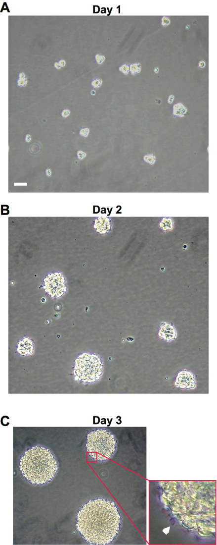
Cultured hNSCs in their proliferative stage will grow as non-adherent neurospheres (Figure 1). Immediately following hNSC passage, there will be many individual cells, which will aggregate and begin to form spheres in the next few days (Figure 1A and 1B). Healthy spheres should be approximately 1-2 mm in diameter 9-10 days following a passage (Figure 1C). Spheres that grow larger than 2 mm will develop a dark center and the growth factors in the medium will not be able to reach the cells in the center of the sphere, resulting in unwanted differentiation. Healthy spheres will appear translucent and display pseudo-cilia around the edges of the sphere (Figure 1C).

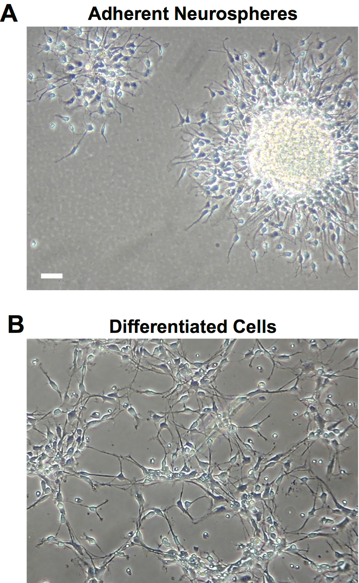
Plating neurospheres on an adherent surface for staining purposes will result in the growth of adherent spheres (Figure 2A). These spheres will touch the surface of the culture vessel and have some projections stretching out on the surface of the vessel, while maintaining a central sphere (Figure 2A). Priming and differentiating the cells require the cells to adhere to a cover slip at the bottom of a cell culture plate, such as a 24-well plate. Following the appropriate priming steps and addition of differentiation medium, the cells will spread out across the surface of the culture vessel and grow as interconnected monolayer of cells (Figure 2B).

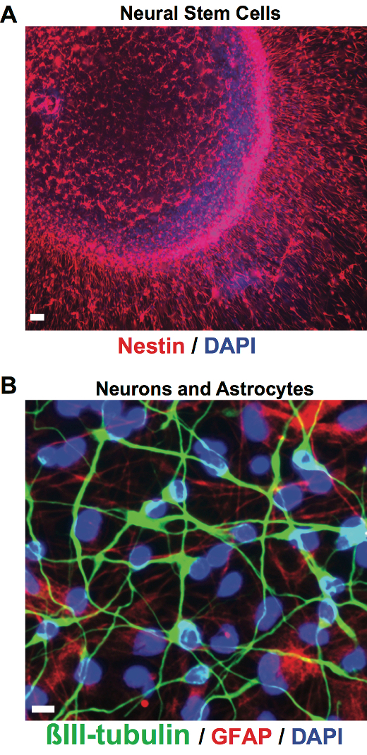
To either confirm the validity of the hNSC culture or phenotype of differentiated cells, fluorescent immunohistochemistry can be used. Nestin is an intermediate filament commonly expressed in NSCs and can be used to verify hNSC culture (Figure 3A). To verify the presence of neurons following differentiation, class III β-tubulin can generally be used to label neurons (Figure 3B). Although the priming step is used to achieve a higher percentage of neurons following differentiation, there will still be astrocytes in the culture. Glial fibrillary acidic protein (GFAP) can be used to label astrocytes in order to quantify which percentage of differentiated cells became astrocytes or neurons.
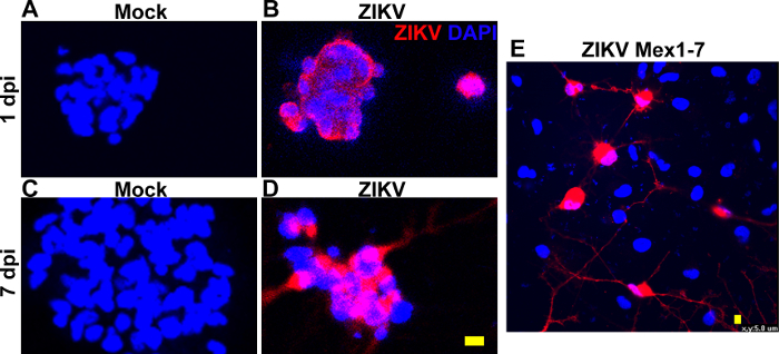
We found that the K048 line of NSCs was infected by both African and Asian strains of ZIKV detected by immunofluorescent staining with a specific ZIKV antibody. The representative images are shown in Figure 4. Mock infection did not elicit a change in hNSC morphology or survival (Figure 4A, 4C). ZIKV tends to stay in the peripheral region of an infected cell one day post a 1-hour infection, but fills the whole cell at 3 to 7 days (Figure 4B, 4D, 4E). Noticeably, with the same MOI (0.1), an estimated 5% infection rate in K048 cells is substantially lower than those recently reported ZIKV infection rate (up to 80%) in human skin-induced NSCs7. This study, reporting 80% infectivity, used the prototype African strain of ZIKV (MR766) that may have adapted a neurotropic phenotype during over 140 passages in mouse brain cells.

Figure 1: Bright field images of hNSC culture. (A-C) Representative bright field images taken at 10X magnification of hNSC culture 1, 4 and 9 days after passaging, respectively. Scale bars: 60 µm. Please click here to view a larger version of this figure.
Figure 2: Bright field images of adherent cultures. (A) Representative bright field images taken at 10X magnification of adherent hNSC culture, 7 days after plating. (B) Representative bright field images taken at 10X magnification of adherent differentiated cells following ELL priming and 9 days of differentiation. Scale bars: 60 µm. Please click here to view a larger version of this figure.
Figure 3: Fluorescent images of hNSCs and differentiated cultures. (A) Representative fluorescent images taken at 20X magnification of adherent hNSCs stained with nuclear marker, DAPI (blue), and stem cell marker Nestin (red). Scale bar: 60 µm (B) Representative fluorescent images taken at 60X magnification of differentiated cells stained with neural marker βIII-tubulin (green), astrocyte maker GFAP (red), and nuclear marker DAPI (blue). Scale bars: 10 µm. Please click here to view a larger version of this figure.
Figure 4: ZIKV infection in human fetal brain-derived NSCs. Human NSCs were either Mock treated (A and C), inoculated for 1 hour with an African lineage strain of ZIKV at MOI of 0.1 (B and D), or with an Asian strain of ZIKV recently isolated from mosquitoes in Mexico in 2016 (E). Immunofluorescent staining detects labeling of ZIKV antibodies in NSCs at one to seven days post inoculation (1 dpi in B, 7dpi in D, and 3 dpi in E). Scale bars 10 µm in A-D, and 5 µm in E. Please click here to view a larger version of this figure.
| DFHGPS media for one T75 | 10 mL |
| TPPS* | 173 µL |
| 200 mM L-Glutamin | 50 µL |
| 10 mg/mL Insulin** | 25 µL |
| 20 µg/mL Epidermal growth factor | 10 µL |
| 20 µg/mL Basic fibroblast growth factor | 10 µL |
| 10 µg/mL Leukemia inhibitor factor | 10 µL |
| 5 mg/mL Heparin | 10 µL |
| * Mixture containing 100 µg/mL Transferrin, 100 µM putresine, 20 nM progesterone, and 30 nM sodium selenite. The mixture is made from concentrated stocks, and aliquots are stored at -80 °C and preferablly used within 6 weeks preparation. The concentrated stocks are 10 mg/mL transferrin, 30 mM putrescine, 10 µM progesterone and 15 µM sodium selenite stored at -80 ˚C. | |
| ** Insulin is dissolved in 0.01 N hydroen chloride, filtered through 0.2 µM low protein binding filter, and stored at 4 ˚C for up to 6 weeks. |
Table 1: Growth factors for New Proliferation Medium.
| dPBS* | 1 mLa | 3 mLb |
| 10% glucose | 60 µL | 180 µL |
| 2.5% Trypsin | 10 µL | 30 µL |
| Dnase** | 5 µL | 15 µL |
| * Dulbecco's phosphate-buffered saline | ||
| ** Deoxyribonuclease | ||
| a for 10 million cells | ||
| b for 30 million cells |
Table 2: Preparation of trypsin.
| Conditioned media | 1 mLa | 3 mLb |
| Trypsin Inhibitor | 10 µL | 30 µL |
| a for 10 million cells | ||
| b for 30 million cells |
Table 3: Preparation of trypsin inhibitor.
| DFHGPS* | 0.7 mLa | 2.1 mLb |
| FBS** 20% | 0.2 mL | 0.6 mL |
| DMSO*** 10% | 0.1 mL | 0.3 mL |
| * DMEM, F12, HEPES, glucose, penicillin-streptomycin | ||
| ** Fetal bovine serum | ||
| *** Dimethyl sulfoxide | ||
| a for 5 million cells in one vial | ||
| b for three vials, 5 million cells/vial |
Table 4: Preparation of freezing medium.
| DFGHPS* for one well in a 24-well plate | 1 mL |
| TPPS** | 17.3 µL |
| 200 mM L-glutamine | 5 µL |
| 10 mg/mL Insulin | 2.5 µL |
| 20 µg/mL Epidermal growth factor | 1 µL |
| 10 µg/mL Leukemia inhibitory factor | 1 µL |
| 1 mg/mL Laminin | 1 µL |
| * Containing DMEM, F12, HEPES, glucose, penicillin-streptomycin | |
| ** Containing 100 µg/mL Transferrin, 100 µM putresine, | |
| 20 nM progesterone, and 30 nM sodium selenite |
Table 5: Preparation of ELL priming medium.
| DFGHPS* for one well in a 24-well plate | 1 mL |
| TPPS** | 17.3 µL |
| 200 mM L-glutamine | 5 µL |
| 10 mg/mL Insulin | 2.5 µL |
| 20 µg/mL basic fibroblast growth factor | 0.5 µL |
| 5 mg/mL Heparin | 0.5 µL |
| 1 mg/mL Laminin | 1 µL |
| * Containing DMEM, F12, HEPES, glucose, penicillin-streptomycin | |
| ** Containing 100 µg/mL Transferrin, 100 µM putresine, | |
| 20 nM progesterone, and 30 nM sodium selenite |
Table 6: Preparation of FHL priming medium.
Discussion
The ability to culture and manipulate hNSCs provides a critical tool that can be used for a variety of purposes from modeling human disease to high throughput drug screening10,11,12,14,15,16,17. Many questions remained to be addressed such as how human fetal brain NSCs or their progeny are susceptible to ZIKV infection, whether different strains of ZIKV infect NSCs with equal efficiency, and how infection during neural development results in microcephaly. We have used this hNSC culture to investigate Zika virus associated neuropathology11,14. By using three strains of cells isolated from three individual donors, we are also able to compare individual differences to susceptibility to the neurological deficits observed following ZIKV infection11. One of the limitations of this technique is the accessibility to hNSC samples and the conditioned medium essential for proper culture. To circumvent this barrier, please contact the corresponding author to arrange a material transfer as the cells used in this protocol are not commercially available.
In vitro studies using hNSCs not only provide unique insight into basic stem cell behavior, they also provide an excellent platform to conduct pre-clinical research. However, the technical challenges of culturing hNSCs can serve as an obstacle in using them as effective and reproducible models. In this protocol, we have outlined key steps and methodologies that can be used to reduce variability in culturing methods and yield a healthy and stable hNSC stem cell culture. A drawback of using hNSC neurosphere culture is that the cells are extremely sensitive to any and all stimuli. Even with the detailed protocol above, it is critical to pay close attention to how much the flasks or dishes of hNSCs are handled, as the subtlest variations on the protocol can result in changes in the neurosphere behavior.
The most crucial part of this methods paper is the cocktail of growth factors and preparation of the different media. If the doubling time of the hNSC culture is very low, or many cells adherie to the culture vessel (when they should be non-adherent), the first step is to ensure that the growth factors being used are the appropriate concentration and not expired. All growth factors in this protocol have a very short shelf life once thawed (5-6 days). Additionally, we have used growth factors from multiple companies and noticed differences in the quality and concentration needed to maintain the culture. Therefore, we highly recommend using the exact growth factors detailed in the Materials Table. The passaging step (steps 3.1-3.22) is also a common source of error. If there are many dead cells following the dissociation process, reduce the number of times of pipetting up and down to dissociate the cells, as too much physical dissociation can result in cell death. Alternatively, if there are many clusters of cells following the dissociation step, increase the vigor or number of times of pipetting up and down, as these clusters will quickly become large spheres that will not remain viable in culture. If the culture is properly maintained, infection with ZIKV is relatively straightforward.
While ZIKV infection is the treatment detailed in this protocol, it is possible to use this hNSC culture system for a wide array of studies. This model system can be used to screen drugs, examine individual susceptibility, and investigate underlying mechanisms of a variety of diseases and developmental disorders14. This system is also ideal for genomic studies since the cultured cells have not been genetically manipulated and show little to no chromosomal abnormalities, even up to 80 passages10,11. We believe this culture system is ideal for modeling in vitro how environmental stimuli such as infection, drugs, alcohol, toxins, etc. can influence development of the nervous system.
Disclosures
The authors have no conflicts of interest to declare.
Acknowledgments
This work was supported by funds from the John S. Dunn Foundation and the Institute for Human Infections and Immunity of the University of Texas Medical Branch (P.W.).
References
- Centers for Disease Control and Prevention. All countries and territories with active Zika virus transmisson. 2016.
- Brasil P, et al. Zika Virus Infection in Pregnant Women in Rio de Janeiro. N Engl J Med. 2016;375(24):2321–2334. doi: 10.1056/NEJMoa1602412. [DOI] [PMC free article] [PubMed] [Google Scholar]
- Hills SL, et al. Transmission of Zika Virus Through Sexual Contact with Travelers to Areas of Ongoing Transmission - Continental United States, 2016. MMWR Morb Mortal Wkly Rep. 2016;65(8):215–216. doi: 10.15585/mmwr.mm6508e2. [DOI] [PubMed] [Google Scholar]
- Centers for Disease Control and Prevention. Facts about microcephaly. 2016.
- Garcez PP, et al. Zika virus impairs growth in human neurospheres and brain organoids. Science. 2016;352(6287):816–818. doi: 10.1126/science.aaf6116. [DOI] [PubMed] [Google Scholar]
- Li C, et al. Zika Virus Disrupts Neural Progenitor Development and Leads to Microcephaly in Mice. Cell Stem Cell. 2016;19(5):672. doi: 10.1016/j.stem.2016.10.017. [DOI] [PubMed] [Google Scholar]
- Tang H, et al. Zika Virus Infects Human Cortical Neural Progenitors and Attenuates Their Growth. Cell Stem Cell. 2016;18(5):587–590. doi: 10.1016/j.stem.2016.02.016. [DOI] [PMC free article] [PubMed] [Google Scholar]
- Wu KY, et al. Vertical transmission of Zika virus targeting the radial glial cells affects cortex development of offspring mice. Cell Res. 2016;26(6):645–654. doi: 10.1038/cr.2016.58. [DOI] [PMC free article] [PubMed] [Google Scholar]
- Jensen JB, Parmar M. Strengths and limitations of the neurosphere culture system. Mol Neurobiol. 2006;34(3):153–161. doi: 10.1385/MN:34:3:153. [DOI] [PubMed] [Google Scholar]
- Wu P, et al. Region-specific generation of cholinergic neurons from fetal human neural stem cells grafted in adult rat. Nat Neurosci. 2002;5(12):1271–1278. doi: 10.1038/nn974. [DOI] [PubMed] [Google Scholar]
- McGrath EL, et al. Differential Responses of Human Fetal Brain Neural Stem Cells to Zika Virus Infection. Stem Cell Reports. 2017;8(3):715–727. doi: 10.1016/j.stemcr.2017.01.008. [DOI] [PMC free article] [PubMed] [Google Scholar]
- Svendsen CN, et al. A new method for the rapid and long term growth of human neural precursor cells. J Neurosci Methods. 1998;85(2):141–152. doi: 10.1016/s0165-0270(98)00126-5. [DOI] [PubMed] [Google Scholar]
- Tarasenko YI, Yu Y, Jordan PM, Bottenstein J, Wu P. Effect of growth factors on proliferation and phenotypic differentiation of human fetal neural stem cells. J Neurosci Res. 2004;78(5):625–636. doi: 10.1002/jnr.20316. [DOI] [PubMed] [Google Scholar]
- Barrows NJ, et al. A Screen of FDA-Approved Drugs for Inhibitors of Zika Virus Infection. Cell Host Microbe. 2016;20(2):259–270. doi: 10.1016/j.chom.2016.07.004. [DOI] [PMC free article] [PubMed] [Google Scholar]
- Jakel RJ, Schneider BL, Svendsen CN. Using human neural stem cells to model neurological disease. Nat Rev Genet. 2004;5(2):136–144. doi: 10.1038/nrg1268. [DOI] [PubMed] [Google Scholar]
- Lopez-Garcia I, et al. Development of a stretch-induced neurotrauma model for medium-throughput screening in vitro: identification of rifampicin as a neuroprotectant. Br J Pharmacol. 2016. [DOI] [PMC free article] [PubMed]
- Mich JK, et al. Prospective identification of functionally distinct stem cells and neurosphere-initiating cells in adult mouse forebrain. Elife. 2014;3:e02669. doi: 10.7554/eLife.02669. [DOI] [PMC free article] [PubMed] [Google Scholar]


