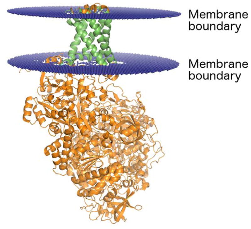
Figure 3.

Ribbon representation of a transmembrane protein (PDB identifier: 1Q16). The membrane boundary planes (displayed in blue) were obtained from the Positioning of Proteins in Membranes (PPM) server [52]. The region of the protein that spans the membrane is shown in green, and the portion of the protein that extends beyond the membrane is shown in orange.
