ABSTRACT
Objectives
The purpose of the present study is to systematically review the accuracy of implant placement with mucosa-supported stereolithographic surgical guide and to find out what factors can influence the accuracy.
Material and Methods
An electronic literature search was performed through the MEDLINE (PubMed) and EMBASE databases. The articles are including human studies published in English from October 2008 to October, 2017. From the examination of selected articles, deviations between virtual planning and actual implant placement were analysed regarding the global apical, global coronal, and angulation position.
Results
A total of 119 articles were reviewed, and 6 of the most relevant articles that are suitable to the criteria were selected. The present data included 572 implants and 93 patients. The result in the present systematic review shows that mean apical global deviation ranges from 0.67 (SD 0.34) mm to 2.19 (SD 0.83) mm, mean coronal global deviation ranges from 0.6 (SD 0.25) mm to 1.68 (SD 0.25) mm and mean angular deviation - from 2.6° (SD 1.61°) to 4.67° (SD 2.68°).
Conclusions
It's clearly shown from most of the examined studies that the mucosa-supported stereolithographic surgical guide, showed not exceeding in apically 2.19 mm, in coronally 1.68 mm and in angular deviation 4.67°. Surgeons should be aware of the possible linear and angular deviations of the system. Accuracy can be influenced by bone density, mucosal thickness, surgical techniques, type of jaw, smoking habits and implant length. Further studies should be performed in order to find out which jaw can have better accuracy and how the experience can influence the accuracy.
Keywords: computer-assisted surgery, dental implant, dimensional measurement accuracy, edentulous jaw, osseointegration, review
INTRODUCTION
Prosthodontic rehabilitation with endosseous dental implants requires precise implant placement for predictable functional, aesthetic and hygienic outcomes [1]. In oral rehabilitation with a traditional surgical approach using osseointegrated implants, the surgeon is often obliged to perform essential muco-periosteal detachments in order to obtain good visibility of the bone structures [2]. A stereolithographic surgical template fabricated using computer assisted planning has been recently introduced in an effort to improve accuracy of implant placement [1]. Stereolithography, a rapid prototyping technology, a newer outcome in dentistry allows the fabrication of surgical guides from three-dimensional computer generated models for precise placement of the implants [3]. The surgical templates fabricated by this technology are preprogramed with individual depth, angulations, mesio-distal and labiolingual positioning of the implant [3]. Among all available cross-sectional radiography, computed tomography (CT) has proven to be an effective diagnostic tool for evaluating bone volume, density, vital anatomical structures, and most importantly in treatment planning and predicting appropriate implant length, size, and position [1]. CT, when coupled with a well-designed radiographic template allows for predictable evaluation of implant sites in relation to available bone, anatomical structures and proposed prosthetic tooth positioning [1]. It offers the opportunity to apply a flapless approach, which on its own may give extra advantages [4]. Shorter surgery duration and less discomfort after surgery were recorded when mucosa-supported surgical guides were used [5]. Therefore, the use of mucosa-supported stereolithographic surgical guides in edentulous patients will increase along with the demand for implant-supported restorations in edentulous patients [4]. Although with all these advantages, everyone must be aware of the fact that there is a variable deviation between the virtual planning and the in vivo position of the implants inserted using computer-aided implantology [6]. The transfer of the virtual three-dimensional implant planning to the surgical field without deviations is unrealistic and it is essential to know the level of accuracy of the method used and the conditions which may influence the degree of accuracy [6].
A previous study reported greater accuracy in the edentulous mandible, which has higher bone density, than in edentulous maxilla [7]. However, in another study, the surgical guide showed higher accuracy in edentulous maxilla because it covered a larger area than the edentulous mandible [8].
To the best of the author's knowledge, present study is the first systematic review about accuracy of stereolithographic surgical guide implant in edentulous patient. The purpose of the present study is to systematically review the accuracy of implant placement with mucosa-supported stereolithographic surgical guide and to find out what factors could influence the accuracy.
MATERIAL AND METHODS
Protocol and registration
The methods of the analysis and inclusion criteria were specified in advance and documented in a protocol. The review was registered in PROSPERO, an international prospective register of systematic reviews [9]. The protocol registration number: CRD42018079905, can be accessed through the following link:
https://www.crd.york.ac.uk/prospero/display_record.php?ID=CRD42018079905
Focus questions
The focus questions were developed according to the problem, intervention, comparison, and outcome (PICO) is presented in Table 1.
Table 1.
PICO table
| Population (P) | Edentulous patients who underwent surgical implant placement using stereolithographic mucosa-supported surgical guide method. |
| Intervention (I) | Implant placement with stereolithographic mucosa-supported surgical guide |
| Comparator or control group (C) | Comparison of planned implant position with actual implant position after surgical implant placement |
| Outcomes (O) | Deviation (distance in mm) between virtual planning and actual implant surgical placement according to global apical, global coronal, and angulation position |
| Focus questions |
Does stereolithographic mucosa-supported surgical guide ensure accurate enough implant placement? What factors could affect the accuracy of implant placement with mucosa-supported stereolithographic surgical guide? |
Types of publications
The review included studies on human published in the English language.
Types of studies
The review included all human prospective studies, retrospective studies and a randomized controlled pilot study published between October 2008 and October 2017, that reported on accuracy of surgical dental implant placement into edentulous jaw using stereolithographic mucosa-supported surgical guide.
Information sources
Information sources were PubMed/Medline and EMBASE databases.
Literature search strategy
To identify the relevant studies, a detailed electronic search was carried out according to PRISMA guidelines [10] within a PubMed/Medline and EMBASE databases using different combinations of the following keywords: "Implant", "Surgical guide", "Edentulous", "Accuracy", and "Stereolithographic". The search details are as followed: implant [All Fields] AND ("surgical procedures, operative" [MeSH Terms] OR ("surgical" [All Fields] AND "procedures"[All Fields] AND "operative" [All Fields]) OR "operative surgical procedures" [All Fields] OR "surgical" [All Fields]) AND guide [All Fields] AND ("mouth, edentulous" [MeSH Terms] OR ("mouth" [All Fields] AND "edentulous" [All Fields]) OR "edentulous mouth" [All Fields] OR "edentulous" [All Fields]) AND accuracy [All Fields] AND stereolithographic [All Fields]. The search was performed only for English articles which were published from October 2007 to October 2016.
Selection of studies
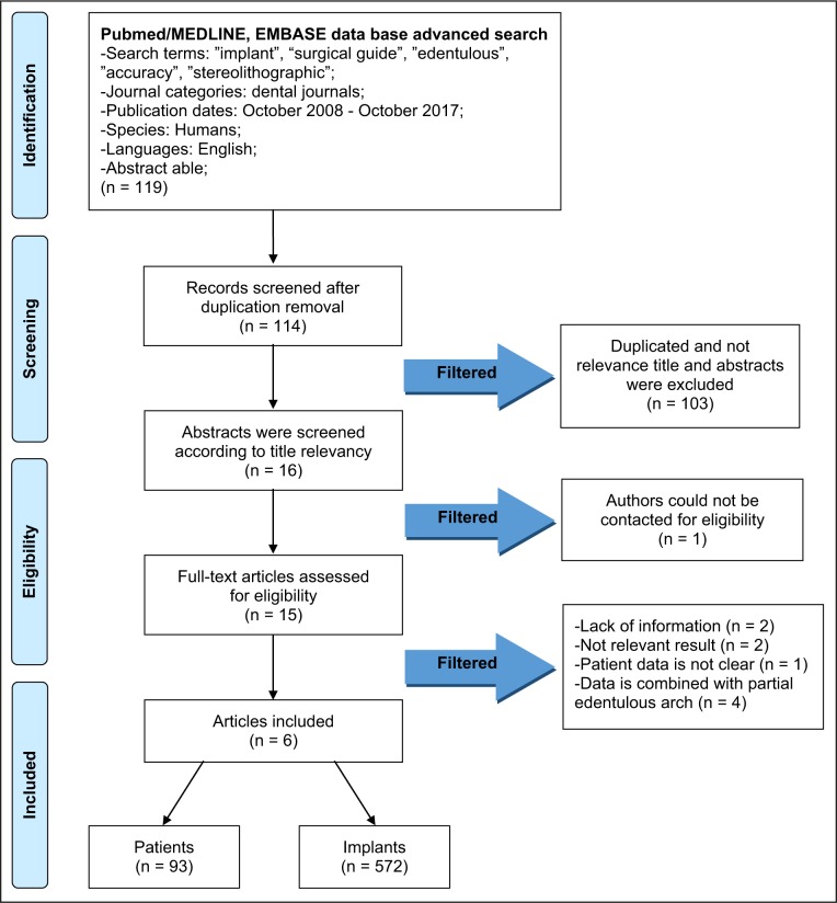
The resulting articles were independently subjected to clarify inclusion and exclusion criteria by two reviewers. First titles and abstracts were screened and finally full reports were obtained for all the studies that were deemed eligible for inclusion in this paper (Figure 1).
Figure 1.
PRISMA flow diagram.
Population
Edentulous patients who underwent surgical implant placement using stereolithographic mucosa-supported surgical guide method.
Inclusion and exclusion criteria
Inclusion criteria for the selection were:
Human studies analysing accuracy of surgical dental implant placement into edentulous jaw using stereolithographic mucosa-supported surgical guide.
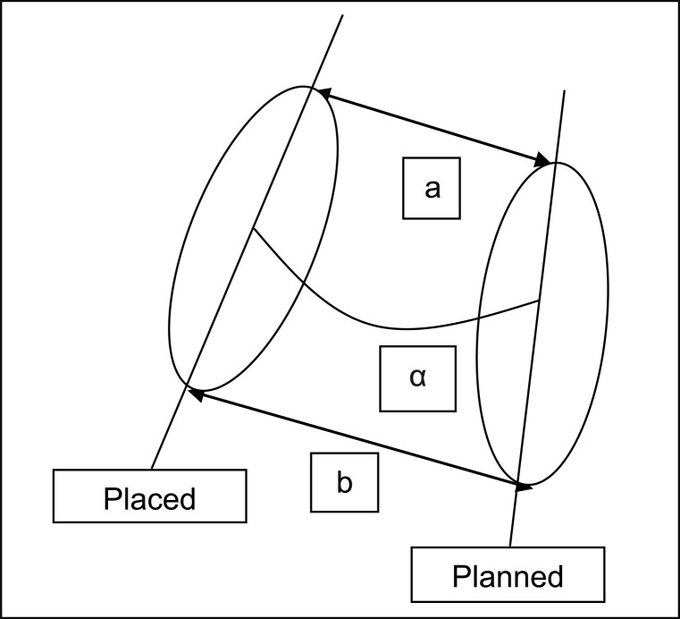
Surgical implant placement accuracy was evaluated according to global deviation which is defined as the three-dimensional distance between the coronal and apical centres of the planned and placed implants.
Exclusion criteria for the selection were:
In vitro studies using mucosa-supported surgical guide implant system.
Studies that placing implants using mucosa-supported surgical guide implant system was done to partial-edentulous jaw.
Studies that placing implants was done by using tooth-supported surgical guide.
Studies that placing implants was done by using bone-supported surgical guide.
Sequential search strategy
The selected articles were subjected independently to clear inclusion and exclusion criteria. The reviewer resolved the ambiguous point by taking advice from an experienced senior reviewer. Following the initial literature search, all articles were chosen according title relevancy, considering the exclusion criteria. Following, studies were excluded based on irrelevant data obtained from the abstracts. The final stage of screening involved reading the full texts and confirming each study's eligibility based on the inclusion criteria.
Data collection process
Data were independently extracted from articles in the form of variables according to the aim and themes of the present review as listed shown below.
Data items
The following data were obtained from the included articles:
"Author(s)" - revealed the author.
"Year of publication" - revealed the year of publication.
"Patients" - describes the patients who were treated by mucosa-supported surgical guide implant system.
"Implant number" - indicates the implant numbers which were placed by mucosa-supported surgical guide implant system to patient.
"Jaw" - describes the jaw of patient that were placed implant by using mucosa-supported surgical guide implant system.
"Smoking habit" - relevant to smoking habit.
"Surgical technique" - indicates surgical technique that clinician is using fixation screw to fix surgical template.
"Deviation according to global apical, global coronal, and angulation" - indicates deviation of placed implant which were placed by using mucosa-supported surgical guide implant system.
Risk of bias assessment
The risk of bias assessment was conducted using Cochrane Risk of Bias Tool [11] for randomized clinical trial and The Newcastle-Ottawa Scale: Cohort Studies [12] for nonrandomized included studies independently.
Synthesis of results
Relevant data of interest on the previously stated variables were collected and organised into table.
Statistical analysis
No meta-analyses could be performed due to the heterogeneity between the studies (different study designs, control groups, and observation periods). Parametric data were expressed as mean and standard deviation (M [SD]).
RESULTS
Study selection
Total of 119 publications were retrieved and 16 articles were reviewed in fully. Initial exclusion was done by duplication and not relevant titles and abstracts. Among them, six publications were selected for the purpose of the systematic review. One publication was unable to screen. According to inclusion and exclusion criteria, finally six publications were included, in which have had evaluated a total of 572 implants from 93 patients.
Exclusion of studies
Exclusion of articles are as followed: articles [1,13] that lack of information of deviation, articles [14,15] with not relevant result and article that patient data is not clear [16]. Articles [17-20] that data is combined with partial edentulous arch.
Risk of bias within studies
The risk of bias that indicated within this article is collected and organised in Table 2 and Table 3. Table 2 shows evaluation of the risk of bias for nonrandomized included studies, using The Newcastle-Ottawa Scale: Cohort Studies Tool [12]. Two studies [4,5] were evaluated with score 6 and three [6,21,22] with score 7. The risk of bias assessment for randomized clinical trial [23] conducted using Cochrane Risk of Bias Tool [11] revealed low risk study judgement.
Table 2.
Evaluation of the risk of bias for nonrandomized included studies, using The Newcastle-Ottawa Scale: Cohort Studies Tool [12]
| Studies | Selection | Comparability | Outcome | Total score |
|---|---|---|---|---|
| Van et al. [4] | ★★★ | ★ | ★★ | 6 |
| Ochi et al. [5] | ★★★★ | ★ | ★ | 6 |
| Cassetta et al. [6] | ★★★★ | ★ | ★★ | 7 |
| Cassetta et al. [21] | ★★★★ | ★ | ★★ | 7 |
| D'haese et al. [22] | ★★★ | ★ | ★★★ | 7 |
Table 3.
The risk of bias assessment for randomized clinical trial [24] conducted using Cochrane Risk of Bias Tool [11]
| Study |
Year of publication |
Random sequence generation |
Allocation concealment |
Blinding of participant's and personnel |
Blinding of outcomes assessment |
Incomplete outcome data |
Selective reporting |
Other bias |
|---|---|---|---|---|---|---|---|---|
| Cassetta et al. [23] | 2017 | + | + | + | + | + | + | + |
+ = low risk; ? = unclear risk; - = high risk.
Study characteristics
Van et al. [4] compared accuracy of surgical dental implant placement into edentulous jaw using stereolithographic mucosa-supported surgical guide by surgeons who do not have experience in guided surgery and surgeons who were experienced (Table 4). Experienced surgeons data were retrieved for comparison from Vercruyssen et al. [24] study. The group of surgeons who did not have experience in surgical guide were those who followed a 3-year post- graduate training in periodontology. They all had a limited surgical experience in implant therapy (30 - 80 implants placed). They had placed implants strictly series of steps (preparation of scan prosthesis, scanning procedure, pre-surgical planning, surgery, accuracy analysis) from November 2011 to January 2014. Procedures were approved under supervision of experienced surgeons. For example, in pre-surgical planning procedure, pre-surgical planning was confirmed by both of experienced periodontologist and prosthodontist. They both used same treatment and accuracy analysis protocol. The result showed that for inexperienced group, the mean apical deviation was 1.102 (0.531) mm, mean coronal deviation was 0.871 (0.495) mm, mean angular deviation was 2.788° (1.475°). However for experienced group, the result of mean apical deviation was 1.598 (0.701) mm, mean coronal deviation was 1.384 (0.643) mm, and mean angular deviation was 2.705° (1.358°). Only in angulation, the inexperienced group scored less than the experienced clinician [4]. The conclusion of the article implies that the surgical experience had no major impact on accuracy of implant placement and inaccuracy was mainly resulted from mal positioning of the guide.
Table 4.
Characteristics of included studies
| Study | Patient | Implant no. |
Guide system |
Rx | Jaw |
Smoking habit |
Surgical technique |
Deviation |
Dev |
Deviation according to jaw (mean [SD]) |
Deviation according to surgical technique (mean [SD]) |
Deviation according to smoking habit (mean [SD]) |
|||||
|---|---|---|---|---|---|---|---|---|---|---|---|---|---|---|---|---|---|
|
Apical deviation (mm) |
Coronal deviation (mm) |
Ang (°) |
|||||||||||||||
| Mean (SD) | Maxilla | Mandible | Fixed | Non-fixed | Yes | No | |||||||||||
| Van et al. [4] | 16 | 75 | Simplant | CBCT |
Maxilla and mandible |
- | Fixed | 1.1 (0.53) | 0.87 (0.5) | 2.79 (1.47) | - | - | - | - | - | - | - |
| Ochi et al. [5] | 15 | 30 | Nobelbiocare | CBCT | Mandible | - | Fixed | 1.08 (0.47) | 0.89 (0.44) | - | - | - | - | - | - | - | - |
| Cassetta et al. [6] | 11 | 95 | Simplant | CT | Maxilla |
Yes (6)/ no (5) |
Fixed (8)/ non-fixed (3) |
2.15 (0.81) | 1.65 (0.56) | 4.62 (2.74) | - | - | 2.11 (0.75) | 2.22 (0.91) | 2.28 (0.84) | 2.04 (0.79) | 2.11 (0.75) |
| - | - | 1.66 (0.57 | 1.64 (0.56) | 1.8 (0.51) | 1.52 (0.58 | 1.66 (0.57) | |||||||||||
| - | - | 4.1 (2.43) | 5.44 (3.02) | 4.41 (3) | 4.79 (2.52) | 4.1 (2.43) | |||||||||||
| Cassetta et al. [21] | 28 | 225 | Simplant | CT |
Maxilla and mandible |
Yes (14)/ no (14) |
Fixed (18)/ non-fixed (10) |
2.19 (0.83) | 1.68 (0.6) | 4.67 (2.68) |
AD (mm) |
2.12 (0.78) | 2.25 (0.88) | 2.09 (0.75) | 2.26 (0.89) | 2.27 (0.83) | 2.08 (0.8) |
|
CD (mm) |
1.68 (0.51) | 1.64 (0.71) | 1.66 (0.58) | 1.68 (0.6) | 1.83 (0.58) | 1.54 (0.56) | |||||||||||
|
Ang (°) |
4.36 (2.9) | 5.46 (2.03) | 4.09 (2.4) | 5.62 (2.8) | 4.5 (2.87) | 4.92 (2.52) | |||||||||||
| D'haese et al. [22] | 13 | 77 | Astra Tech AB | CT | Maxilla |
Yes (5)/ no (8) |
Fixed | 1.13 (0.52) | 0.91 (0.44) | 2.6 (1.61) | - | - | - | - | - | - | - |
| Cassetta et al. [23] |
5 exp |
37 | 3Diemme | CT |
Maxilla and mandible |
No | Fixed | 0.67 (0.34) | 0.6 (0.25) | 3.21 (1.57) | - | - | - | - | - | - | - |
|
5 inexp |
33 | 1.02 (0.44) | 0.75 (0.18) | 3.07 (2.7) | - | - | - | - | - | - | - | ||||||
Ang = angulation; Dev = deviation; exp = inexperienced; inexp = inexperienced; Rx = radiological technique; CT = computer topography; CBCT = cone-beam computer topography; SD = standard deviation; AD = apical deviation; CD = coronal deviation.
Similar pilot study was taken place in 2017 by Cassetta et al. [23], the aim of this research was to compare the accuracy between group of experienced surgeons (expert in computer-guided implantology) and group of inexperienced surgeons (none the less expert in standard implantology). Ten of healthy edentulous (maxilla and mandible) patients were enrolled, then divided into two different groups. Group I was comprised of experienced surgeons who had experienced in placing at least 500 implants using computer-guided implantology. Group II was comprised of inexperienced surgeons who had no experience of computer-guided implantology, but at least 500 were implanted inserted using conventional implantology. Unlike study of Van et al. [4], the result came out to be very different. For inexperienced group, the mean apical deviation was 1.02 (0.44) mm, mean coronal deviation was 0.75 (0.18) mm, mean angulation was 3.07° (2.7°). For experienced group, the mean apical deviation was 0.67 (0.34) mm, mean coronal deviation was 0.6 (0.25) mm, mean angular deviation was 3.21° (1.57°). The inexperienced group had performed better only in terms of the angular deviation results [23]. Perhaps there had been difference in result between Cassetta et al. [23] and Van et al. [4]. The result of Cassetta et al. [23] had some limits though in which a pilot study was performed for a few patients only giving less creditability. Cassetta et al. [23] concluded in his article that inexperienced group's resulting in higher coronal and apical deviation were due to the error from position.
Cassetta et al. [6,21] analysed factors that can influence the accuracy of mucosa supported surgical guide technique. In the article [21], the author compared the results of accuracy according to surgical technique, jaw, and smoking habits. Also, author took measurement of mucosal thickness to find out the relation between mucosal thickness and smoking habits. According to the result, accuracy of implant insertion using a mucosa-supported stereolithographic surgical guide in completely edentulous patients was mainly affected by variable factors such as surgical technique, jaw, and smoking habits. The result also showed that a high level of accuracy was obtained when fixed mucosa-supported stereolithographic surgical guides were used in the maxilla. This was most likely attributable to the fact that fixation screws and greater surface support reduced any possible displacement of the guide during surgery [21]. In addition, the mucosal thickness also affected accuracy, thicker the mucosa, higher the deviation values were. Similar research was done by Cassetta et al. [6], but this study was constricted to only maxilla. It is interesting to know that study showed a significant difference when comparing the global coronal deviations among smokers and non-smokers.
Ochi et al. [5] in their study discussed more precisely factors that could affect accuracy of implant placement with mucosa-supported stereolithographic surgical guides in edentulous mandibles. The study covered both of the model and the patient study and yet only the patient study was used for this research after considering inclusion and exclusion criteria: 30 implants were placed among the 15 patients, global deviation in the neck was 0.89 (0.44) mm and for the apex, it was 1.08 (0.47) mm. According to Spearman's rank correlation coefficients for each deviation and bone density, mucosal thickness, and area of supporting mucosa, had showed that there were significant negative correlations between bone density and depth deviations at the implant neck and apex. Author assumed that when surgical guides were used, the implants tended to be placed more superficially. Closer to the planned position when they were deeply inserted on lower density bone sites. Also there was a significant positive correlation between mucosal thickness and the global deviation at implant apex. Study had showed that increased mucosal thickness led to higher global deviation at the implant apex. This was due to thick mucosa causing movement of the guide and resulting in positional discrepancy between CT scan of the radiographic guide and surgical guide during surgery.
D'haese et al. [22] performed a prospective study on the accuracy of mucosally supported stereolithographic surgical guide in fully edentulous maxilla. Seventy eight implants were installed in 13 edentulous maxilla, and 77 implants were analysed among 78 implants. One of the implant was lost shortly after the insertion due to abscess formation which caused by remnants of impression material. The result showed that mean global coronal deviation of 0.91 (0.44) mm, mean global apical deviation of 1.13 (0.52) mm, and mean angular deviation of 2.6° (1.61°). And the study included result of differences in coronal and apical inter implant position, comparing the virtual distance with the in vivo inter implant distance after surgery on a patient level. The result showed that the mean coronal deviation of 0.18 (0.15) mm mean apical deviation was 0.33 (0.28) mm which was substantially lower than the global coronal and apical deviation. The significant difference was observed when comparing the global apical deviation of short and long implants. Shorter implants (8 mm) showed mean global coronal deviation lower than 0.75 mm while longer implants (15 mm) showed about 1 mm. It was similar in mean global apical deviation as well; shorter implants (8 mm) showed less than 1mm while longer implants (15 mm) showed more than 1.75 mm. Shorter implants showed significantly lower apical deviations compared to that of longer ones. Author found that optimal positioning of the fixation screws and support and stability of the guide on the mucosa are very important issue to obtain better accuracy.
DISCUSSION
In the current systematic review, mucosa-supported stereolithographic surgical guide's accuracy and factors which could affect guided surgery were assessed.
In order to evaluate the accuracy of placing implant using mucosa-supported stereolithographic surgical guide, as a parameter, the global deviation is defined as the three-dimensional distance between the coronal and apical centres of the planned and placed implants (Figure 2). The angular deviation is calculated as the three-dimensional angle between the longitudinal axes of both. In Van et al. [4], other deviation values were included which are as follows: apical depth, coronal depth, bucco-lingual, mesio-distal. In D'haese et al. [22] study inter implant deviation was included. But in order to standardize the results, it was omitted.
Figure 2.
Three-dimensional evaluation of planned and placed implant positions.
a = global apical; α = angle; b = global coronal.
The result in the present systematic review showed that mean apical global deviation was from 0.67 (0.34) mm to 2.19 (0.83) mm, mean coronal global deviation was from 0.6 (0.25) mm to 1.68 (0.25) mm and mean angular deviation was from 2.6° (1.61°) to 4.67° (2.68°). According to current data, apical deviation was higher than coronal deviation. It can be explained by the fact that implant guidance is optimal in the coronal part of the prepared osteotomies because of the limited effect of the angular deviation on the global deviation, which increases at a larger distance, that is, further into the bone [22].
Guided surgery could be very helpful for those of surgeons who do not have much of surgical experience of dental implant treatment. Van et al. [4] made a study about how possibly experience of placing implant could affect the accuracy of guided surgery via mucosa-supported stereolithographic surgical guide. According to current data, only in angulation, the inexperienced group scored less than that of the experienced surgeon's. And author had concluded that surgical experience had no major influence on accuracy of implant placement and malpositioning of the guide was the main factor of inaccuracy. This result could be achieved from each step in the procedures for planning and surgery were done under supervision of trained surgeons. In contrast, Cassetta et al. [23] had their randomized controlled pilot study demonstrated that the inexperienced group performed better only in the angular deviation results. And experienced group showed better accuracy in global apical, coronal deviation. The major difference was observed in the positioning error between experienced and inexperienced group. In the aspect of how the experience could affect the accuracy, this present review included limited number of studies. Therefore, a further research study should be conducted.
The factors retrieved from independent studies that could influence the accuracy of mucosa-supported stereolithographic surgical guides suggested are as follows: (1) bone density, (2) mucosal thickness, (3) surgical techniques, (4) the jaw, (5) smoking habits, and (6) implant length.
Ochi et al. [5] suggested that there was negative correlations between bone density and depth deviations. Of the 30 implants placed, 21 were placed more superficially and 9 were placed more deeply, yet there was depth control of the drills. It can be assumed that when surgical guides are used, the implants tend to be placed more superficially than planned and they come close to the planned position when inserted more deeply at bone sites with lower bone density [5]. Also, Ochi et al. [5] suggested that there is positive correlation between mucosal thickness and the global deviation at the implant apex implied that an increased mucosal thickness led to higher global deviation at the implant apex. As the mucosal thickness gets thicker, the global deviation at the apex would increase. This suggestion is supported by Cassetta et al. [6,21] studies. Authors [6,21] had found that smokers tends to have more deviation than non-smokers and this was because smokers had a significantly thick mucosal biotype compared to nonsmokers. Cassetta et al. [6,21] also suggested that surgical technique affected accuracy, as the fixed surgical guides provided a higher level of accuracy. This was a result of the reduced possibility of displacement of the guide. D'haese et al. [22] demonstrated that control of the proper fit of the surgical guide was achieved by optimal positioning of the fixation screws and this would decrease the value of deviation. In the Cassetta et al. [21] study, mandible may allow a greater possibility of guide displacement than maxilla since the reduced area of support. D'haese et al. [22] found that statistically significant difference was found when comparing the global apical deviation of short versus long implants, as implant length got longer, the global apical deviation got bigger. This could have clinical consequences when more implants are installed in anatomically compromised regions [22]. There were also more factors could influence the deviation of mucosa-supported stereolithographic surgical guide. However these factors needed to be carefully studied further to find correlation with deviation. Virtual dental implant operation planning and template-guided surgery has gained attention as a method of improving the predictability of dental implant placement. Nokar et al. [25] determined and compared the accuracy of an advanced surgical template based on computer-aided design/computer-assisted manufacture (CAD/CAM) with the conventional surgical template. It is interesting to note, that the average differences between the planned and actual entry points in the mesiodistal and buccolingual directions, lengths, and angles of the implants and the osteotomy showed a considerable reduction in the CAD/CAM group versus the conventional group (P < 0.005). Authors concluded that the accuracy of implant placement was improved using an innovative CAD/CAM surgical template. However implant position depend on the individual anatomy of the jaws and the ability to place the CAD/CAM-guided surgical template in the proper position [26]. Zhou et al. [27] compared how radiology method could affect the accuracy of guided surgery in his systematic review and meta-analysis. Based on his study result, he concluded that there is no such significant difference in the accuracy between CT and CBCT.
CONCLUSIONS
It's clearly shown from most of the examined studies that the mucosa-supported stereolithographic surgical guide, showed not exceeding in apically 2.19 mm, in coronally 1.68 mm and in angular deviation 4.67°. Surgeons should be aware of the possible linear and angular deviations of the system. Accuracy can be influenced by bone density, mucosal thickness, surgical techniques, type of jaw, smoking habits and implant length. Further studies should be performed in order to find out which jaw can have better accuracy and how the experience can influence the accuracy.
Acknowledgments
ACKNOWLEDGMENTS AND DISCLOSURE STATEMENTS
The authors confirm that this article content has no conflict of interest with any financial organization regarding the material discussed in the article.
REFERENCES
- 1.Al-Harbi SA, Sun AY. Implant placement accuracy when using stereolithographic template as a surgical guide: preliminary results. Implant Dent. 2009 Feb;18(1):46-56. [DOI] [PubMed]
- 2.Giordano M, Ausiello P, Martorelli M, Sorrentino R. Reliability of computer designed surgical guides in six implant rehabilitations with two years follow-up. Dent Mater. 2012 Sep;28(9):e168-77. [DOI] [PubMed]
- 3.Ramasamy M, Giri, Raja R, Subramonian, Karthik, Narendrakumar R. Implant surgical guides: From the past to the present. J Pharm Bioallied Sci. 2013 Jun;5(Suppl 1):S98-S102. [DOI] [PMC free article] [PubMed]
- 4.Van de Wiele G, Teughels W, Vercruyssen M, Coucke W, Temmerman A, Quirynen M. The accuracy of guided surgery via mucosa-supported stereolithographic surgical templates in the hands of surgeons with little experience. Clin Oral Implants Res. 2015 Dec;26(12):1489-94. [DOI] [PubMed]
- 5.Ochi M, Kanazawa M, Sato D, Kasugai S, Hirano S, Minakuchi S. Factors affecting accuracy of implant placement with mucosa-supported stereolithographic surgical guides in edentulous mandibles. Comput Biol Med. 2013 Nov;43(11): 1653-60. [DOI] [PubMed]
- 6.Cassetta M, Pompa G, Di Carlo S, Piccoli L, Pacifici A, Pacifici L. The influence of smoking and surgical technique on the accuracy of mucosa-supported stereolithographic surgical guide in complete edentulous upper jaws. Eur Rev Med Pharmacol Sci. 2012 Oct;16(11):1546-53. [PubMed]
- 7.Ozan O, Orhan K, Turkyilmaz I. Correlation between bone density and angular deviation of implants placed using CT-generated surgical guides. J Craniofac Surg. 2011 Sep;22(5):1755-61. [DOI] [PubMed]
- 8.Pettersson A, Kero T, Gillot L, Cannas B, Fäldt J, Söderberg R, Näsström K. Accuracy of CAD/CAM-guided surgical template implant surgery on human cadavers: Part I. J Prosthet Dent. 2010 Jun;103(6):334-42. [DOI] [PubMed]
- 9.Chien PF, Khan KS, Siassakos D. Registration of systematic reviews: PROSPERO. BJOG. 2012 Jul;119(8):903-5. [DOI] [PubMed]
- 10.Moher D, Liberati A, Tetzlaff J, Altman DG. PRISMA Group. Preferred reporting items for systematic reviews and meta-analyses: the PRISMA statement. Int J Surg. 2010;8(5):336-41. [DOI] [PubMed]
- 11.Higgins JPT, Green S. Cochrane Handbook for Systematic Reviews of Interventions. URL: http://www.cochrane.org/cochrane-interventions-handbook.
- 12.Wells GA, Shea B, O'Connell D, Peterson J, Welch V, Losos M, Tugwell P. The Newcastle-Ottawa Scale (NOS) for assessing the quality of nonrandomised studies in meta-analyses. URL: http://www.ohri.ca/programs/clinical_epidemiology/oxford.asp.
- 13.Giordano M, Ausiello P, Martorelli M. Accuracy evaluation of surgical guides in implant dentistry by non-contact reverse engineering techniques. Dent Mater. 2012 Sep;28(9):e178-85. [DOI] [PubMed]
- 14.Cassetta M. Immediate loading of implants inserted in edentulous arches using multiple mucosa-supported stereolithographic surgical templates: a 10-year prospective cohort study. Int J Oral Maxillofac Surg. 2016 Apr;45(4): 526-34. [DOI] [PubMed]
- 15.Cassetta M, Di Mambro A, Giansanti M, Stefanelli LV, Barbato E. How does an error in positioning the template affect the accuracy of implants inserted using a single fixed mucosa-supported stereolithographic surgical guide? Int J Oral Maxillofac Surg. 2014 Jan;43(1):85-92. [DOI] [PubMed]
- 16.Ozan O, Turkyilmaz I, Ersoy AE, McGlumphy EA, Rosenstiel SF. Clinical accuracy of 3 different types of computed tomography-derived stereolithographic surgical guides in implant placement. J Oral Maxillofac Surg. 2009 Feb;67(2): 394-401. [DOI] [PubMed]
- 17.Valente F, Schiroli G, Sbrenna A. Accuracy of computer-aided oral implant surgery: a clinical and radiographic study. Int J Oral Maxillofac Implants. 2009 Mar-Apr;24(2):234-42. [PubMed]
- 18.Cassetta M, Stefanelli LV, Giansanti M, Calasso S. Accuracy of implant placement with a stereolithographic surgical template. Int J Oral Maxillofac Implants. 2012 May-Jun;27(3):655-63. [PubMed]
- 19.Cassetta M, Stefanelli LV, Giansanti M, Di Mambro A, Calasso S. Depth deviation and occurrence of early surgical complications or unexpected events using a single stereolithographic surgi-guide. Int J Oral Maxillofac Surg. 2011 Dec;40(12):1377-87. [DOI] [PubMed]
- 20.Arisan V, Karabuda ZC, Ozdemir T. Accuracy of two stereolithographic guide systems for computer-aided implant placement: a computed tomography-based clinical comparative study. J Periodontol. 2010 Jan;81(1):43-51. [DOI] [PubMed]
- 21.Cassetta M, Giansanti M, Di Mambro A, Stefanelli LV. Accuracy of positioning of implants inserted using a mucosa-supported stereolithographic surgical guide in the edentulous maxilla and mandible. Int J Oral Maxillofac Implants. 2014 Sep-Oct;29(5):1071-8. [DOI] [PubMed]
- 22.D'haese J, Van De Velde T, Elaut L, De Bruyn H. A prospective study on the accuracy of mucosally supported stereolithographic surgical guides in fully edentulous maxillae. Clin Implant Dent Relat Res. 2012 Apr;14(2):293-303. [DOI] [PubMed]
- 23.Cassetta M, Bellardini M. How much does experience in guided implant surgery play a role in accuracy? A randomized controlled pilot study. Int J Oral Maxillofac Surg. 2017 Jul;46(7):922-930. [DOI] [PubMed]
- 24.Vercruyssen M, De Laat A, Coucke W, Quirynen M. An RCT comparing patient-centred outcome variables of guided surgery (bone or mucosa supported) with conventional implant placement. J Clin Periodontol. 2014 Jul;41(7):724-32. [DOI] [PubMed]
- 25.Nokar S, Moslehifard E, Bahman T, Bayanzadeh M, Nasirpouri F, Nokar A. Accuracy of implant placement using a CAD/CAM surgical guide: an in vitro study. Int J Oral Maxillofac Implants. 2011 May-Jun;26(3):520-6. [PubMed]
- 26.Kero T, Pettersson A, Fäldt J, Andersson M, Gillot L, Cannas B, Näsström K, Söderberg R. Virtual variation simulation of CAD/CAM template-guided surgeries performed on human cadavers: Part II. J Prosthet Dent. 2010 Jul;104(1):48-55. [DOI] [PubMed]
- 27.Zhou W, Liu Z, Song L, Kuo CL, Shafer DM. Clinical Factors Affecting the Accuracy of Guided Implant Surgery-A Systematic Review and Meta-analysis. J Evid Based Dent Pract. 2018 Mar;18(1):28-40. [DOI] [PubMed]


