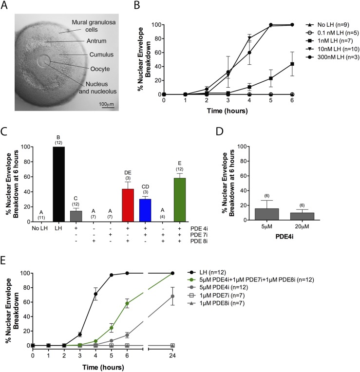
Figure 2.
PDE4, PDE7, and PDE8 act together to suppress premature NEBD in follicle-enclosed oocytes. (A) A follicle before exposure to LH or a PDE inhibitor; an intact nucleus is visible in the oocyte. (B) Time course of NEBD in response to various concentrations of LH. (C) Percent NEBD at 6 hours after treatment of follicles with a saturating concentration of LH (300 nM), individual PDE inhibitors, pairs of inhibitors, or all three inhibitors together (5 μM rolipram, 1 μM PDE7i, 1 μM PDE8i). (D) Similar percent NEBD in response to 5 and 20 μM rolipram. (E) Time course of NEBD in response to LH (300 nM), individual PDE inhibitors [concentrations as in (C)], or a mixture of the three inhibitors. Numbers in parentheses indicate the number of independent experiments. Different letters indicate significant differences (P < 0.05).

