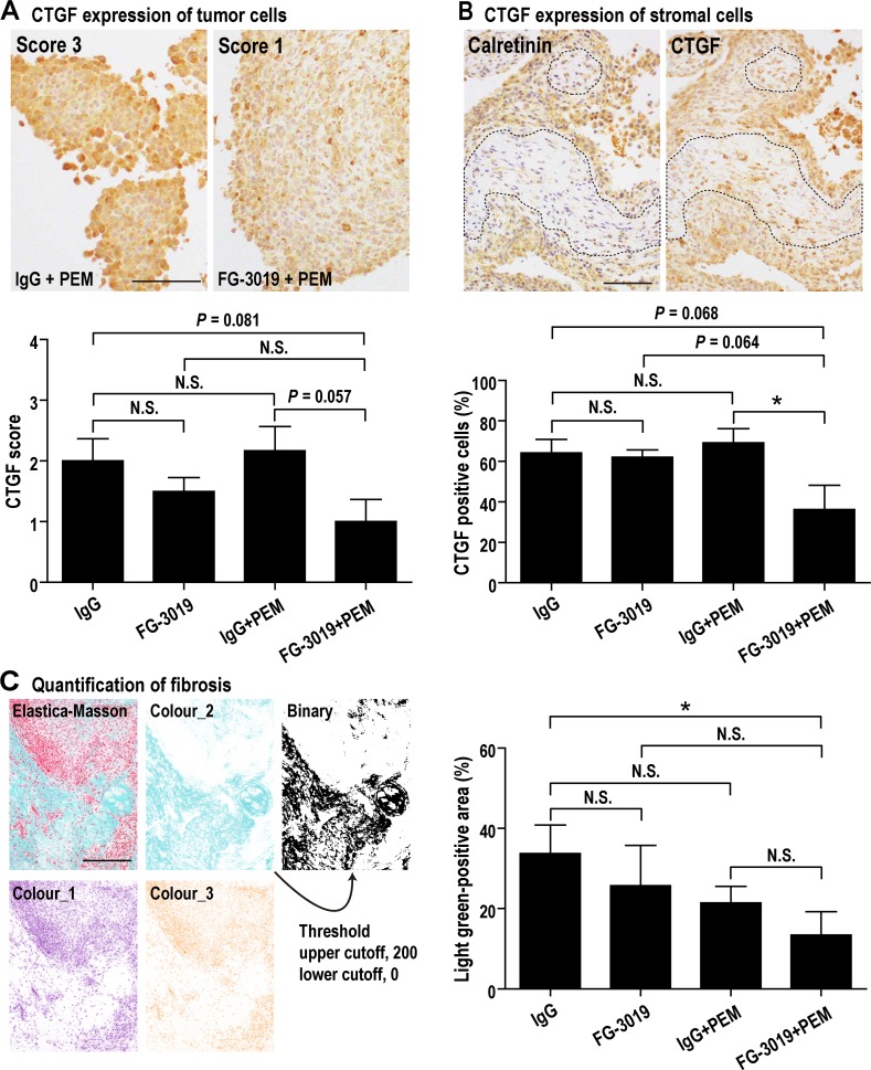
Figure 5. Effects of FG-3019 on CTGF expression and fibrosis.
(A) CTGF expression of tumor cells. Tumor (mesothelioma) cells were diffusely positive for CTGF. Degree of CTGF immunostaining in tumor cells (CTGF score) was evaluated as follows: 0, no staining or no mesothelioma cells; 1, weak; 2, moderate; 3, strong. CTGF score of FG-3019 + PEM group was lower than IgG + PEM group. (B) CTGF expression of stromal cells. For judging stromal portion in tumor (dotted line), we used H&E staining and immunohistochemical staining of calretinin (calretinin is a positive marker for mesothelioma). Stromal cells were negative for calretinin and spindle shaped (mesothelioma cells were cuboidal). Stromal cells were sporadically positive for CTGF, so we calculated the percentage of CTGF-positive stromal cells. The percentage of FG-3019 + PEM group was lower than the other group. (C) Tumor fibrosis (Elastica-Masson staining). Fibrosis, which is shown as light green positive, was quantified with ImageJ as described in Materials and Methods. FG-3019 + PEM significantly decreased the fibrosis of tumor. N = 6 (IgG), N = 6 (FG-3019), N = 6 (IgG + PEM), N = 6 (FG-3019 + PEM) in (A)–(C); means ± SEM, *P < 0.05. N is the number of mice used. Pathological specimens generated for each mouse and analyzed. Bar; 50 μm in (A), 100 μm in (B), 200 μm in (C). N.S., not significant.

