History
Birkett was the first to discover and document femoral head fractures in 1869 while performing a postmortem dissection [1]. These high-energy injuries are infrequent and occur in conjunction with 5% to 15% of all posterior hip dislocations [3, 5, 11]. Femoral head fractures pose a management challenge; these can be technically difficult to address.
In 1954 Stewart and Milford described four grades of dislocations of the hip; dislocations with a fracture of the head or neck of the proximal femur were classified as Grade IV [14].
In 1957, Garrett Pipkin, an orthopaedic surgeon from Kansas City, Missouri, further subclassified Stewart and Milford Grade IV injuries. This classification system of femoral head fractures came to be known as the Pipkin classification system [10]. Pipkin developed this classification system based on his observations of 24 patients (25 fractures). Twenty-two of the 25 fractures were attributable to motor vehicle collisions [10].
Purpose
As noted above, the rationale for development of Pipkin’s classification system was to subclassify Grade IV fracture-dislocations of the hip as classified by Stewart and Milford [14].
Pipkin was hopeful that his classification system would shine further light on Grade IV injuries, as there had been little published regarding the outcomes and sequelae of these injuries. Highlighted sequelae include posttraumatic arthritis, osteonecrosis, heterotopic ossification, and sciatic nerve injury. Additionally, although not the primary purpose, he was able to provide a management scheme for these injuries with the use of his classification.
Description
Pipkin classified these injuries as one of four types [10]: Type 1 is defined as a hip dislocation with a femoral head fracture caudad to the fovea capitis femoris (Fig. 1); Type 2 is defined as a hip dislocation with a femoral head fracture cephalad to the fovea capitis femoris (Fig. 2); Type 3 fractures are a Type I (Fig. 3) or Type II (Fig. 4) femoral head fracture with an associated femoral neck fracture; and Type 4 fractures are defined as a Type 1 or 2 with an associated acetabular rim fracture (Fig. 5).
Fig. 1.
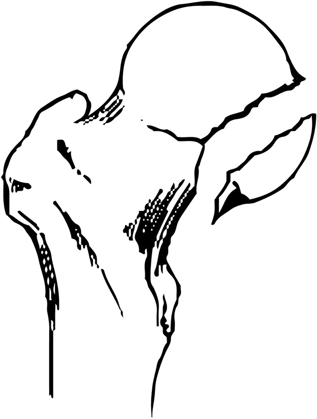
A Pipkin Type I fracture occurs caudal to the fovea capitis. (Published with permission from Jason Black, Web Media Specialist, Department of Orthopaedics and Sports Medicine, University of Washington, Seattle, WA, USA.)
Fig. 2.
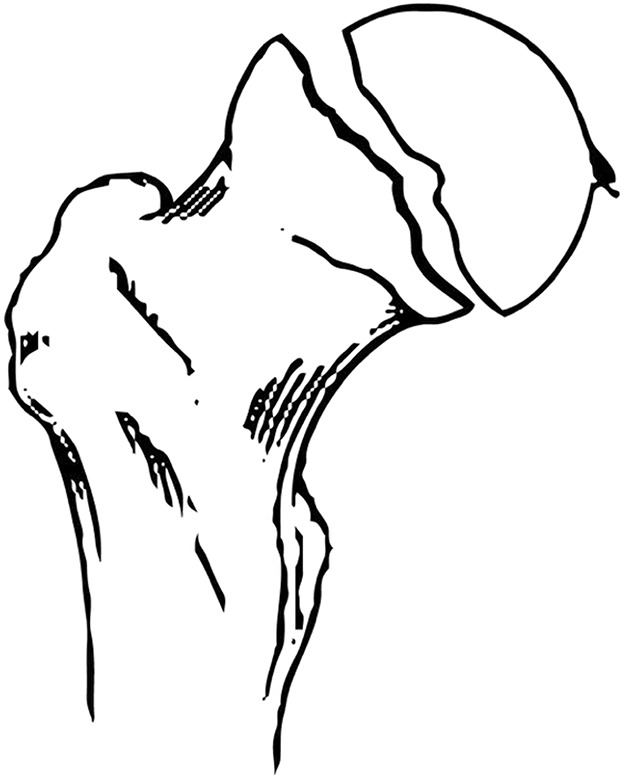
This illustration shows a Pipkin Type II fracture of the femoral head cephalad to the fovea capitis. (Published with permission from Jason Black, Web Media Specialist, Department of Orthopaedics and Sports Medicine, University of Washington, Seattle, WA, USA.)
Fig. 3.

A Pipkin Type III femoral head fracture inferior to the fovea centralis and femoral neck fracture is shown. (Published with permission from Jason Black, Web Media Specialist, Department of Orthopaedics and Sports Medicine, University of Washington, Seattle, WA, USA.)
Fig. 4.
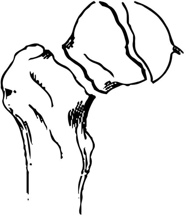
A Pipkin Type III femoral head fracture superior to the fovea centralis and femoral neck fracture is shown in this illustration. (Published with permission from Jason Black, Web Media Specialist, Department of Orthopaedics and Sports Medicine, University of Washington, Seattle, WA, USA.)
Fig. 5.
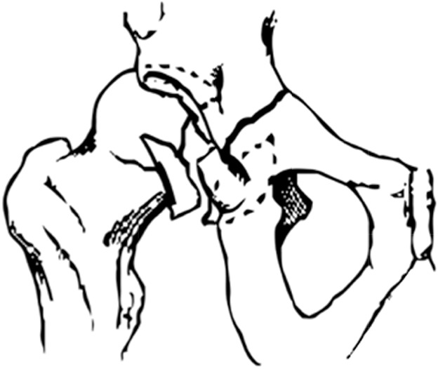
The illustration shows a Pipkin Type IV femoral head fracture in addition to an acetabular fracture. (Published with permission from Jason Black, Web Media Specialist, Department of Orthopaedics and Sports Medicine, University of Washington, Seattle, WA, USA.)
Pipkin’s basis for using the fovea capitis as a division between Types 1 and 2 fractures is that the ligamentum teres remains attached to the inferior fragment in a Type 2 injury, often resulting in substantial rotation of this fragment. The rotated caudal segment of the femoral head with the ligamentum attached could prevent a concentric reduction of the cranial head segment. Furthermore, he theorized that rotation of the caudal segment with the ligamentum attached is difficult to correct by closed means, providing a basis to consider open reduction and internal fixation for patients with Type 2 injuries and who are fit for surgery; by contrast, Type 1 fractures may be successfully treated more frequently by closed means alone. Outcomes were reported in relation to the Thompson and Epstein criteria [15], which uses a combination of radiographic and clinical information to provide an outcome of poor to excellent. Pipkin stated that no patients in his series fulfilled criteria to be classified as having an ‘excellent’ outcome as all patients had some degree of radiographic degenerative changes.
Pipkin preferred closed treatment for these injuries, as the patients in his series who received closed treatment had better outcomes that those who underwent open reduction. He attributed inferior outcomes in those who required surgical treatment to a combination of repeated attempts at closed reduction, delay in treatment, and the trauma of surgery. Pipkin reported that surgery was indicated when reduction of the dislocation and/or fracture was not achievable by closed measure, if an obstructive fragment was present, or if there was comminution of the fracture fragment [10].
For Types 1 and 2 injuries and the femoral head component of Type 4 injuries, he recommended attempting closed reduction as the primary means of management. Additionally, he recommended that the acetabular rim component of Type 4 injuries be managed with reduction and fixation. For Type 3 injuries, Pipkin stated that closed management may be possible, but that open treatment of at least the neck fracture component is more practical owing to the substantial forces that would prevent closed reduction of the head and neck components in combination with dislocation [10].
Validation
The Pipkin classification is relatively simple from a radiographic standpoint, but to our knowledge, no studies have reported on the interobserver and intraobserver reliability of his classification system. This likely is because the majority of the available studies regarding femoral head fractures are limited to small series owing to the infrequency of this injury.
However, several studies have evaluated prognosis after surgical and nonsurgical treatment of patients whose fractures were graded using the Pipkin classification. In general, they show better results with Pipkin Types 1 and 2 fractures than with Pipkin Types 3 or 4 fractures, which provides some face validity to the classification scheme. However, results are somewhat mixed.
Marchetti et al. [7] found that patients with Pipkin Types 1 and 2 fractures had better outcome scores on the Thompson and Epstein scale [15] after a mean followup of 49 months than did patients with Types 3 or 4 fractures (76% versus 56% good results, respectively).
In a study with a mean followup of nearly 7 years, 76% of the patients who sustained Pipkin Types 1, 2, and 4 fractures had excellent or good clinical outcomes when evaluated according to Thompson and Epstein scale [15]. Patients with Pipkin Types 1 and 2 fractures did better clinically than those with Type 4 fractures [9, 15]. There were no patients with Type 3 fractures in the series of Oransky et al. [9], thus limiting the study’s evaluation of the Pipkin system in relation to outcomes.
A systematic review of 155 patients with femoral head fractures in 11 studies found no statistical difference in outcomes among Pipkin types when using Thompson and Epstein criteria alone. [4].
In contrast to the above outcomes using the Thomas and Epstein scale, Stannard et al. [13] evaluated outcomes using the Short Form Heath Survey-12 (SF-12). They found that the physical component scores were lower in patients with Pipkin Type 2 fractures compared with those with Type 1 or Type 4 fractures.
Limitations
The primary limitation of the Pipkin classification is the lack of interobserver and intraobserver validation. To our knowledge, this validation has yet to be performed. Without this validation the classification is very limited to serve as a trustworthy classification system. This lack of validation may be attributable to the infrequency of these injuries, with data limited to small series.
In our opinion, the Pipkin classification system does not serve as a sufficient guide for surgical treatment of femoral head fractures. Several factors not included in this classification system must be considered when determining surgical treatment. These factors include the ability to obtain and maintain a concentric reduction, size of the femoral head fracture, displacement of the femoral head fracture, and the characteristics of the associated acetabular fracture in Type 4 injuries.
A systematic review of femoral head fractures found that Pipkin Type 1 fractures were the most likely treated nonoperatively, with 21.1% of these fractures undergoing nonoperative treatment, consistent with Pipkin’s belief that Type 1 fractures are able to be treated more frequently by closed means [4]. Additionally, Type 3 fractures were found to be the most-frequent type to be treated with arthroplasty, with 38.9% of these injuries treated in this manner [4]. Although Types 2 and 3 injuries are more likely to be treated with open reduction and internal fixation or arthroplasty, there is variability in the management of Types 1 and 4 fractures [4], with the aforementioned factors playing a role in decision making.
The sequelae of these injuries, including posttraumatic arthritis, osteonecrosis, heterotopic ossification, and sciatic nerve injury have been reported in several series [4, 7, 12]. However, no correlation has been shown between Pipkin type and risk of development of these sequelae [4, 7, 12]. As mentioned by Letournel and Judet [6], the magnitude of force required to cause fracture of the acetabulum can cause a substantial degree of injury to the femoral head cartilage and to vascularity of the femoral head. This degree of injury is difficult to appreciate radiographically alone. Thus this lends to why the development of posttraumatic arthritis and osteonecrosis of the femoral head are difficult to predict based on the Pipkin classification system alone.
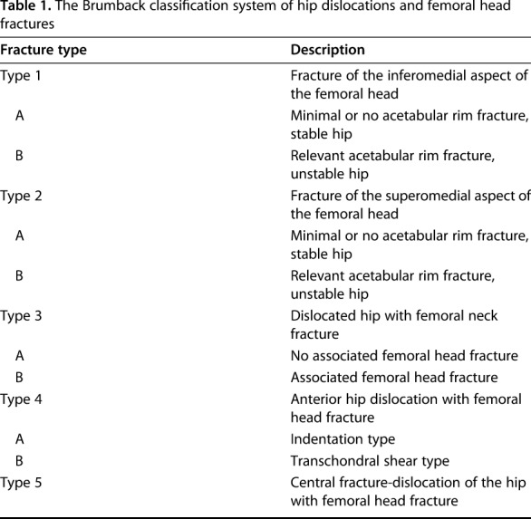
Alternative classification systems for femoral head fractures have since been developed, including those described by Brumback et al. [2], Yoon et al. [16], and the AO/OTA classification system as reported by Marsh et al. [8]. The classification system of Brumback et al. [2] is more comprehensive than the Pipkin classification system, taking into account the direction of dislocation and joint stability (Table 1). This system appears to provide prognostic value, with patients sustaining Type 3B and Type 5 injuries faring the worst, and patients with Type 2B fractures having the best physical outcomes [13]. As the Brumback system highlights the importance of joint instability, direction of dislocation, and acetabular fracture severity in the prediction of a poorer outcome [2], some consider that it may be a more-accurate classification system [4]. However, until intraobserver and interobserver reliability of the Brumback classification are validated in a robust way, we recommend readers use it only with caution.
Table 1.
The Brumback classification system of hip dislocations and femoral head fractures
Yoon et al. [16] developed a modification of Pipkin’s classification system to help guide treatment. A Type I fracture is a small fracture of the femoral head distal to the fovea centralis, too small or too fragmented to be fixed with screws. Type II is a larger fracture of the head distal to the fovea centralis. Type III is a large fracture of the head proximal to the fovea centralis, and Type IV is a comminuted fracture of the head. They concluded that Type I fractures were best treated with fragment excision, Types II and III by reduction and fixation, and Type IV by arthroplasty, specifically hemiarthroplasty [16]. However, their classification system is limited as it is subjective and also has not been validated; therefore we recommend readers use this system only with caution.
Conclusion
Although the Pipkin system is the most-frequently used system for classification of femoral head fractures [4], it is not comprehensive; it does not take into account the degree of comminution of the fractured fragments or the size of the head fracture, size of the acetabular fracture, or joint stability in Type 4 injuries. Thus this classification system is lacking in its abilities to serve as a guide for operative intervention. However, mid- and long-term studies that have evaluated the prognosis of patients with femoral head fractures found that Pipkin's classification is prognostically useful, in that patients with Types 1 and 2 fractures have better outcomes, as defined by Thompson and Epstein [15], than patients with Types 3 and 4 fractures [4, 7]. Finally, as the interobserver and intraobserver reliability of the Pipkin classification are unknown, it is substantially limited in its abilities as a reliable classification system.
Acknowledgements
We thank Jason Black, Web Media Specialist (Department of Orthopaedics and Sports Medicine, University of Washington) for creation of the figures contained in this article.
Footnotes
Each author certifies that he has no commercial associations (eg, consultancies, stock ownership, equity interest, patent/licensing arrangements, etc) that might pose a conflict of interest in connection with the submitted article.
All ICMJE Conflict of Interest Forms for authors and Clinical Orthopaedics and Related Research® editors and board members are on file with the publication and can be viewed on request.
References
- 1.Birkett J. Description of a dislocation of the head of the femur, complicated with its fracture; with remarks by John Birkett (1815–1904). 1869. Clin Orthop Relat Res. 2000;377:4–6. [DOI] [PubMed] [Google Scholar]
- 2.Brumback RJ, Kenzora JE, Levitt LE, Burgess AR, Poka A. Fractures of the femoral head. Proceedings of the Hip Society, 1986. St Louis, MO: CV Mosby; 1987:181–206. [PubMed] [Google Scholar]
- 3.Epstein HC, Wiss DA, Cozen L. Posterior fracture-dislocation of the hip with fractures of the femoral head. Clin Orthop Relat Res. 1985;201:9–17. [PubMed] [Google Scholar]
- 4.Giannoudis PV, Kontakis G, Christoforakis Z, Akula M, Tosounidis T, Koutras C. Management, complications and clinical results of femoral head fractures. Injury. 2009;40:1245–1251. [DOI] [PubMed] [Google Scholar]
- 5.Hougaard K, Thomsen PB. Traumatic posterior fracture dislocation of the hip with fracture of the femoral head or neck, or both. J Bone Joint Surg Am.1988;70:233–239. [PubMed] [Google Scholar]
- 6.Letournel E, Judet R. Fractures of the Acetabulum. 2nd ed. Berlin Germany: Springer-Verlag; 1993. [Google Scholar]
- 7.Marchetti ME, Steinberg GG, Coumas JM. Intermediate-term experience of Pipkin fracture-dislocations of the hip. J Orthop Trauma. 1996;10:455–461. [DOI] [PubMed] [Google Scholar]
- 8.Marsh JL, Slongo TF, Agel J, Broderick JS, Creevey W, DeCoster TA, Prokuski L, Sirkin MS, Ziran B, Henley B, Audigé L. Fracture and dislocation classification compendium -2007: Orthopaedic Trauma Association classification, database and outcomes committee. J Orthop Trauma. 2007;21 (suppl):S1–133. [DOI] [PubMed] [Google Scholar]
- 9.Oransky M, Martinelli N, Sanzarello I, Papapietro N. Fractures of the femoral head: a long-term follow-up study. Musculoskelet Surg. 2012;96:95–99. [DOI] [PubMed] [Google Scholar]
- 10.Pipkin G. Treatment of grade IV fracture dislocation of the hip. J Bone Joint Surg Am. 1957;39:1027–1042. [PubMed] [Google Scholar]
- 11.Sahin V, Karakas ES, Aksu S, Atlihan D, Turk CY, Halici M. Traumatic dislocation and fracture-dislocation of the hip: a long-term follow-up study. J Trauma. 2003;54:520–529. [DOI] [PubMed] [Google Scholar]
- 12.Scolaro JA, Marecek G, Firoozabadi R, Krieg JC, Routt ML. Management and radiographic outcomes of femoral head fractures. J Orthop Traumatol. 2017 Feb 10. [Epub ahead of print] 10.1007/s10195-017-0445-z. [DOI] [PMC free article] [PubMed] [Google Scholar]
- 13.Stannard JP, Harris HW, Volgas DA, Alonso JE. Functional outcome of patients with femoral head fractures associated with hip dislocations. Clin Orthop Relat Res. 2000; 377:44–56. [DOI] [PubMed] [Google Scholar]
- 14.Stewart MJ, Milford LW. Fracture-dislocation of the hip: an end-result study. J Bone Joint Surg Am. 1954;36:315–342. [PubMed] [Google Scholar]
- 15.Thompson VP, Epstein HC. Traumatic dislocation of the hip: a survey of two hundred and four cases covering a period of twenty-one years. J Bone Joint Surg Am. 1951;33:746–778. [PubMed] [Google Scholar]
- 16.Yoon TR, Rowe SM, Chung JY, Song EK, Jung ST, Anwar IB. Clinical and radiographic outcome of femoral head fractures: 30 patients followed for 3–10 years. Acta Orthop Scand 2001;72:348–353. [DOI] [PubMed] [Google Scholar]


