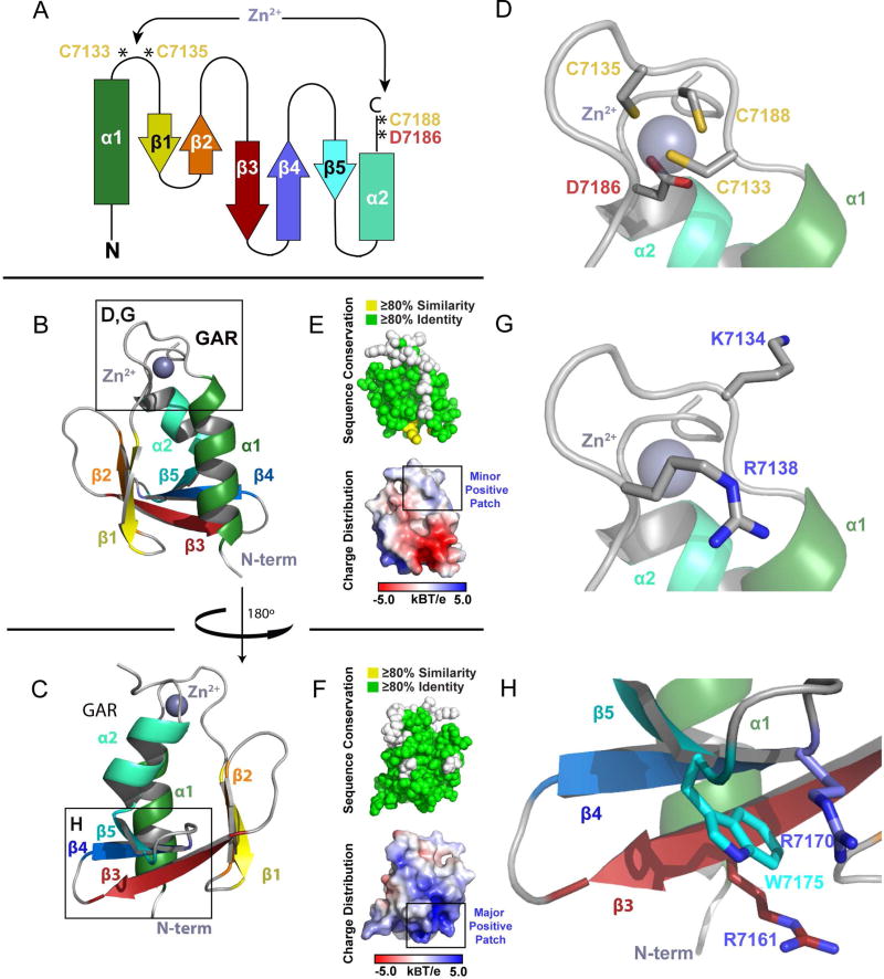
Figure 3. The GAR Domain is a Novel α/β Sandwich that Coordinates Zinc.
(A) Secondary structure topology of the GAR domain α/β sandwich showing the location of the residues that coordinate zinc. (B) Ribbon diagram of the GAR domain as colored in A. (C) The GAR domain as shown in (B), rotated 180° about the y-axis. (D) Zoom view of the zinc binding site boxed in (B), showing the Cys2-Asp-Cys residues that coordinate zinc. (E,F) The GAR domain illustrating cross-species conservation as delineated in Figure 1C (above, spherical representation) and displaying electrostatic surface potential (below, surface representation). Domain orientation in (E) and (F) correspond to the orientation in (B) and (C) respectively. (G,H) Zoom view of two positively charged regions on the GAR domain, corresponding to the boxed regions in (B) and (C) respectively. See also Figure S2.

