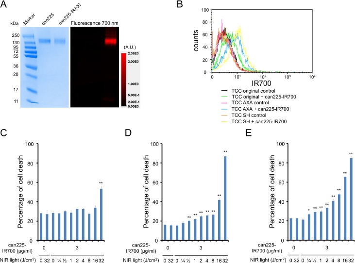
Figure 1. Confirmation of EGFR expression as a target for NIR-PIT in three TCC cell lines, and evaluation of in vitro NIR-PIT.
(A) Validation of can225-IR700 by SDS-PAGE (left: Colloidal Blue staining, right: fluorescence). Diluted can225IgG was used as a control. (B) Expressions of EGFR in three TCC cell lines were evaluated by flow cytometry. After 6 h of can225-IR700 incubation, TCC SH cells showed high fluorescence signal, TCC AXA cells showed intermediate fluorescence signal, and TCC original cells showed low fluorescence signal. Fluorescence in three TCC cell lines was completely blocked by adding excess can225IgG. (C) Membrane damage of TCC original cells induced by NIR light exposure was measured with the dead cell count using propidium iodide (PI) staining. No membrane damage was observed in TCC original cells after 0.25–16 J/cm2 of NIR light exposure. Membrane damage was only shown after 32 J/cm2 of NIR light exposure (n = 5, **p < 0.01, vs. untreated control, by Student’s t test). (D) Membrane damage of TCC AXA cells induced by NIR-PIT was measured with the dead cell count using PI staining, which increased in a light dose dependent manner (n = 5, **p < 0.01, vs. untreated control, by Student’s t test). There was no significant cytotoxicity associated with NIR light exposure alone in the absence of can225-IR700 and with can225-IR700 alone without NIR light exposure. (E) Membrane damage of TCC SH cells induced by NIR-PIT was measured with the dead cell count using PI staining, which increased in a light dose dependent manner (n = 5, **p < 0.01, vs. untreated control, by Student’s t test). The NIR-PIT effects in TCC SH cells were higher than TCC original and TCC AXA cells. There was no significant cytotoxicity associated with NIR light exposure alone in the absence of can225-IR700 and with can225-IR700 alone without NIR light exposure.

