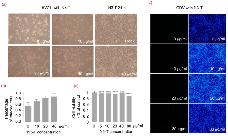
Figure 6.
N3-T Promotes Viral Replication in a Dose-Dependent Manner. (a,b) RD cells were mock-infected or exposed to EV71 of 1 TCID50 for 24 h in the presence of PBS or N3-T at the indicated concentrations. The cells were imaged by light microscopy using an Olympus microscope (magnificence 100×) (a); and infected cells were quantified from three randomly selected images (b); Shown are the means with SD, (c) Cells were treated with various concentrations of N3-T for 24 h, and cell viability was determined by the lactate dehydrogenase (LDH) assay; (d) Vero cells were exposed to CDV of 0.01 MOI for 36 h in the presence of PBS or N3-T at 0, 10, 20, or 30 µg/mL. The infected cells were imaged by fluorescence microscopy using an Olympus IX73 microscope (magnification of 100×) equipped with a DP73 camera (left) and assessed under light microscopy (panels c,d). Each image is representative of three experiments (right).

