

Abstract
A large body of evidence now indicates that the amount of mercury released from dental amalgam fillings can be significantly accelerated by exposure to radiofrequency electromagnetic fields (RF-EMFs) such as common mobile phones and magnetic resonance imaging (MRI). Studies performed on the increased microleakage of dental amalgam restorations after exposure to RF-EMFs have further supported these findings. Although the accelerated microleakage induced by RF-EMFs is clinically significant, the entire mechanisms of this phenomenon are not clearly understood. In this paper, we introduce “Triple M” effect, a new evidence-based theory which can explain the accelerated microleakage of dental amalgam fillings after exposure to different sources of electromagnetic radiation. Based on this theory, there are saliva-filled tiny spaces between amalgam and the tooth. Exposure of the oral cavity to RF-EMFs increases the energy of these small amounts of saliva. Due to the small mass of saliva in these tiny spaces, a small amount of energy will be required for heating. Moreover, reflection of the radiofrequency radiation on the inner walls of the tiny spaces causes interference which in turn produces some “hot spots” in these spaces. Finally, formation of gas bubbles in response to increased temperature and very rapid expansion of these bubbles will accelerate the microleakage of amalgam. Experiments that confirm the validity of this theory are discussed.
Keywords: Microleakage , Dental Amalgam , Electromagnetic Fields , Triple M Effect
Introduction
Over the last 150 years, dental amalgam, a combination of metals (about 50% mercury in elemental form and other metals such as silver, tin and copper) has been used in restorative dentistry due to its outstanding mechanical properties, ease of manipulation as well as its low cost. Substantial evidence now indicates that the level of mercury released from dental amalgam fillings can be significantly accelerated by exposure to different sources of radiofrequency electromagnetic fields (RF-EMFs) such as common mobile phones and magnetic resonance imaging (MRI) [1-3]. Studies performed on the microleakage of dental amalgam restorations have further supported these findings [4,5]. Kursun et al. in 2014 showed that exposure to X-rays, a high energy ionizing component of the wide spectrum of electromagnetic radiation, can accelerate the release of mercury from dental amalgam fillings [6]. These findings have recently been reviewed by Mortazavi and Mortazavi [7]. Over the past several years, our laboratories at the Ionizing and Non-ionizing Radiation Protection Research Center (INIRPRC) have expanded their focus on studying the health effects of exposure to some common and/or occupational sources of electromagnetic fields (EMFs) such as cellular phones [8-16], mobile base stations [17], mobile phone jammers [18-20], laptop computers [21], radars [9], dentistry cavitrons [22], MRI [2,23], Wi-Fi routers [24] and different coils [25,26]. In this paper, we propose a new mechanism that effectively explains the accelerated microleakage of dental amalgam fillings after exposure to different sources of electromagnetic radiation.
Amalgam Microleakage
Microleakage can be defined as a clinically detectable passage of bacteria, fluids, molecules or ions between the walls of a cavity and the filling material [27]. The issue of microleakage is believed to be a major challenge in clinical dentistry. Statistically significant differences in microleakage between the extracted teeth samples exposed to MRI and controls have been reported by Yilmaz and Misirlioglu in 2013 [4]. Moreover, Shahidi et al. who have evaluated the microleakage of amalgam fillings following MRI, reported that MRI is not an absolutely safe technique in patients with amalgam fillings [5].
Introducing “Triple M” Effect
This effect is titled “Triple M” effect because this mechanism is proposed by Mehdizadeh AR, Mortazavi G and Mortazavi SAR. The “Triple M” effect is based on these physical facts:
1. Due to shrinkage, there are spaces between amalgam and the tooth, a phenomenon which is usually called “marginal micro leakage”.
2. These small spaces are filled with very small amounts of saliva (Figure 1).
Figure1.

Reflection of radiofrequency radiation on the inner walls of small spaces between the amalgam and tooth causes interference which produces “hot spots”. Then, formation of gas bubbles in response to increased temperature and expansion of these bubbles increases the microleakage of amalgam.
3. Exposure of oral cavity to RF-EMFs (MRI or mobile phone radiation), raises the energy of these small amounts of saliva. The heat energy required in any material to increase the temperature from t1 to t2 can be calculated as follows:
Q = mc ΔT (1)
Where Q in this equation is the heat energy (J), m and C are mass (kg) and specific heat (J/kg °C), respectively. ΔT is the temperature change (°C). Considering Equation 1, when there is a very small mass (e.g. the mass of saliva in nanospaces created by marginal micro leakage), a small amount of energy will be required for heating.
4. Reflection of the radiofrequency radiation on the inner walls of the above-mentioned small spaces causes interference which in turn produces some “hot spots” in these spaces. It is worth mentioning that this effect is similar to the issue of inhomogeneity of radiation intensity and formation of hot spots in commercial microwave ovens (manufacturers of microwave ovens have solved this problem by using a rotating glass plate in the oven) [28]. It should be noted that bone (or tooth) reflects microwave and this reflection is reported to cause overheating in the adjacent regions [29].
5. Formation of gas bubbles in response to increased temperature and very rapid expansion of these bubbles will accelerate the microleakage of amalgam.
“Triple M” Effect Verification
Paknahad et al. have recently evaluated the effect of pulsed electromagnetic fields (PEMF) generated by a pair of Helmholtz coils (50 Hz) on microleakage of amalgam restorations [26]. In this study, 46 non-carious extracted human premolars with identical amalgam fillings were randomly divided into exposed (Helmholtz coils) and non-exposed control groups. These researchers could not find any statistically significant difference between the microleakage scores of the exposure and control groups. As the frequency in this experiment was only 50 Hz, it can explain the reason for major differences in the findings of Paknahad and those obtained by Yilmaz and Misirlioglu [4] and Shahidi et al. who found increased microleakage of amalgam fillings following MRI [5]. We believe that the microleakage dependency of the frequency of EMFs has originated from the cardinal role of frequency of EMFs in the absence of energy in water as heat (Figure 2).
Figure2.
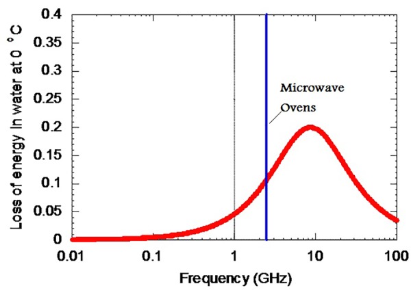
The frequency of EMFs affects the magnitude of the loss of energy in water as heat.
Conclusion
Dental amalgam fillings have been used in restorative dentistry for more than 150 years. There are reports which indicate that exposure of amalgam to RF-EMFs can lead to accelerated microleakage of amalgam. Despite the undeniable importance of this effect in clinical dentistry, its mechanisms are not fully understood yet. In this paper, we introduced “Triple M” effect, which explains the reason for increased microleakage of dental amalgam fillings after exposure to different sources of electromagnetic radiation. According to this theory, absorption of the energy of RF-EMFs in the saliva-filled tiny spaces between amalgam and the tooth increases the energy of the small amounts of saliva in these tiny spaces. Furthermore, reflection of the radiofrequency radiation on the inner walls of the tiny spaces makes interferences which in turn produce some “hot spots” in such tiny spaces. At the next stage, formation of gas bubbles in response to increased temperature and very rapid expansion of these bubbles increases the microleakage of amalgam. Further experiments are needed to verify the validity of this theory for other sources of RF-EMFs.
Acknowledgement
This study was supported by the Ionizing and Non-ionizing Radiation Protection Research Center (INIRPRC), Shiraz University of Medical Sciences (SUMS), Shiraz, Iran.
Conflict of Interest:None declared.
References
- 1.Mortazavi SM, Neghab M, Anoosheh SM, Bahaeddini N, Mortazavi G, Neghab P, et al. High-field MRI and mercury release from dental amalgam fillings. Int J Occup Environ Med. 2014;5:101–5. [PMC free article] [PubMed] [Google Scholar]
- 2.Mortazavi SM, Daiee E, Yazdi A, Khiabani K, Kavousi A, Vazirinejad R, et al. Mercury release from dental amalgam restorations after magnetic resonance imaging and following mobile phone use. Pak J Biol Sci. 2008;11:1142–6. doi: 10.3923/pjbs.2008.1142.1146. [DOI] [PubMed] [Google Scholar]
- 3.WHO. Promoting the phase down approach of dental amalgam in developing countries. Geneva: World Health Organization; 2014. [Google Scholar]
- 4.Yilmaz S, Misirlioglu M. The effect of 3 T MRI on microleakage of amalgam restorations. Dentomaxillofac Radiol. 2013;42:20130072. doi: 10.1259/dmfr.20130072. [ PMC Free Article] [DOI] [PMC free article] [PubMed] [Google Scholar]
- 5.Shahidi SH, Bronoosh P, Alavi AA, Zamiri B, Sadeghi AR, Bagheri MH, et al. Effect of magnetic resonance imaging on microleakage of amalgam restorations: an in vitro study. Dentomaxillofac Radiol. 2009;38:470–4. doi: 10.1259/dmfr/30077669. [DOI] [PubMed] [Google Scholar]
- 6.Kursun S, Öztas B, Atas H, Tastekin M. Effects of X-rays and magnetic resonance imaging on mercury release from dental amalgam into artificial saliva. Oral Radiology. 2014;30:142–6. doi: 10.1007/s11282-013-0154-0. [DOI] [Google Scholar]
- 7.Mortazavi G, Mortazavi SM. Increased mercury release from dental amalgam restorations after exposure to electromagnetic fields as a potential hazard for hypersensitive people and pregnant women. Rev Environ Health. 2015;30:287–92. doi: 10.1515/reveh-2015-0017. [DOI] [PubMed] [Google Scholar]
- 8.Mortazavi SM, Motamedifar M, Namdari G, Taheri M, Mortazavi AR, Shokrpour N. Non-linear adaptive phenomena which decrease the risk of infection after pre-exposure to radiofrequency radiation. Dose Response. 2014;12:233–45. doi: 10.2203/dose-response.12-055.Mortazavi. [ PMC Free Article] [DOI] [PMC free article] [PubMed] [Google Scholar]
- 9.Mortazavi SM, Taeb S, Dehghan N. Alterations of visual reaction time and short term memory in military radar personnel. Iran J Public Health. 2013;42:428–35. [ PMC Free Article] [PMC free article] [PubMed] [Google Scholar]
- 10.Mortazavi SM, Rouintan MS, Taeb S, Dehghan N, Ghaffarpanah AA, Sadeghi Z, et al. Human short-term exposure to electromagnetic fields emitted by mobile phones decreases computer-assisted visual reaction time. Acta Neurol Belg. 2012;112:171–5. doi: 10.1007/s13760-012-0044-y. [DOI] [PubMed] [Google Scholar]
- 11.Mortazavi S, Mosleh-Shirazi M, Tavassoli A, Taheri M, Mehdizadeh A, Namazi S, et al. Increased Radioresistance to Lethal Doses of Gamma Rays in Mice and Rats after Exposure to Microwave Radiation Emitted by a GSM Mobile Phone Simulator. Dose Response. 2013;11:281–92. doi: 10.2203/dose-response.12-010.Mortazavi. [ PMC Free Article] [DOI] [PMC free article] [PubMed] [Google Scholar]
- 12.Mortazavi S, Mosleh-Shirazi M, Tavassoli A, Taheri M, Bagheri Z, Ghalandari R, et al. A comparative study on the increased radioresistance to lethal doses of gamma rays after exposure to microwave radiation and oral intake of flaxseed oil. Iranian Journal of Radiation Research. 2011;9:9–14. [Google Scholar]
- 13.Mortavazi S, Habib A, Ganj-Karami A, Samimi-Doost R, Pour-Abedi A, Babaie A. Alterations in TSH and Thyroid Hormones following Mobile Phone Use. Oman Med J. 2009;24:274–8. doi: 10.5001/omj.2009.56. [DOI] [PMC free article] [PubMed] [Google Scholar]
- 14.Mortazavi SM, Daiee E, Yazdi A, Khiabani K, Kavousi A, Vazirinejad R, et al. Mercury release from dental amalgam restorations after magnetic resonance imaging and following mobile phone use. Pak J Biol Sci. 2008;11:1142–6. doi: 10.3923/pjbs.2008.1142.1146. [DOI] [PubMed] [Google Scholar]
- 15.Mortazavi SM, Ahmadi J, Shariati M. Prevalence of subjective poor health symptoms associated with exposure to electromagnetic fields among university students. Bioelectromagnetics. 2007;28:326–30. doi: 10.1002/bem.20305. [DOI] [PubMed] [Google Scholar]
- 16.Mortazavi S, Motamedifar M, Namdari G, Taheri M, Mortazavi A. Counterbalancing immunosuppression-induced infections during long-term stay of humans in space. Journal of Medical Hypotheses and Ideas. 2013;7:8–10. [Google Scholar]
- 17.Mortazavi S. Safety issue of mobile phone base stations. J Biomed Phys Eng. 2013;3:1–2. [PMC free article] [PubMed] [Google Scholar]
- 18.Mortazavi S, Parsanezhad M, Kazempour M, Ghahramani P, Mortazavi A, Davari M. Male reproductive health under threat: Short term exposure to radiofrequency radiations emitted by common mobile jammers. J Hum Reprod Sci. 2013;6:124–8. doi: 10.4103/0974-1208.117178. [ PMC Free Article] [DOI] [PMC free article] [PubMed] [Google Scholar]
- 19.Rafati A, Rahimi S, Talebi A, Soleimani A, Haghani M, Mortazavi SM. Exposure to Radiofrequency Radiation Emitted from Common Mobile Phone Jammers Alters the Pattern of Muscle Contractions: an Animal Model Study. J Biomed Phys Eng. 2015;5:133–42. [ PMC Free Article] [PMC free article] [PubMed] [Google Scholar]
- 20.Shekoohi Shooli F, Mortazavi SA, Jarideh S, Nematollahii S, Yousefi F, Haghani M, et al. Short-Term Exposure to Electromagnetic Fields Generated by Mobile Phone Jammers Decreases the Fasting Blood Sugar in Adult Male Rats. J Biomed Phys Eng. 2016;6:27–32. [ PMC Free Article] [PMC free article] [PubMed] [Google Scholar]
- 21.Mortazavi SMJ, Tavassoli A, Ranjbari F, Moammaiee P. Effects of laptop computers’ electromagnetic field on sperm quality. Journal of Reproduction & Infertility. 2010;11(4) [Google Scholar]
- 22.Mortazavi SM, Vazife-Doost S, Yaghooti M, Mehdizadeh S, Rajaie-Far A. Occupational exposure of dentists to electromagnetic fields produced by magnetostrictive cavitrons alters the serum cortisol level. J Nat Sci Biol Med. 2012;3:60–4. doi: 10.4103/0976-9668.95958. [ PMC Free Article] [DOI] [PMC free article] [PubMed] [Google Scholar]
- 23.Mortazavi G, Haghani M, Rastegarian N, Zarei S, Mortazavi SMJ. Increased Release of Mercury from Dental Amalgam Fillings due to Maternal Exposure to Electromagnetic Fields as a Possible Mechanism for the High Rates of Autism in the Offspring: Introducing a Hypothesis. Journal of Biomedical Physics & Engineering. 2016;6(1):41–46. [PMC free article] [PubMed] [Google Scholar]
- 24.Mahmoudi R, Mortazavi S, Safari S, Nikseresht M, Mozdarani H, Jafari M, et al. Effects of microwave electromagnetic radiations emitted from common Wi-Fi routers on rats’ sperm count and motility. Int J Radiat Res. 2015;13:363–8. [ PMC Free Article] [Google Scholar]
- 25.Haghnegahdar A, Khosrovpanah H, Andisheh-Tadbir A, Mortazavi G, Saeedi Moghadam M, Mortazavi S, et al. Design and fabrication of helmholtz coils to study the effects of pulsed electromagnetic fields on the healing process in periodontitis: preliminary animal results. J Biomed Phys Eng. 2014;4:83–90. [ PMC Free Article] [PMC free article] [PubMed] [Google Scholar]
- 26.Paknahad M, Shahidi S, Mortazavi SMJ, Mortazavi G, Moghadam MS, Nazhvani AD. The Effect of Pulsed Electromagnetic Fields on Microleakage of Amalgam Restorations: An in Vitro Study. Shiraz E-Medical Journal. 2016;17(2) [Google Scholar]
- 27.Vanishree HS, Shanthala BM, Bobby W. The comparative evaluation of fracture resistance and microleakage in bonded amalgam, amalgam, and composite resins in primary molars. Indian J Dent Res. 2015;26:446–50. doi: 10.4103/0970-9290.172019. [DOI] [PubMed] [Google Scholar]
- 28.Kappe C, Dallinger D, Murphree S. Practical Microwave Synthesis for Organic Chemists 2009. Germany, Wiley-VCH: Weinheim; 2009. [Google Scholar]
- 29.Regier M, Schubert H, Knoerzer K. The microwave processing of foods. Toronto: Elsevier; 2005. [Google Scholar]