Abstract
Proprioception may be the least well measured of all contributors to the neural control of movement. New precise, reliable measures of proprioception are needed for clinical diagnosis of impairment, and to measure outcomes of proprioceptive training. The purpose of this simple, non-invasive method is to temporarily knockdown upper limb proprioception in healthy adults, to an extent that would be useful in the development and testing of upper limb proprioception measures. Knockdown models have two main advantages over studying humans with impaired proprioception: participant availability and the ability to control the extent of impairment across participants. Current published methods of temporary proprioception knockdown of the upper limb, such as ischemic nerve blocks and cryotherapy, are invasive, impractical, or uncomfortable for the participant. Here, vibration over the ulnar groove was used to reduce upper limb proprioception. High frequency vibration may reduce proprioceptive acuity by inhibiting pacinian corpuscle-induced input. The effect of vibration used in this protocol was confirmed using two quantitative measures. This method was simple to administer, comfortable for participants, and practical.
Keywords: Behavior, Issue 133, Kinesthesia, Upper Extremity, Protocol, Measurement, Somatosensation, High-frequency Vibration, Non-Invasive
Introduction
Of all contributors to the neural control of movement, proprioception may be the least well measured. Research measures of proprioception using specialized equipment have recently achieved reliability, validity, and precision;1,2,3 in contrast, clinical measures of proprioception, the most common being limb position sense testing,4 have low resolution, are contaminated by other sensory modalities,4 and have poor or no published psychometric properties.5 New precise, reliable measures of proprioception are needed to elucidate peripheral and central mechanisms of proprioceptive control,3 for clinical diagnosis of impairment, and to measure outcomes of proprioceptive training.2,5,6,7 Toward this end, a simple, non-invasive method to temporarily impair or 'knockdown' proprioception is needed.
Proprioceptive knockdown in healthy humans allows researchers to infer the role of proprioceptive function in a sensorimotor task, which is useful to inform the development and validation of standardized measures. Knockdown models have two main advantages over studying humans with impaired proprioception. The first is participant availability; individuals with proprioception impairment are not easily accessible to many researchers. Second, unlike in vivo proprioception impairment, knockdown models may allow the ability to control the extent of impairment across participants.
Current published methods of temporary proprioception knockdown of the upper limb are invasive, impractical, or uncomfortable for the participant. Anesthetic injections, while relatively safe, require technical expertise and may be considered invasive by some research participants. Ischemic nerve blocks cause discomfort and a blood test to screen for clotting disorders prior to their application is practiced.8 Cryotherapy also causes discomfort. The average time of application for cryotherapy to impact proprioception is 20.3 ± 5.3 min.9 Once cryotherapy is removed, a brief window in which to measure proprioception prior to rewarming remains, which may contribute to the inconsistent effect of cryotherapy on joint position sense.10 High frequency (300 Hz) vibration was used successfully to reduce proprioceptive acuity in a finger movement detection task; the mechanism was reported to be pacinian corpuscle-induced inhibition of input from other vibration sensitive cutaneous receptors.11 Recently, soleus muscle vibration (80 Hz) was found to decrease isometric force production accuracy by distorting proprioceptive information.12 However, a simple non-invasive method for temporary knockdown of upper limb proprioception has not been published.
The purpose of this method is to use high frequency vibration to temporarily knockdown upper limb proprioception in healthy adults. Knockdown was confirmed using two measures, the Vibration Detection Threshold (VDT) and the tablet version of the Brief Kinesthesia Test (tBKT). The VDT is a psychophysical measure of sensitivity thought to reflect Aα afferent axon transmission.13 Proprioceptive performance was quantified using the tBKT that is under development in our lab. The Brief Kinesthesia Test (BKT), based on the work of Ayres,14 is an experimental instrument that was tested for but not included in the National Institutes of Health (NIH) Toolbox core batteries.15,16 The BKT includes three reaching trials for each upper limb. The tBKT includes 20 reaching trials per upper limb and is being developed with the goal of improving the psychometric properties over the original test. The tBKT involves a sensory input (examiner guidance of upper limb to target), central processing (remember the spatial location of the target) and a motor output (attempting to locate the target after guidance is removed), thought to be necessary in a measure of overall proprioceptive performance.17 The VDT and the tBKT measurements, represent low and higher levels, respectively, in the somatosensory hierarchy,18 and thus should provide a more comprehensive quantification of proprioception than either measure used alone.
Two neural mechanisms relate most closely to the reduced proprioceptive acuity caused by high frequency vibration. First, Pacinian corpuscles are the cutaneous mechanoreceptor most commonly associated with vibration detection. The continuous vibration used in this protocol likely raises the receptor tuning threshold of vibration detection based on the neural mechanism of short-term habituation in the Aα and β fiber group associated with Pacinian corpuscles.19 The physiologic result is that a vibration of the same intensity and frequency, such as 128 Hz used in the VDT test, is felt for a shorter duration. Second, it is thought that muscle spindles, via Aα afferent fibers, code muscle length inaccurately following high frequency vibration resulting in distorted proprioceptive information as evidenced by reduced accuracy during force reproduction,12 illusion of movement,20,21 and reduced kinesthesia.22
Protocol
The Institutional Review Board at the College of St. Scholastica has approved the study under which this protocol was developed and tested.
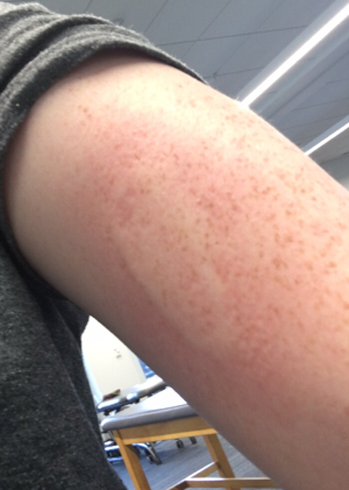
NOTE: The manufacturer's specifications of the vibrator used in this protocol indicated that the frequency on 'high' was 11,00 rpm (183.3 Hz). This frequency was confirmed using a sample of vibration data collected through one input of a differential amplifier sampled at 2 kHz. The mean period of the signal was 5.56 x 10⁻³ s, which is equivalent to 180 Hz. In individuals with Raynaud's disease, vibration may cause Raynaud's phenomenon, a temporary vasoconstriction at the site of application, which may last minutes to hours, and may be accompanied by numbness, itching or pain.23 Figure 1 shows an example of Raynaud's phenomenon caused by vibration similar to that used in this protocol. Potential participants should be screened for Raynaud's prior to participating in this protocol.
1. Proprioception Knockdown Protocol:
Gather pen, 183 Hz vibrator, 2.5-inch wide strap and a rubber band (see the Table of Materials).
- Palpate the ulnar nerve within the ulnar groove of the upper extremity being tested.
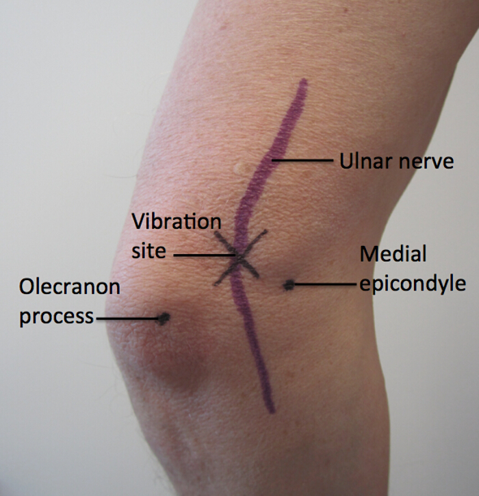
- Place an "x" mark on the skin over the ulnar groove just superior to the line between the olecranon process of the humerus and the medial epicondyle of the ulna. NOTE: Figure 2 shows this location on the dorsum of the elbow .
- Place head of the vibrator onto marked location with handle of massager superior to head.
- Apply the wide strap around handle portion of vibrator and participant's arm.
- Place rubber band around neck of vibrator head and participant's arm to hold vibrator head into place and maintain contact between participant's skin and the vibrator head, ensuring that the rubber band is a proper size to retain the head against the skin but not to restrict movement or circulation.
Ask the participant to bend and straighten his/her elbow. He/she should feel no interference from the straps. If he/she reports interference, adjust the straps and rubber band so that freedom of movement is ensured.
- Turn vibrator on high, wait for 2 min (based on unpublished data).
- Conduct a measure of proprioception performance (results from the tablet version of the BKT are presented here) with the vibrator running on high for the duration of the test
- Conduct the BKT. Seat the participant in a standard height chair (18-inch seat height), at a standard height table (29-inch height) with their vision occluded from the tablet using a curtain. Sit across from the participant.
- Guide the participants index finger, held at the distal phalanx only, from the start of the line to the end of the line (target) and back to the start of the line. Release the participant's finger and instruct them to touch the target. Complete 20 trials with each upper limb.
- Assign the participants' scores for each upper limb as the average absolute error (distance from the target) in centimeters.
- Conduct psychophysical measures (such as the VDT) immediately upon removal of the vibration stimulus. NOTE: In the results presented, the duration of the vibration stimulus prior to the VDT was 5 ± 1 minute.
2. Vibration Detection Threshold (adapted from13)
- Gather 128 Hz tuning fork, a stopwatch, textbook (provides a firm yet compliant surface on which to strike the tuning fork), table (29-inch height), standard chair (18-inch seat height), masking tape, and pen.
- Create a 1 ½ inch x 1 ½ inch square on the textbook using masking tape to be used as a target for consistency in striking the tuning fork.
- Using a permanent marker, color the bottom 1 mm of the stem of the tuning fork (this mark will be used to standardize the pressure of tuning fork application during the test).
- Seat the participant in the chair at the table. Ask her/him to extend the forearm of the extremity being tested supinated and completely rest it on the table, including the elbow. Ask them to relax the extremities. NOTE: The examiner should be seated across from the participant.
- Mark a dot on the skin over the distal biceps tendon, approximately 1 cm superior to the elbow crease.
- Place the book at the edge of table closest to participant, between participant's elbows.
- Say: "This is a test of your ability to detect vibration. Now I will put this tuning fork on your biceps tendon. Please tell me if you feel any vibration, then say 'Now' immediately when the sensation of vibration disappears. This procedure will be repeated 3 times on each arm."
- Ensure that the discs at the ends of each prong of the tuning fork are tight prior to beginning. If they are loose they will rattle and reduce the duration of the tuning fork's resonance making the test unreliable.
- Hold the stem of the tuning fork loosely between thumb and index finger and strike it on the book inside of the square target with enough force to produce resonance.
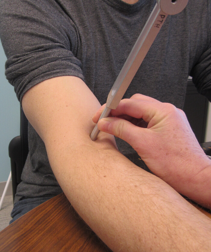
- Immediately after striking, place tuning fork on the test location, using enough pressure to depress the skin and conceal the 1 mm band on the tuning fork from vision. NOTE: Figure 3 shows the tuning fork on the distal biceps tendon test location used in this protocol.
- Using the stopwatch, quantify the time from placement of the tuning fork onto participant's skin until the participant no longer feels vibration (says 'Now').
- Repeat two more times on the same arm for a total of three trials.
- Calculate the mean time to disappearance of vibration stimulus across the three trials to determine the vibration detection threshold (VDT).
Representative Results
Using the protocol presented here, we tested 20 healthy adults, 8 were male (mean age (SD)= 32.5 (12.5) years; 19 right-, and 1 left- handed). The participants had no known pathology involving the upper extremities. Handedness was assessed using the Edinburgh Handedness Inventory.24 Study participants reported no adverse events.
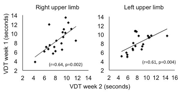
Both upper limbs of each participant were tested using the VDT and the tBKT at two separate sessions, one week apart. VDT, as described in the protocol, was quantified at the distal biceps tendon. The mean of three trials was used for analysis. For the tBKT, mean absolute error across 20 trials was used for analysis. At test session two, participants completed the same measures under the condition of temporary proprioception knockdown. ThePearson product-moment correlation (r) evaluated the test-retest reliability of the Vibration Detection Threshold (VDT). A statistically significant moderate to good relationship was found between the testing time points for the right (r = 0.64, p = 0.002, n = 20) and left (r = 0.61, p = 0.004, n = 20) upper limbs (Figure 4). The intraclass correlation coefficient (ICC) for the right VDT was (ICC 1, 3) = 0.77, n = 20, for the left VDT was (ICC 1, 3) = 0.76, n = 20. The standard error of measurement for the right VDT was 0.96 seconds, 95% CI = 6.5-10.3 s, the Minimum Detectable Difference (MDD) was 2.2 s. For the left VDT, the standard error of measurement was 0.83 s, 95% CI = 6.7-9.9 s, the MDD was 1.9 s. The intraclass correlation coefficient (ICC) for the right tBKT was (ICC 1, 3) = 0.55, n = 20, for the left tBKT was (ICC 1, 3) = 0.72, n = 20.
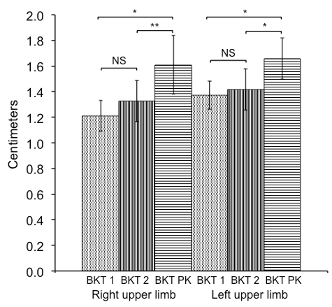
To test the directional hypothesis that proprioception knockdown (PK) using vibration would result in proprioception impairment, one-tailed paired t-tests were used to compare mean error between week 1, week 2 and the PK conditions for the VDT and BKT. The proprioception knockdown protocol resulted in statistically poorer scores on the VDT and the tBKT for both upper limbs, while the control conditions were not statistically different (Figures 5 and 6).
The extent of proprioception knockdown that resulted from the protocol was quantified by calculating the effect size (Cohen's d). The effect size on the VDT was d = 4.04 for the right upper limb and d = 4.24 for the left. For the tBKT d= 0.68 and 0.56 for the right and left, respectively.
Figure 1: Raynaud's phenomenon. An example of Raynaud's phenomenon caused by vibration. Please click here to view a larger version of this figure.
Figure 2: Vibration site for proprioception knockdown. The location of vibration in this protocol is on the dorsum of the elbow within the ulnar groove just superior to a line between the olecranon process and the medial epicondyle. Please click here to view a larger version of this figure.
Figure 3: Site for placement of the tuning fork for the Vibration Detection Test. For this study the Vibration Detection Threshold of the distal biceps tendon was quantified using a 128 Hz tuning fork. Please click here to view a larger version of this figure.
Figure 4: Correlation of VDT between week 1 and 2. Using data from participants tested under normal conditions one week apart the Pearson product-moment correlation (r) was used to evaluated test-retest reliability of the Vibration Detection Threshold (VDT). A statistically significant moderate to good relationship was found between the testing time points for the right (r = 0.64, p = 0.002) and left (r = 0.61, p = 0.004) upper limbs.
Figure 5: Comparison in VDT between week 1, 2 and PK conditions. One-tailed, paired t-tests were used to compare the mean time in seconds on the Vibration Detection Threshold (VDT) between week 1, week 2 and the proprioception knockdown (PK) conditions (n=20). Error bars show the standard deviation. (*) p-value < 0.001.
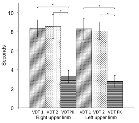
Figure 6: Comparison in BKT between week 1, 2 and PK conditions. One-tailed, paired t-tests were used to compare mean error in centimeters on the tablet version of the Brief Kinesthesia Test (BKT) between week 1, week 2 and the proprioception knockdown (PK) conditions (n = 20). Error bars show the standard deviation. (*) p-value < 0.001; (**) p-value = 0.001; NS = Not statistically significant.
Discussion
This protocol provides a method to knock down human proprioception in the upper limb. Across 20 healthy participants the effect of proprioceptive knockdown was large as measured by VDT a psychophysical measure of sensitivity thought to reflect Aα afferent axon transmission. The VDT was measured as quickly as possible after removal of vibration, when Aα afferent discharge is reduced.25 The effect of this protocol on error in reaching to a target with visual occlusion (tBKT) was moderate. This may reflect that the protocol involves only a single vibration stimulus at the elbow, one joint in the multi-joint task of reaching. With respect to other available methods, such as ischemic nerve block,8 the method described here has two noteworthy differences. First, the application of vibration to the skin is well tolerated by participants and easy to apply. Second, this method results in reduced, but not absent, proprioceptive sense. This more closely parallels proprioceptive impairment in neurologic conditions such as stroke,26 compared to methods which produce total deafferentation.
Based on this work, and that of other researchers, three factors appear critical to proprioceptive knockdown using vibration: the frequency of vibration, timing, and location. First, higher frequency vibration is most commonly associated with proprioception impairment. Proprioception distortion by vibration was greater at 80 Hz than at 30 Hz in a force production task.12 Higher frequency vibration has been associated with error in proprioceptive signals,27 kinesthetic illusion (80 Hz),25,28,29 and reduced ability to detect movement (300 Hz).11 Second, while there is some aftereffect of vibration on kinesthetic illusion,25,30 the duration of effect of high frequency vibration on proprioceptive impairment is poorly described; therefore, most protocols apply high frequency vibration before (0.1 s,12 30 s,11 and in this protocol 2 min) and during tests that involve movement. Finally, maintaining contact of the vibration source over nerve, tendon, or muscle belly11,12 during movement without impeding movement is imperative, thus a small lightweight source of vibration is optimal. The ulnar groove was chosen for this protocol due to the superficial position of the ulnar nerve and triceps tendon. If participants report inconsistent sensation of vibration modification to the protocol may be necessary to ensure continuous contact of the vibrator with the skin, such as by using alternative strapping methods.
A minor limitation of this method is that in individuals with Raynaud's disease, vibration may cause Raynaud's phenomenon. Researchers should screen participants for Raynaud's when using this protocol. Another limitation is that the duration of the impairment caused by vibration is unknown. Although the head of the vibrator in this protocol was over the ulnar nerve in the ulnar groove, it is unknown whether the changes in proprioception resulted from vibration of the ulnar nerve or to the adjacent distal triceps tendon, or other structures.
In the future, methods could be developed and tested in which high frequency vibration is applied to more than one structure at a time to attempt to achieve a larger effect. The duration of the knockdown effect is of interest, as is a greater understanding of the neural mechanisms, which underlie the effect of vibration on proprioception. As it is, this protocol will facilitate the testing and refinement of quantitative clinical measures of proprioception, which are greatly needed.
Disclosures
The authors have nothing to disclose.
Acknowledgments
The authors would like to acknowledge Jon Nelson PhD, PT, for conducting the analysis to confirm the vibration frequency of the vibrator used in this protocol.
References
- Dukelow SP, et al. Quantitative assessment of limb position sense following stroke. Neurorehabilitation and neural repair. 2010;24:178. doi: 10.1177/1545968309345267. [DOI] [PubMed] [Google Scholar]
- Cappello L, et al. Robot-aided assessment of wrist proprioception. Frontiers in human neuroscience. 2015;9 doi: 10.3389/fnhum.2015.00198. [DOI] [PMC free article] [PubMed] [Google Scholar]
- Han J, Waddington G, Adams R, Anson J, Liu Y. Assessing proprioception: a critical review of methods. Journal of Sport and Health Science. 2016;5:80–90. doi: 10.1016/j.jshs.2014.10.004. [DOI] [PMC free article] [PubMed] [Google Scholar]
- Goble DJ. Proprioceptive acuity assessment via joint position matching: from basic science to general practice. Physical therapy. 2010;90:1176–1184. doi: 10.2522/ptj.20090399. [DOI] [PubMed] [Google Scholar]
- Meyer S, Karttunen AH, Thijs V, Feys H, Verheyden G. How do somatosensory deficits in the arm and hand relate to upper limb impairment, activity, and participation problems after stroke? A systematic Review. Physical Therapy. 2014;94 doi: 10.2522/ptj.20130271. [DOI] [PubMed] [Google Scholar]
- Elangovan N, Herrmann A, Konczak J. Assessing proprioceptive function: evaluating joint position matching methods against psychophysical thresholds. Physical therapy. 2014;94:553. doi: 10.2522/ptj.20130103. [DOI] [PMC free article] [PubMed] [Google Scholar]
- Aman JE, Elangovan N, Yeh IL, Konczak J. The effectiveness of proprioceptive training for improving motor function: a systematic review. Frontiers in human neuroscience. 2014;8 doi: 10.3389/fnhum.2014.01075. [DOI] [PMC free article] [PubMed] [Google Scholar]
- Thiemann U, et al. Cortical post-movement and sensory processing disentangled by temporary deafferentation. Neuroimage. 2012;59:1582–1593. doi: 10.1016/j.neuroimage.2011.08.075. [DOI] [PubMed] [Google Scholar]
- Furmanek MP, Słomka K, Juras G. The effects of cryotherapy on proprioception system. BioMed research international. 2014;2014 doi: 10.1155/2014/696397. [DOI] [PMC free article] [PubMed] [Google Scholar]
- Costello JT, Donnelly AE. Cryotherapy and joint position sense in healthy participants: a systematic review. Journal of athletic training. 2010;45:306–316. doi: 10.4085/1062-6050-45.3.306. [DOI] [PMC free article] [PubMed] [Google Scholar]
- Weerakkody N, Mahns D, Taylor J, Gandevia S. Impairment of human proprioception by high-frequency cutaneous vibration. The Journal of physiology. 2007;581:971–980. doi: 10.1113/jphysiol.2006.126854. [DOI] [PMC free article] [PubMed] [Google Scholar]
- Boucher JA, Normand MC, Boisseau É, Descarreaux M. Sensorimotor control during peripheral muscle vibration: An experimental study. Journal of manipulative and physiological therapeutics. 2015;38:35–43. doi: 10.1016/j.jmpt.2014.10.013. [DOI] [PubMed] [Google Scholar]
- Rolke R, et al. Quantitative sensory testing: a comprehensive protocol for clinical trials. European Journal of Pain. 2006;10:77–77. doi: 10.1016/j.ejpain.2005.02.003. [DOI] [PubMed] [Google Scholar]
- Ayres AJ. Sensory integration and praxis test (SIPT) Los Angeles, Western Psychological Services. 1989.
- Dunn W, et al. Somatosensation assessment using the NIH Toolbox. Neurology. 2013;80:S41–S44. doi: 10.1212/WNL.0b013e3182872c54. [DOI] [PMC free article] [PubMed] [Google Scholar]
- Dunn W, et al. Measuring Change in Somatosensation Across the Lifespan. American Journal of Occupational Therapy. 2015;69:6903290020p6903290021–6903290020p6903290029. doi: 10.5014/ajot.2015.014845. [DOI] [PMC free article] [PubMed] [Google Scholar]
- Witchalls J, Blanch P, Waddington G, Adams R. Intrinsic functional deficits associated with increased risk of ankle injuries: a systematic review with meta-analysis. Br J Sports Med. 2012;46:515–523. doi: 10.1136/bjsports-2011-090137. [DOI] [PubMed] [Google Scholar]
- Borstad AL, Nichols-Larson D. Assessing and treating Higher-level Somatosensory Impairments Post Stroke. Topics in Stroke Rehabilitation. 2014;21:290–295. doi: 10.1310/tsr2104-290. [DOI] [PubMed] [Google Scholar]
- Kandel ER, Schwartz JH, Jessell TM, Siegelbaum SA, Hudspeth AJ. Principles of neural science. Vol. 4. New York: McGraw-hill; 2000. [Google Scholar]
- Goodwin GM, Mccloskey DI, Matthews P. The contribution of muscle afferents to keslesthesia shown by vibration induced illusionsof movement and by the effects of paralysing joint afferents. Brain. 1972;95:705–748. doi: 10.1093/brain/95.4.705. [DOI] [PubMed] [Google Scholar]
- Burke D, Hagbarth KE, Löfstedt L, Wallin BG. The responses of human muscle spindle endings to vibration of non-contracting muscles. The Journal of physiology. 1976;261:673–693. doi: 10.1113/jphysiol.1976.sp011580. [DOI] [PMC free article] [PubMed] [Google Scholar]
- Roll J, Vedel J. Kinaesthetic role of muscle afferents in man, studied by tendon vibration and microneurography. Experimental Brain Research. 1982;47:177–190. doi: 10.1007/BF00239377. [DOI] [PubMed] [Google Scholar]
- Wigley FM. Raynaud's phenomenon. New England Journal of Medicine. 2002;347:1001–1008. doi: 10.1056/NEJMcp013013. [DOI] [PubMed] [Google Scholar]
- Oldfield RC. The assessment and analysis of handedness: the Edinburgh inventory. Neuropsychologia. 1971;9:97–113. doi: 10.1016/0028-3932(71)90067-4. [DOI] [PubMed] [Google Scholar]
- Kito T, Hashimoto T, Yoneda T, Katamoto S, Naito E. Sensory processing during kinesthetic aftereffect following illusory hand movement elicited by tendon vibration. Brain research. 2006;1114:75–84. doi: 10.1016/j.brainres.2006.07.062. [DOI] [PubMed] [Google Scholar]
- Carey LM, Oke LE, Matyas TA. Impaired limb position sense after stroke: a quantitative test for clinical use. Arch.Phys.Med.Rehabil. 1996;77:1271–1278. doi: 10.1016/s0003-9993(96)90192-6. [DOI] [PubMed] [Google Scholar]
- Roll J, Vedel J, Ribot E. Alteration of proprioceptive messages induced by tendon vibration in man: a microneurographic study. Experimental brain research. 1989;76:213–222. doi: 10.1007/BF00253639. [DOI] [PubMed] [Google Scholar]
- Calvin-Figuière S, Romaiguère P, Roll J-P. Relations between the directions of vibration-induced kinesthetic illusions and the pattern of activation of antagonist muscles. Brain research. 2000;881:128–138. doi: 10.1016/s0006-8993(00)02604-4. [DOI] [PubMed] [Google Scholar]
- Fallon JB, Macefield VG. Vibration sensitivity of human muscle spindles and Golgi tendon organs. Muscle & nerve. 2007;36:21–29. doi: 10.1002/mus.20796. [DOI] [PubMed] [Google Scholar]
- Seizova-Cajic T, Smith JL, Taylor JL, Gandevia SC. Proprioceptive movement illusions due to prolonged stimulation: reversals and aftereffects. PloS one. 2007;2:e1037. doi: 10.1371/journal.pone.0001037. [DOI] [PMC free article] [PubMed] [Google Scholar]


