Abstract
Exhaled volatile organic compounds (VOCs) have aroused considerable interest, since they can serve as biomarkers for disease diagnosis and environmental exposure in a non-invasive manner. In this work, we present a protocol to characterize the exhaled VOCs in real time by using secondary nanoelectrospray ionization coupled to high resolution mass spectrometry (Sec-nanoESI-HRMS). The homemade Sec-nanoESI source was readily set up based on a commercial nanoESI source. Hundreds of peaks were observed in the background-subtracted mass spectra of exhaled breath, and the mass accuracy values are -4.0-13.5 ppm and -20.3-1.3 ppm in the positive and negative ion detection modes, respectively. The peaks were assigned with accurate elemental composition according to the accurate mass and isotopic pattern. Less than 30 s is used for one exhalation measurement, and it takes approximately 7 min for six replicated measurements.
Keywords: Chemistry, Issue 133, volatile organic compounds, breath analysis, secondary nanoelectrospray ionization, high resolution mass spectrometry, real time
Introduction
With fast development of modern analytical techniques, hundreds of volatile organic compounds (VOCs) have been identified in human exhaled breath1. These VOCs mostly result from alveolar air (~350 mL for a healthy adult) and anatomical dead space air (~150 mL)2, which are affected by body metabolism3,4,5,6,7,8 and environmental pollution9, respectively. As a result, if identified, these VOCs are promising to be used as biomarkers for disease diagnosis and environmental exposure in a non-invasive manner.
Though gas chromatography mass spectrometry (GC-MS) is the most widely used technique for qualitative and quantitative analysis of exhaled VOCs2, direct MS techniques, which have been developed for real-time breath analysis, have the advantages of high time resolution and simple sample pre-preparation. Direct MS techniques, such as proton transfer reaction MS (PTR-MS)10, selected ion flow tube MS (SIFT-MS)11, secondary electrospray ionization MS (SESI-MS)12,13 (also named as extractive electrospray ionization MS, EESI-MS14,15), trace atmospheric gas analyzer (TAGA)16 and plasma ionization MS (PI-MS)17 have been investigated in recent years.
Among all the direct MS techniques, SESI is well-known as a universal soft ionization technique19,20,21; and the source is easy to be customized and coupled to different types of mass spectrometers, e.g., time of flight mass spectrometer8,15, ion trap mass spectrometer14 and orbitrap mass spectrometer12,18. Up to now, SESI-MS has been successfully used in diagnosing respiratory diseases22, gauging circadian rhythm3,6,23, pharmacokinetics7,8, and revealing metabolic pathways4, etc. Most recently, a commercial SESI source has become available.
In this study, a facile and compact secondary nanoelectrospray ionization source (Sec-nanoESI) was set up and coupled to a high-resolution mass spectrometer. Real-time measurements of exhaled VOCs in breath were presented.
Protocol
Caution: Please consult all relevant material safety data sheets (MSDS) before use. Please use appropriate personal protective equipment, e.g., lab coat, gloves, goggles, full length pants and closed-toe shoes).
1. Set up the Sec-nanoESI source
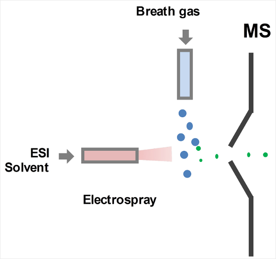
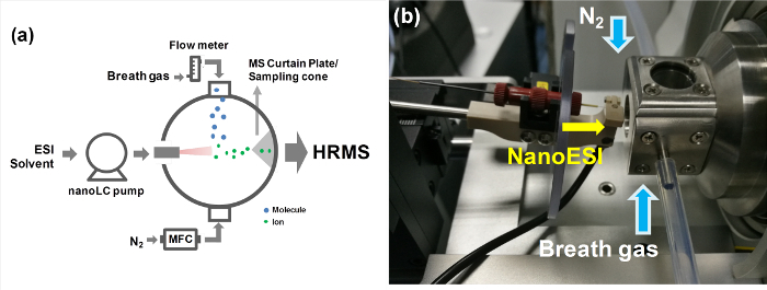
Set up a Sec-nanoESI source according to the SESI process, i.e., the breath gas is introduced to intersect an electrospray plume and ionized by the charged droplets (Figure 1). The sources built in individual labs depend on the interface of the mass spectrometer used24,25. Here, set up the Sec-nanoESI source based on a commercial nanoESI source (Figure 2) and implement to a benchtop quadrupole orbitrap mass spectrometer. NOTE: The main body of the source is a cubic stainless-steel chamber (Length 25 mm, I.D. 13 mm) (Figure 2b) with an inlet (I.D. 4 mm) to introduce the nanoESI capillary into the chamber. Thus, the chamber is not completely sealed (Figure 2b).
Install two stainless-steel tubes (Length 8 mm, O.D. 5 mm, I.D. 3 mm) on each side of the chamber for gas delivery.
Equip two quartz windows (I.D. 14 mm) on the top and bottom of the chamber to check the position of the tip of the nanoESI capillary and nanoESI spray either by eyes or a digital microscope.
Weld the chamber to the sweep cone of the mass spectrometer. NOTE: The design may change depending on the particular geometry of the atmospheric pressure interface of the mass spectrometer used in individual labs.


2. Instrument optimization
- Calibrate the mass spectrometer in both positive and negative ion detection modes according to manufacturer's instructions. By applying calibration, mass spectrometer parameters, such as lens potentials and detection conditions, are optimized to give good sensitivity and peak shape at a specified resolution value. The mass resolution of 70000 is used here.
- Perform complete Q Exactive calibrations by using the commercial ESI source; however, mass calibration can be performed with any compatible sources, including customized ones.
Set the temperature of the ion transfer tube (ITT) of the mass spectrometer > 100 °C. Though the highest temperature can be set at 350 °C, it may result in the decomposition of some compounds. Thus, 150°C is used in this experiment. NOTE: For the mass spectrometers featuring a sampling orifice instead of ITT, the temperature of the sampling orifice is set > 100 °C.
For the ESI solvent and flow rate, select the appropriate ESI solvent on the basis of properties of solvent (e.g., polarity and volatility) and targeted compounds (e.g., proton affinity). A mixture of water and methanol in various ratios has been commonly used as ESI solvent25. In this experiment, use water containing 0.1% (v/v) formic acid, for high ionization efficiency has been reported this solvent13,19,23. Set the flow rate of ESI solvent in the range of 0-1.5 μL/min and 200 nL/min. NOTE: Degas ESI solvent for 30 min before use.
Optimize Sec-nanoESI source parameters, mainly nanoESI voltage and nanoESI capillary tip position. The voltage commonly ranges from 2.0 to 4.5 kV. Use 2.5 kV here. NOTE: Higher ESI voltage is applied as the flow rate increases. The distance between the tip and the mass spectrometer orifice can be adjusted from 1 to 5 mm. After optimization, the normalized intensity level (NL) observed in a mass spectrum should be > 1x10⁶ and the variation of total ion chromatogram (TIC) should be < 10% in both positive and negative ion detection modes. The mass spectrum and TIC are obtained in the mass range of m/z 50-750.
Apply pure gas to the source. This is an optional step, aiming at reducing the influence of VOCs from indoor air. High purity nitrogen gas (N2, 99.99%) or pure air can be used. With the presence of pure gas, the NL observed in a mass spectrum should be > 1x10⁵ and the variation of TIC should be < 10% in both positive and negative ion detection modes. High purity N2 is used here and delivered at 0.8 L/min. NOTE: The total flow rate of pure gas and breath gas should be higher than the flow rate through the orifice of the mass spectrometer.
3. Measurement of exhaled breath
- Inhale the indoor air and perform a normal exhalation to breathe out all the air in the lungs at a constant flow rate. Monitor the exhalation flow rate either by a manometer or a flow meter visible to the subject. Use Teflon (PTFE) tubing to deliver the breath gas23.
- Connect the inlet of the flow meter to a Nafion tubing (Length 60 cm) to remove the water vapor in the exhaled breath and connect the outlet of the flow meter to a PTFE tubing (length 13 cm, I.D. 4 mm). It takes < 30 s for one exhalation measurement.
- To minimize confounding effects, have participants from eating, drinking, and brushing their teeth at least 30 min prior to the measurements23. NOTE: To minimize the influence of VOCs from indoor air, it has been reported to inhale pure gas instead of indoor air26. When a Nafion tubing is used, some polar compounds may be lost.
- During measurement, keep checking if the ion intensity exceeds the linear detection limit of the instrument or not. The saturation of signal can lead to an artifact peak that does not practically result from the compound in the sample. By inhaling through the nose, part of ambient VOCs and particles would be removed; however, it is noteworthy that compounds in the nasal passages may also be detected.
4. Obtain a breath fingerprint and a time trace of a compound
Obtain chromatograms and mass spectra. Use software (e.g., Xcalibur) to record chromatograms and mass spectra. Because this is direct MS analysis and no chromatographic separation is performed, the total ion chromatogram (TIC) actually indicates the time trace of all the signals detected in the mass spectra and the extracted ion chromatogram (EIC) shows the time trace of a specified compound. NOTE: For other commercial mass spectrometers, the chromatograms and mass spectra can be obtained by the corresponding data acquisition software.
- Obtain an exhaled breath fingerprint by selecting a number of scans in the TIC when exhaled breath is measured. Obtain a mass spectrum representing an average of these scans by the software.
- To eliminate background peaks from the breath fingerprint, use the Subtract Background tool in the software. Please refer to the user guide provided by the manufacturer. In brief, select the same number of scans when no breath sample is introduced, and subtract the background mass spectrum from the breath fingerprint. NOTE: In this method, the threshold to identify the features in the breath fingerprint is defined as three times the standard deviation of the background signal. For other commercial mass spectrometers, background subtraction can be performed by the corresponding data acquisition software.
Obtain a time trace of a specified compound. Select the peak of a targeted compound in the breath fingerprint, and the time trace of the compound is acquired subsequently by the software.
Representative Results
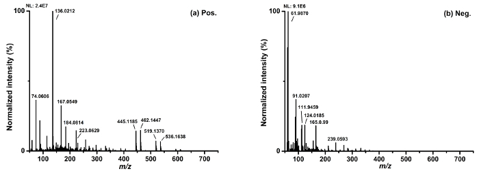
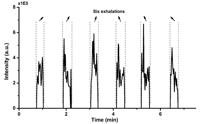
Figure 3 shows the breath fingerprints in the mass range of m/z 50-750 recorded under both positive and negative ion detection modes. 291 peaks (peak intensity > 5.0x10⁴) and 173 peaks (peak intensity > 3.0x10⁴) have been observed in background-subtracted breath fingerprints in the positive and negative ion detection modes, respectively. To identify peaks in the mass spectra, please refer to prior publications for details12,18,24,29. In brief, both volatile metabolites and VOCs from indoor air have been detected. For example, the peak at m/z 74.0606 (Figure 3a) results from exhaled N,N-dimethylformamide or aminoactone according to the Human Metabolome Database (HMDB); peaks at m/z 462.1447 and m/z 536.1638 (Figure 3a) are from the adducts of exhaled ammonia and polysiloxanes (laboratory contaminants)12. The typical mass accuracy values in positive and negative ion detection modes are -4.0-13.5 ppm and -20.3-1.3 ppm, respectively. Figure 4 presents the time trace of indole, a typical endogenous compound, which is detected by six replicated measurements of exhaled breath from one subject. It takes less than 7 min for all six measurements.


Figure 1. Schematic for SESI-MS analysis of VOCs in exhaled breath. Please click here to view a larger version of this figure.
Figure 2. (a) A schematic diagram and (b) a photo of the Sec-nanoESI source used in this experiment. Please click here to view a larger version of this figure.
Figure 3. Background-subtracted breath fingerprints obtained in (a) positive and (b) negative ion detection modes in the mass range of m/z 50-750. Please click here to view a larger version of this figure.
Figure 4. Time trace of indole detected by six replicated measurements of exhaled breath from one subject. Please click here to view a larger version of this figure.
Discussion
Constructing the Sec-nanoESI source based on a commercial nanoESI source, the ionization efficiency is higher than that of using an ESI source30. In addition, the ionization efficiency is further improved in a closed chamber, as it isolates the process from the ambient background air, and at the same time facilitates the mixing between the gas sample and the spray plume. By using a Sec-nanoESI, less parameters need to be optimized compared to an ESI source, making it easier for installation, application and maintenance.
If no signal is observed or sensitivity decreases significantly when performing breath analysis by Sec-nanoESI-MS, one should check the position of the spray capillary tip and also the formation of droplets at the tip of the capillary. Align the tip with the orifice of the mass spectrometer. Change the spray capillary to a new one if the spray capillary is blocked or the tip is contaminated. Otherwise, check whether the ITT of the instrument is blocked or contaminated. Replace or clean the ITT if necessary. Turn off ESI voltage before checking the spray capillary. Set the temperature of ITT at room temperature and wait until the temperature drops down.
SESI-HRMS has been demonstrated to be a sensitive and selective technique for real-time breath analysis4,6,12. In the past few years, this technique has been successfully applied to gauging circadian variation3,6, monitoring pharmacokinetics7,8, identifying metabolic pathways5, etc. Lately, amino acids in human breath have been successfully quantified by SESI-MS for the first time, which is remarkable progress in quantitative analysis5. With further investigations, SESI-HRMS could establish itself as a useful and efficient noninvasive clinical method.
Disclosures
The authors have nothing to disclose.
Acknowledgments
This work has been financially supported by National Natural Science Foundation of China (No. 91543117).
References
- De Lacy Costello B, et al. A review of the volatiles from the healthy human body. J. Breath Res. 2014;8(1):014001–014030. doi: 10.1088/1752-7155/8/1/014001. [DOI] [PubMed] [Google Scholar]
- Phillips M, Greenberg J. Ion-trap detection of volatile organic compounds in alveolar breath. Clin. Chem. 1992;38(1):60–65. [PubMed] [Google Scholar]
- Martínez-Lozano Sinues P, et al. Circadian variation of the human metabolome captured by real-time breath analysis. PLoS One. 2014;9(12):0114422–0114438. doi: 10.1371/journal.pone.0114422. [DOI] [PMC free article] [PubMed] [Google Scholar]
- Garcia-Gomez D, et al. Secondary electrospray ionization coupled to high-resolution mass spectrometry reveals tryptophan pathway metabolites in exhaled human breath. Chem. Common. 2016;52(55):8526–8528. doi: 10.1039/c6cc03070j. [DOI] [PubMed] [Google Scholar]
- Garcia-Gomez D, et al. Real-time quantification of amino acids in the exhalome by secondary electrospray ionization-mass spectrometry: A proof-of-principle Study. Clin. Chem. 2016;62(9):1230–1237. doi: 10.1373/clinchem.2016.256909. [DOI] [PubMed] [Google Scholar]
- Martínez-Lozano Sinues P, Kohler M, Brown SA, Zenobia R, Dallmann R. Gauging circadian variation in ketamine metabolism by real-time breath analysis. Chem. Common. 2017;53(14):2264–2267. doi: 10.1039/c6cc09061c. [DOI] [PubMed] [Google Scholar]
- Gamez G, et al. Real-time, in vivo monitoring and pharmacokinetics of valproic acid via a novel biomarker in exhaled breath. Chem. Common. 2011;47(17):4884–4886. doi: 10.1039/c1cc10343a. [DOI] [PubMed] [Google Scholar]
- Li X, et al. Drug pharmacokinetics determined by real-time analysis of mouse breath. Angew. Chem. Int. Ed. 2015;54(27):7815–7818. doi: 10.1002/anie.201503312. [DOI] [PubMed] [Google Scholar]
- Amorim LLA, Cardeal ZL. Breath air analysis and its use as a biomarker in biological monitoring of occupational and environmental exposure to chemical agents. J. Chromatogr. B. 2007;853(1-2):1–9. doi: 10.1016/j.jchromb.2007.03.023. [DOI] [PubMed] [Google Scholar]
- Bajtarevic A, et al. Noninvasive detection of lung cancer by analysis of exhaled breath. BMC Cancer. 2009;9:348. doi: 10.1186/1471-2407-9-348. [DOI] [PMC free article] [PubMed] [Google Scholar]
- Smith D, Wang TS, Pysanenko A, Španěl P. A selected ion flow tube mass spectrometry study of ammonia in mouth- and nose-exhaled breath and in the oral cavity. Rapid Commun. Mass Spectrom. 2008;22(6):783–789. doi: 10.1002/rcm.3434. [DOI] [PubMed] [Google Scholar]
- Li X, Huang L, Zhu H, Zhou Z. Direct human breath analysis by secondary nano-electrospray ionization ultrahigh resolution mass spectrometry: Importance of high mass resolution and mass accuracy. Rapid Commun. Mass Spectrom. 2017;31(3):301–308. doi: 10.1002/rcm.7794. [DOI] [PubMed] [Google Scholar]
- Martínez-Lozano P, Fernandez de la Mora J. Electrospray ionization of volatiles in breath. Int. J. Mass Spectrom. 2007;265(1):68–72. [Google Scholar]
- Zeng Q, et al. Detection of creatinine in exhaled breath of humans with chronic kidney disease by extractive electrospray ionization mass spectrometry. J. Breath Res. 2016;10(1):016008–016015. doi: 10.1088/1752-7155/10/1/016008. [DOI] [PubMed] [Google Scholar]
- Chen HW, Wortmann A, Zhang WH, Zenobi R. Rapid in vivo fingerprinting of nonvolatile compounds in breath by extractive electrospray ionization quadrupole time-of-flight mass spectrometry. Angew. Chem. Int. Ed. 2007;46(4):580–583. doi: 10.1002/anie.200602942. [DOI] [PubMed] [Google Scholar]
- Benoi FM, Davldson WR, Lovett AM, Nacson S, Ngo A. Breath analysis by atmospheric pressure ionization mass spectrometry. Anal. Chem. 1983;55(4):805–807. doi: 10.1021/ac00255a053. [DOI] [PubMed] [Google Scholar]
- Bregy L, Martínez-Lozano Sinues P, Nudnova MM, Zenobi R. Real-time breath analysis with active capillary plasma ionization-ambient mass spectrometry. J. Breath Res. 2014;8(2):027102–027110. doi: 10.1088/1752-7155/8/2/027102. [DOI] [PubMed] [Google Scholar]
- Gaugg MT, et al. Expanding metabolite coverage of real-time breath analysis by coupling a universal secondary electrospray ionization source and high resolution mass spectrometry-a pilot study on tobacco smokers. J. Breath Res. 2016;10(1):016010–016020. doi: 10.1088/1752-7155/10/1/016010. [DOI] [PubMed] [Google Scholar]
- Martínez-Lozano P, Zingaro L, Finiguerra A, Cristoni S. Secondary electrospray ionization-mass spectrometry: breath study on a control group. J. Breath Res. 2011;5(1):016002–016012. doi: 10.1088/1752-7155/5/1/016002. [DOI] [PubMed] [Google Scholar]
- Martínez-Lozano Sinues P, Zenobi R, Kohler M. Analysis of the exhalome a diagnostic tool of the future. Chest. 2013;144(3):746–749. doi: 10.1378/chest.13-1106. [DOI] [PubMed] [Google Scholar]
- Martínez-Lozano Sinues P, Fernandez de la Mora J. Direct analysis of fatty acid vapors in breath by electrospray ionization and atmospheric pressure Ionization-Mass Spectrometry. Anal. Chem. 2008;80(21):8210–8215. doi: 10.1021/ac801185e. [DOI] [PubMed] [Google Scholar]
- Martínez-Lozano Sinues P, et al. Breath analysis in real time by mass spectrometry in chronic obstructive pulmonary disease. Respiration. 2014;87(4):301–310. doi: 10.1159/000357785. [DOI] [PubMed] [Google Scholar]
- Martínez-Lozano Sinues P, Kohler M, Zenobi R. Monitoring diurnal changes in exhaled human breath. Anal. Chem. 2013;85(1):369–373. doi: 10.1021/ac3029097. [DOI] [PubMed] [Google Scholar]
- Chen HW, Zenobi R. Neutral desorption sampling of biological surfaces for rapid chemical characterization by extractive electropray ionization mass spectrometry. Nat. Protoc. 2008;3(9):1467–1475. doi: 10.1038/nprot.2008.109. [DOI] [PubMed] [Google Scholar]
- Li X, Hu B, Ding J, Chen HW. Rapid characterization of complex viscous samples at molecular levels by neutral desorption extractive electrospray ionization mass spectrometry. Nat. Protoc. 2011;7(6):1010–1025. doi: 10.1038/nprot.2011.337. [DOI] [PubMed] [Google Scholar]
- Gordon SM, Szidon JP, Krotoszynski BK, Gibbons RD, O'Neill HJ. Volatile organic compounds in exhaled air from patients with lung cancer. Clin. Chem. 1985;31(8):1278–1282. [PubMed] [Google Scholar]
- Ding JH, et al. Development of extractive electrospray ionization ion trap mass spectrometry in vivo breath analysis. Analyst. 2009;134(10):2040–2050. doi: 10.1039/b821497b. [DOI] [PubMed] [Google Scholar]
- Basum G, Dahnke H, Halmer D, Hering P, Mürtz M. Online recording of ethane trances in human breath via infrared laser spectroscopy. J. Appl. Physiol. 2003;95(6):2583–2590. doi: 10.1152/japplphysiol.00542.2003. [DOI] [PubMed] [Google Scholar]
- Tøien Ø. Automated open flow respirometry in continuous and long-term measurements: design and principles. J. Appl. Physiol. 2013;114(8):1094–1107. doi: 10.1152/japplphysiol.01494.2012. [DOI] [PubMed] [Google Scholar]
- Huang L, Li X, Xu M, Huang ZX, Zhou Z. Identification of relatively high molecular weight compounds in human breath using secondary nano electrospray ionization ultrahigh resolution mass spectrometry. Chem. J. Chinese U. 2017;38(5):752–757. [Google Scholar]


