This study defines the clinical, biomarker, and molecular spectrum and provides genotype-phenotype correlations among patients with ataxia with oculomotor apraxia type 1 from 18 countries.
Key Points
Questions
What are the clinical, biomarker, and molecular delineations and genotype-phenotype correlations of ataxia with oculomotor apraxia type 1?
Findings
In this analysis of 80 patients with ataxia with oculomotor apraxia type 1, levels of α-fetoprotein were slightly elevated. The p.Trp279* mutation was the most frequent APTX mutation in the white population.
Meaning
Increased α-fetoprotein levels may constitute a new biomarker of ataxia with oculomotor apraxia type 1, and oculomotor apraxia may be correlated with more severe disease.
Abstract
Importance
Ataxia with oculomotor apraxia type 1 (AOA1) is an autosomal recessive cerebellar ataxia due to mutations in the aprataxin gene (APTX) that is characterized by early-onset cerebellar ataxia, oculomotor apraxia, axonal motor neuropathy, and eventual decrease of albumin serum levels.
Objectives
To improve the clinical, biomarker, and molecular delineation of AOA1 and provide genotype-phenotype correlations.
Design, Setting, and Participants
This retrospective analysis included the clinical, biological (especially regarding biomarkers of the disease), electrophysiologic, imaging, and molecular data of all patients consecutively diagnosed with AOA1 in a single genetics laboratory from January 1, 2002, through December 31, 2014. Data were analyzed from January 1, 2015, through January 31, 2016.
Main Outcomes and Measures
The clinical, biological, and molecular spectrum of AOA1 and genotype-phenotype correlations.
Results
The diagnosis of AOA1 was confirmed in 80 patients (46 men [58%] and 34 women [42%]; mean [SD] age at onset, 7.7 [7.4] years) from 51 families, including 57 new (with 8 new mutations) and 23 previously described patients. Elevated levels of α-fetoprotein (AFP) were found in 33 patients (41%); hypoalbuminemia, in 50 (63%). Median AFP level was higher in patients with AOA1 (6.0 ng/mL; range, 1.1-17.0 ng/mL) than in patients without ataxia (3.4 ng/mL; range, 0.8-17.2 ng/mL; P < .01). Decreased albumin levels (ρ = −0.532) and elevated AFP levels (ρ = 0.637) were correlated with disease duration. The p.Trp279* mutation, initially reported as restricted to the Portuguese founder haplotype, was discovered in 53 patients with AOA1 (66%) with broad white racial origins. Oculomotor apraxia was found in 49 patients (61%); polyneuropathy, in 74 (93%); and cerebellar atrophy, in 78 (98%). Oculomotor apraxia correlated with the severity of ataxia and mutation type, being more frequent with deletion or truncating mutations (83%) than with presence of at least 1 missense variant (17%; P < .01). Mean (SD) age at onset was higher for patients with at least 1 missense mutation (17.7 [11.4] vs 5.2 [2.6] years; P < .001).
Conclusions and Relevance
The AFP level, slightly elevated in a substantial fraction of patients, may constitute a new biomarker for AOA1. Oculomotor apraxia may be an optional finding in AOA1 and correlates with more severe disease. The p.Trp279* mutation is the most frequent APTX mutation in the white population. APTX missense mutations may be associated with a milder phenotype.
Introduction
Autosomal recessive cerebellar ataxias (ARCAs) consist of a heterogeneous group of neurodegenerations due to aprataxin mutations, including ataxia with oculomotor apraxia type 1 (AOA1). Ataxia with oculomotor apraxia type 1 has been described as an early-onset ataxia with cerebellar atrophy, oculomotor apraxia (OMA), choreodystonia, and peripheral neuropathy. Although controversial, OMA may be defined as horizontal saccades of elevated latencies commonly associated with hypometric staircase saccades, oculocephalic dissociation, and/or head thrust. Ataxia with oculomotor apraxia type 1, the most frequent ARCA in Japan, has also been found in other countries. During the course of AOA1, decreased serum albumin levels and elevated serum total cholesterol levels may be observed.
The prevalence of OMA in AOA1 remains debatable. Whether the α-fetoprotein (AFP) serum level is a biomarker of AOA1 has not been elucidated, and genotype-phenotype correlations need to be strengthened. In this study, we sought to better define the clinical, biological (especially regarding biomarkers of the disease), and molecular spectrum of AOA1 and to provide genotype-phenotype correlations.
Methods
We performed a retrospective analysis of the clinical, biological, electrophysiologic, imaging, and molecular data of all patients consecutively diagnosed with AOA1 in our genetics laboratory at Institut de Génétique et de Biologie Moléculaire et Cellulaire, Institut National de la Santé et de la Recherche Medicale–U964/ Centre National de la Recherche Scientifique–Unité Mixte de Recherche 7104/Université de Strasbourg, Illkirch, France, from January 1, 2002, through December 31, 2014. Sequencing of the APTX gene (OMIM 606350) was performed on request for cases of cerebellar ataxia with hypoalbuminemia and/or early-onset cerebellar ataxia combined with peripheral neuropathy and/or cerebellar atrophy using brain magnetic resonance imaging. The local ethics committee of Hôpitaux Universitaires de Strasbourg, Strasbourg, France, approved the study. All participants provided written informed consent.
Clinical Analysis
Age at onset of the disease, duration of disease (DD), and age at last examination were recorded, as well as sex, consanguinity, and geographic origin. Patients underwent neurologic examination, motor and sensory nerve conduction studies, and brain magnetic resonance imaging. Exhaustive abnormal findings on the clinical examination included oculomotor signs (strabismus, saccadic pursuit, OMA, oculocephalic dissociation, and head thrust), movement disorders (chorea, dystonia, parkinsonism, tremor, and myoclonus), pyramidal signs, areflexia, muscle wasting, ataxia, and intellectual disability.
Oculocephalic dissociation was defined as dissociation between tandem head and eye movement, with the eyes lagging during head rotation. Peripheral neuropathy was defined by reduced deep tendon reflexes or areflexia, distal loss of vibration sense, and electrophysiologic evidence of nerve conduction abnormalities. Possible cerebellar atrophy was evaluated on sagittal and axial magnetic resonance images by a neuroradiologist and a neurologist.
A spinocerebellar degeneration functional score (SDFS) was used to evaluate the disability stage from 1 to 7, with higher scores representing severe impairment. The SDFS corrected for DD (SDFS:DD ratio) was used to evaluate the progression rate of the disease. A scale of assessment and rating of ataxia (SARA) was used to evaluate spinocerebellar degeneration.
Biological Analysis
We measured serum levels of albumin, total cholesterol, low-density lipoprotein cholesterol, creatine kinase, and AFP. For some patients, several measurements of AFP and albumin levels were obtained during the course of the disease.
Mutational Analysis
We obtained DNA from total blood samples from patients using standard procedures. Genetic analysis of APTX was performed using conventional Sanger sequencing. Exons 1 to 6 of APTX were amplified as previously described. For exon 7, a new pair of intronic flanking primers was designed (forward, 5′-TAT TGG TTT TCC AGA GTA GC-3′; reverse, 5′-GAA TAG GTG CCA GCA GTT T-3′) and used for polymerase chain reaction and amplification (10-second denaturation at 94°C, 10” annealing at 64°C, and 10” elongation at 72°C for 35 cycles). Polymerase chain reaction products were purified with a commercially available kit (NucleoSpin Extract 2; Macherey-Nagel GmbH) and sequenced from the forward and reverse strands. Sequencing was performed using a Taq cycle kit (Applied Biosystems). Reaction products were run on an automated DNA sequencer (model 3100; Applied Biosystems). Homozygous exon deletions were readily detected by absence of polymerase chain reaction amplification product and were verified with polymerase chain reaction tests using a different set of primers. In 1 case, a single heterozygous mutation was identified, and the copy number variation was excluded by multiplex ligation-dependent probe amplification analysis (SALSA MLPA kit P316-A1 for recessive ataxias; MRC-Holland). This case was considered to be a heterozygous carrier by chance, and a diagnosis of AOA1 was excluded.
One patient was diagnosed using targeted exon capture strategy coupled with multiplexing and high-throughput sequencing of 57 genes causing ataxia when mutated, including APTX. When available, parents underwent sequencing to verify segregation in trans of the mutations. For consistency with most publications on APTX mutation identification, the RefSeq gene reference NM_175073, encoding a 342–amino acid isoform of APTX, was used for mutation report.
Serum AFP Level Measurement in Control Groups
Measurement of serum AFP levels in control individuals was performed using the immunoanalysis Kryptor Brahms method as established in the Laboratoire de Biochimie Générale et Spécialisée in the University Hospital of Strasbourg, France. The AFP level assessment using this method with 100 healthy controls provided a median value of 3.2 ng/mL with a 97.5th percentile at 7 ng/mL, which was retained as the upper limit of normal in our study (to convert AFP levels to micrograms per liter, multiply by 1.0). All healthy controls had an AFP level from 0.5 to 15.7 ng/mL. Levels of AFP were assessed in 102 patients who had no cerebellar ataxia but were affected by Parkinson disease (n = 37), atypical parkinsonism (n = 13), Huntington disease (n = 3), dystonia (n = 1), Alzheimer disease (n = 11), multiple sclerosis (n = 11), amyotrophic lateral sclerosis (n = 2), peripheral neuropathy (n = 16), and myopathy (n = 6). Levels of AFP were assessed in 74 controls with AOA2 described previously and 76 patients with sporadic late-onset cerebellar ataxia (SLOCA), with a large variety of cerebellar variants of multiple system atrophy (n = 29), dominant or recessive ataxia (n = 9), immune-mediated causes or malformations or toxic or other acquired causes (n = 6), or undetermined (n = 32).
Genotype-Phenotype Correlation Studies
For genotype-phenotype correlations, 2 different classification schemes were used. Because missense mutations are often hypomorphic and may cause a milder phenotype, patients with AOA1 were divided into groups with (1) at least 1 missense APTX mutation and (2) 2 truncating APTX mutations. To test whether hypomorphic missense mutations are clustered in a specific part of the histidine triad (HIT) domain, we performed a second analysis in which patients were divided into the following 4 groups according to the type of mutation and its location in APTX: (1) homozygous p.Trp279*; (2) two truncating mutations with at least 1 different from p.Trp279*; (3) at least 1 missense mutation in the first part of HIT domain, before the HIT motif (β sheets 2 and 3 and α helix 2, containing the missense mutations p.Lys197Gln, p.Ala198Val, Pro206Leu, and Val230Gly); and (4) at least 1 missense mutation in the second part of the HIT domain at or after the part that includes the HIT motif (β sheets 4 and 5 and α helix 3, containing the missense mutations p.Leu261Phe, p.Asp267Gly, and p.Ser270Phe). The 2 groups and then the 4 groups were compared regarding their clinical, electrophysiologic, imaging, and biochemical features. The second analysis of the 4 groups was performed when all the mutations of 80 patients with AOA1 were known.
Statistical Analysis
Data were analyzed from January 1, 2015, through January 31, 2016. All statistical analyses were performed with SAS software for Windows (release 9.3; SAS Institute). The χ2 and the Fisher exact tests were applied to reveal differences in proportions between groups or associations between category variables. We used nonparametric statistical tests for the analysis of quantitative data because the variable distribution was not normal. We applied the Kruskall-Wallis test for comparisons of quantitative variables across 3 or more independent groups. In case of statistically significant results, groups underwent pairwise comparison, and the P values were adjusted using the Bonferroni-Holm method. The linear associations between quantitative variables were assessed using the Spearman correlation procedure. For all statistical tests, we considered P < .05 as statistically significant.
Results
Eighty patients (46 men [58%] and 34 women [42%]; mean [SD] age, 28.5 [13.7] years) from 51 families were diagnosed with AOA1. Fifty-seven patients were new, and 23 had been described previously.
Clinical Data
Table 1 and Table 2 summarize the main features of the patients with AOA1 (2 patients are shown in Video 1 and Video 2). Patients originated from France (n = 23), Algeria (n = 11), Morocco (n = 8), Tunisia (n = 4), Brazil (n = 3), Switzerland (n = 3), India (n = 2), Belgium (n = 2), the United States (n = 2), Ireland (n = 2), Israel (n = 2), Italy (n = 2), Lebanon (n = 2), Rwanda (n = 2), Turkey (n = 2), the Netherlands (n = 1), Syria (n = 1), Portugal (n = 1), and North Africa (without geographic precision) (n = 7). Oculomotor apraxia correlated positively with the SARA score; the higher the SARA score, the more frequent the OMA (z = 1.739; P = .04). The progression rate of the disease (SDFS:DD ratio) correlated positively with the presence of movement disorder; the faster the disease progression, the more intense the movement disorder (z = 1.839; P = .03).
Table 1. Main Qualitative Variables of Patients With AOA1 .
| Qualitative Variable | No. (%) of Patients With AOA1 (N = 80) |
|---|---|
| Consanguinity | 48 (60) |
| Oculomotor apraxia | 49 (61) |
| Hypometric saccades | 59 (74) |
| Nystagmus | 49 (61) |
| Saccadic pursuit | 57 (71) |
| Horizontal strabismus | 20 (25) |
| Oculocephalic dissociation | 40 (50) |
| Head thrust | 36 (45) |
| Pes cavus | 30 (38) |
| Scoliosis | 27 (34) |
| Pyramidal signs | 9 (11) |
| Intellectual disability | 42 (53) |
| Current chorea | 32 (40) |
| Chorea disappearing during the course of the disease | 6 (8) |
| Dystonia | 20 (25) |
| Parkinsonism | 2 (3) |
| Tremor | 52 (65) |
| Myoclonus | 8 (10) |
| Peripheral neuropathy | 75 (94) |
| Cerebellar atrophy | 79 (99) |
| Elevated AFP level (<7 ng/mL) | 33 (41) |
| Decreased albumin level (3.5-4.8 g/dL) | 50 (63) |
| Elevated total cholesterol level (0.5 -2 g/L) | 45 (56) |
| Elevated LDL cholesterol level (<1.60 g/L) | 34 (43) |
| Elevated CK level (10-200 U/L) | 23 (29) |
Abbreviations: AFP, α-fetoprotein; AOA1, ataxia with oculomotor apraxia type 1; CK, creatine kinase; LDL, low-density lipoprotein.
SI conversion factors: To convert AFP to micrograms per liter, multiply by 1.0; total and LDL cholesterol to millimoles per liter, multiply by 0.259; and CK to microkatals per liter, multiply by 0.0167.
Table 2. Main Quantitative Variables of Patients With AOA1.
| Quantitative Variables (n = 80) |
Median (Range) | Mean (SD) |
|---|---|---|
| Age at onset, y | 6 (2-40) | 7.7 (7.4) |
| Age at examination, y | 26 (9-59) | 28.5 (13.7) |
| Duration of disease, y | 17 (4-52) | 20.9 (12.6) |
| Age at wheelchair use (n = 40), y | 18 (4-57) | 20 (11.4) |
| Duration of disease until wheelchair use (n = 40), y | 11.8 (2-38) | 13.6 (8.6) |
| SARA score at last examinationa | 28 (6-37) | 26.4 (7.6) |
| SDFS score at last examinationb | 6 (3-7) | 5.5 (0.9) |
| Progression rate, SDFS:DD ratio | 0.3 (0.1-1.2) | 0.3 (0.2) |
| AFP level, ng/mL | 6.0 (1.1-17.0) | 6.6 (4) |
| Albumin level, g/dL | 3.25 (1.78-4.59) | 3.32 (0.65) |
| Total cholesterol level, g/L | 2.1 (1.0-3.8) | 2.2 (0.7) |
| LDL cholesterol level, g/L | 1.7 (0.8-3.2) | 1.7 (0.6) |
| CK level, U/L | 185 (32-360) | 168.4 (101.5) |
Abbreviations: AFP, α-fetoprotein; AOA1, ataxia with oculomotor apraxia type 1; CK, creatine kinase; DD, disease duration; LDL, low-density lipoprotein; SARA, scale for the assessment and rating of ataxia; SDFS, spinocerebellar degeneration functional score.
SI conversion factors: To convert AFP to micrograms per liter, multiply by 1.0; albumin to grams per liter, multiply by 10.0; total and LDL cholesterol to millimoles per liter, multiply by 0.259; and CK to microkatals per liter, multiply by 0.0167.
Score range from 0 to 40, with higher scores indicating more more severe ataxia.
Scores range from 1 to 7, with higher scores indicating severe impairment.
Video 1. Patient 72.
The patient was homozygous for the Ala198Val mutation in the APTX gene at 28 years of age. Dysmetria during the finger-nose test and oculocephalic dissociation (dissociation of eyes-head when looking toward a lateral target) with hypometric saccades during the head movement task were noted.
Video 2. Patient 76.
The patient was heterozygous for the Trp279* and Lys197Gln mutation in the APTX gene at 28 years of age. Cerebellar ataxia at gait and dysmetria during the finger-nose test were noted.
Biological Data
Biological data regarding the biomarkers are presented in Table 2. Longer DD was correlated with lower albumin serum levels (ρ = −0.532; P < .01); the longer the DD, the higher the serum AFP level (ρ = 0.637; P < .01) and the higher the serum total cholesterol level (ρ = 0.571; P < .01). The biomarkers measured with lower levels of albumin correlated with increased total cholesterol (ρ = −0.451; P = .02) and AFP (ρ = −0.505; P < .01) levels. We did not find any phenotype-genotype correlation according to AFP level.
Mutation Analysis
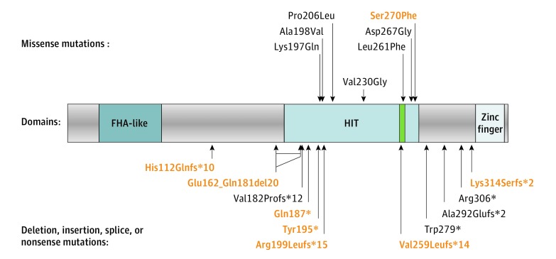
We identified 8 new mutations (Table 3 and Figure 1). Seven of these mutations were estimated to result in a major disruption of aprataxin and included 2 nonsense mutations (p.Gln187* and p.Tyr195*), 1 large in-frame deletion (del exon4), and 4 frameshift mutations (c.336_337delCA, c.774_775insCTTTCAACTA, c.940_956del17, and c.596delG). Only 1 mutation was a missense mutation (p.Ser270Phe), resulting in nonconservative substitution affecting an amino acid residue conserved in all eukaryotes and was predicted to be pathogenic by 2 different algorithms (Sift score, 0.02; PolyPhen2 score, 1.00).
Table 3. Description of the 8 New Mutations.
| Patient No. (Geographic Origin) | Nucleotide Change (Exon) | Amino Acid Change | Mutation Status |
|---|---|---|---|
| 8 (Tunisia) | c.809C>T (exon 6) | p.Ser270Phe | Compound heterozygous (c.875-1G>A (exon 7)) |
| 19 (France) | c.336_337delCA (exon 3) | p.His112Glnfsa10 | Compound heterozygous (c.617C>T (exon 5) p.Pro206Leu) |
| 30 (France) | c.774_775insCTTTCAACTA(exon 6) | p.Val259Leufsa14 | Compound heterozygous (c.837G>A (exon 6) p.Trp279a) |
| 32 (Turkey) | c.940_956del17 (exon 7) | p.Lys314Serfsa2 | Homozygous |
| 41 and 42 (Brazil) | del exon 4 | p.Glu162_Gln181del20 | Homozygous |
| 45 (the Netherlands) | c.559C>T (exon 5) | p.Gln187a | Compound heterozygous (c.837G>A (exon 6) p.Trp279a) |
| 46 and 47 (Morocco) | c.585C>A (exon 5) | p.Tyr195a | Homozygous |
| 48, 49, and 50 (India) | c.596delG (exon 5) | p.Arg199Leufsa15 | Homozygous |
Abbreviations: del, deletion; ins, insertion; fs, frameshift.
Indicates stop codon.
Figure 1. Position of the Mutations With Respect to the Aprataxin (APTX) Protein Domains.
The histidine triad (HIT) motif (HxHxH) is highlighted in green. The deletion, insertion, and splice site mutations are translated into estimated protein consequences. Correspondence for novel mutations (red) is indicated in Table 3; c.544-2A>G (acceptor) and c.770 + 1G>A (donor) exon 5 splice site mutations are estimated to result in frameshift exon 5 skipping p.Val182Profs*12; c.875-1G>A (last) exon 7 acceptor splice site mutation is estimated to result in use of a minor alternative exon 7’ (isoform coded by transcript NM_175069) Ala292Glufs*2. The position of the in-frame exon 4 deletion (Glu162_Gln181del20) is indicated by a double arrow that represents the extent of internal peptide deletion. The large deletion of exons 1 to 4 is not represented because it is estimated to result in complete absence of APTX transcription.
The p.Trp279* mutation, initially only associated with the Portuguese founder haplotype, was found in 53 of 80 patients (66%). Forty-one patients (51%) were homozygous for p.Trp279*.
Genotype-Phenotype Correlation
Patients with AOA1 were divided between those with at least 1 missense mutation in APTX (n = 15) and those with 2 truncating mutations (n = 65). Patients with at least 1 missense mutation had a higher mean (SD) age at onset (17.7 [11.4] vs 5.2 [2.6] years; P < .001) and a less severe mean (SD) SDFS score (2.7 [1.5] vs 4.4 [1.5]; P < .01). Correlations for oculomotor apraxia (83% vs 17%; P < .001), oculocephalic dissociation (82% vs 18%; P = .02), and head thrust (83% vs 17%; P = .01) were found with the second type of mutation, which occurred more frequently in patients with deletion or truncating mutations than in patients with at least 1 missense mutation. We found no difference between groups regarding other qualitative or quantitative variables.
In the second analysis, the patients with AOA1 were divided into the 4 groups described in the Genotype-Phenotype Correlation Studies subsection of the Methods, including 41 (51%) in group 1, 24 (30%) in group 2, 10 (13%) in group 3, and 5 (6%) in group 4. Mean (SD) age at onset was higher for patients in group 4 (25 [10.9] years) compared with group 3 (13.4 [10.1] years; P = .03), group 1 (5.8 [2.5] years; P < .01), and group 2 (2.0 [2.5] years; P < .01).
Comparison of AFP Level With Control Groups
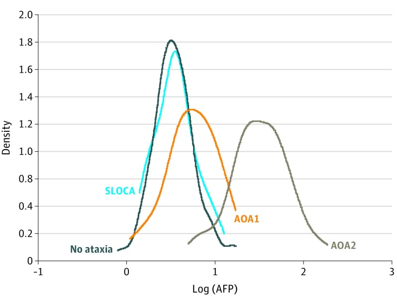
Levels of AFP in controls without ataxia, patients with AOA2, patients with SLOCA, and patients with AOA1 are presented in Figure 2. The median AFP level was significantly higher in patients with AOA1 (6.0 ng/mL; range, 1.1-17.0 ng/mL; mean [SD], 6.6 [4.0] ng/mL) compared with patients without ataxia (3.4 ng/mL; range, 0.8-17.2 ng/mL; mean [SD], 41. [2.8] ng/mL; P < .01) and patients with SLOCA (3.5 ng/mL; range, 1.4-12.7 ng/mL; mean [SD], 4.1 [2.4] ng/mL; P < .01). Median AFP was significantly higher in patients with AOA2 (32.2 ng/mL; range, 5.0-185.0 ng/mL; mean [SD], 42.1 [34.6] ng/mL) compared with patients with AOA1 (P < .01), controls without ataxia (P < .01), and patients with SLOCA (P < .001). The difference in AFP levels between the groups without ataxia and with SLOCA were not significant (P > .99).
Figure 2. Levels of α-Fetoprotein (AFP) .
Distribution of AFP levels in 102 control individuals (no ataxia), 76 patients with sporadic late-onset cerebellar ataxia (SLOCA), 74 patients with ataxia with oculomotor apraxia type 2 (AOA2) described previously, and 80 patients with AOA1 in our study. The density of points is presented as log of the AFP serum level.
Discussion
We herein describe the clinical, biomarker, and molecular findings of the largest international cohort, to our knowledge, of patients with AOA1 reported thus far. The most frequent clinical findings consisted of early-onset cerebellar ataxia with cerebellar atrophy, axonal sensory-motor neuropathy, and eventual OMA (61% of cases). Overall, clinical, electrophysiologic, and imaging features were in accordance with those of previously reported smaller series. Amyotrophy, which should also be included in the evaluation, can belong to the phenoype of AOA1.
The nonsense mutation p.Trp279*, first reported in association with the Portuguese founder haplotype, was the most frequent mutation, representing 66% of all mutated alleles. This mutation was found in patients from several countries, including the Netherlands, Belgium, Ireland, Morocco, Algeria, and Israel, indicating an ancient origin. This finding suggests that mutation p.Trp279* may be the most frequent APTX mutation in the white population.
We found that the mean age at onset was higher for patients with at least 1 missense mutation and therefore manifesting a less severe presentation. Oculomotor apraxia, oculocephalic dissociation, and head thrust were more frequent in patients with deletion or truncating mutations than in patients with at least 1 missense mutation. These results are consistent with a recessively inherited disease due to the loss of protein function, even if such findings were not observed in a large cohort of patients affected with AOA2 due to senataxin loss of function. In our study, OMA, although not a universal finding, was correlated with the SARA score, suggesting that OMA is a marker of an advanced form of AOA1. Oculomotor apraxia is also variably present in patients with AOA2 and ataxia-telangiectasia. The absence of OMA, however, was frequently observed in a significant number of patients, which reinforces the notion that the diagnosis of AOA1 should still be considered in patients with autosomal recessive cerebellar ataxias lacking OMA.
Some missense mutations (p.Lys197Gln, p.Ala198Val, p.Pro206Leu, and p.Val230Gly) appeared to be associated with a severe phenotype, whereas others (p.Leu261Phe, p.Asp267Gly, and p.Ser270Phe) seemed to be linked with a milder presentation. Aprataxin includes the following 3 domains: (1) the PANT domain (PNKP-AOA1 N-terminal domain), also designated as putative forkhead-associated (FHA) domain, corresponding to the N-terminal region of aprataxin; (2) a HIT domain; and (3) a C-terminal domain containing a divergent zinc-finger motif. Aprataxin is involved in DNA single-strand break repair, interactions with several proteins involved in the base excision repair and ribonucleotide excision repair pathways, and actions in RNA-DNA damage response to protect the genome from the accumulation of adenylyl ribosylated 5′ termini. Of note, the milder missense mutations are located within or just downstream of the HIT motif HVHLH of the HIT domain, whereas the other missense mutations are located in the first part of the HIT domain. The HIT motif is the catalytic site for adenosine monophosphate removal activity from the 5′ terminus of aborted ligation products. Missense mutations localized in the HIT domain lead to APTX destabilization with a severe quantitative reduction and alteration of the subcellular distribution, being perinuclear (cytoplasmic) instead of nuclear and nucleolar. Some genotype-phenotype correlations have been previously reported in patients with AOA1. In 15 patients with AOA1, Date et al reported that patients carrying p.Pro206Leu and p.Val263Gly mutations appear to have a later onset and a milder phenotype than do those carrying c.689_690insT or c.del840delT frameshift mutations. In a series of 28 patients with AOA1, Criscuolo et al found that patients carrying p.His201Gln, p.Pro206Leu, and p.Leu223Pro presented with a later age at onset, ranging from 28 to 40 years. In 2011, Yokoseki et al described 58 patients with AOA1 and demonstrated that those homozygous for c.689-690insT (40 patients) had a more severe phenotype than did those with a p.Pro206Leu or p.Val263Gly mutation (9 patients). In our study, the milder presentation associated with the p.Pro206Leu mutation did not reach significance, presumably because the association with severity is small and our study included few patients with p.Pro206Leu. However, we present, to our knowledge for the first time, probably because of the large size of the sample, a genotype-phenotype correlation for the missense APTX mutations according to position relative to the HIT domain.
Results regarding potential biomarkers in AOA1 were interesting. The serum AFP level was elevated in 32 patients (40%). Previous studies have only reported 3 cases of AOA1 with slightly increased AFP levels (7.8, 14, and 14.5 ng/mL). For the first time, to our knowledge, we reveal that the serum AFP level could be a biomarker of AOA1 that increases with DD. Our results further suggest that a slightly increased AFP level in a patient with early-onset ataxia is potentially compatible with AOA1. Hypoalbuminemia was found in 62% of patients, which is comparable with findings in other series. Our biological data confirm that serum albumin levels decrease and cholesterol levels increase during the course of the disease. Hypoalbuminemia and hypercholesterolemia are the most characteristic biochemical findings in AOA1, whereas an elevated AFP level is typical of patients with ataxia-telangiectasia or AOA2 and may be encountered in AOA4 and to some extent in ARCA3.
Modifications of albumin and serum AFP levels were identified in patients with AOA1 in our study, although no alteration of AFP serum level was detected during the clinical course of the disease in a large series of 90 patients with AOA2. Even if AFP is a biomarker of recessive ataxias with peripheral neuropathy and optional OMA (such as AOA1, AOA2, ataxia-telangiectasia, and AOA4), the underlying biological link between AFP level and cerebellar ataxia, peripheral neuropathy, and oculomotor abnormalities is still unknown. In contrast to ataxia-telangiectasia, to our knowledge, sensitivity to ionizing radiation and susceptibility to cancer are not increased in AOA1 and AOA2. Patients with AOA1 presented with normal protein intake and rate of albumin degradation; decreased synthesis of albumin in the liver is suspected to be the cause of hypoalbuminemia. Elevated AFP levels and hypoalbuminemia are found on either side of transcriptional impairment in the liver. In hepatocytes, AFP and albumin genes have opposite transcriptional regulatory mechanisms. Elevated AFP levels in ataxia-telangiectasia and AOA2 likely also proceed from liver transcriptional dysregulation. In the current era of searching for biomarkers, the importance of AFP must be highlighted, especially because its serum level increases progressively during the course of AOA1 in the same way that serum albumin levels decrease progressively. If our data are confirmed by further studies, this increase could be of interest for designing further clinical trials devoted to ARCA with elevated AFP levels.
Movement disorders reported in our series included chorea, dystonia, tremor, and, less frequently, myoclonus. Proportions of movement disorders were similar to the 40% chorea, 16% dystonia, and 8% myoclonus in the study by Yokoseki et al (32 patients [40%], 20 patients [25%], and 8 patients [10%], respectively, in our study). Chorea is classically described in AOA1. Mild to severe dystonia and intentional tremor have already been described in some cases. We found that movement disorders are more frequent in patients with rapid disease progression.
Limitations
The primary limitation of this study is the retrospective nature. Subsequently, repeated measures of biomarkers to better see their progression are lacking.
Conclusions
Our study describes, to our knowledge, the largest series of patients with AOA1 and strengthens the delineation of the clinical, molecular, and biomarker characteristics of this rare form of ARCA. We demonstrate that OMA may be lacking, despite the misleading name of AOA1; AFP serum level may be a new biomarker of AOA1; the p.Trp279* mutation may be the most frequent APTX mutation in the white population; and missense APTX mutations may be associated with a milder phenotype. Given the limitations of a retrospective study, an additional prospective study with repeated measures of biomarkers (albumin, cholesterol, and AFP levels) is required to strengthen our findings.
References
- 1.Anheim M, Tranchant C, Koenig M. The autosomal recessive cerebellar ataxias. N Engl J Med. 2012;366(7):636-646. [DOI] [PubMed] [Google Scholar]
- 2.Moreira MC, Barbot C, Tachi N, et al. The gene mutated in ataxia-ocular apraxia 1 encodes the new HIT/Zn-finger protein aprataxin. Nat Genet. 2001;29(2):189-193. [DOI] [PubMed] [Google Scholar]
- 3.Date H, Onodera O, Tanaka H, et al. Early-onset ataxia with ocular motor apraxia and hypoalbuminemia is caused by mutations in a new HIT superfamily gene. Nat Genet. 2001;29(2):184-188. [DOI] [PubMed] [Google Scholar]
- 4.Aicardi J, Barbosa C, Andermann E, et al. Ataxia-ocular motor apraxia: a syndrome mimicking ataxia-telangiectasia. Ann Neurol. 1988;24(4):497-502. [DOI] [PubMed] [Google Scholar]
- 5.Le Ber I, Moreira M-C, Rivaud-Péchoux S, et al. Cerebellar ataxia with oculomotor apraxia type 1: clinical and genetic studies. Brain. 2003;126(pt 12):2761-2772. [DOI] [PubMed] [Google Scholar]
- 6.Panouillères M, Frismand S, Sillan O, et al. Saccades and eye-head coordination in ataxia with oculomotor apraxia type 2. Cerebellum. 2013;12(4):557-567. [DOI] [PubMed] [Google Scholar]
- 7.Uekawa K, Yuasa T, Kawasaki S, Makibuchi T, Ideta T. A hereditary ataxia associated with hypoalbuminemia and hyperlipidemia: a variant form of Friedreich’s disease or a new clinical entity [in Japanese]? Rinsho Shinkeigaku. 1992;32(10):1067-1074. [PubMed] [Google Scholar]
- 8.Shimazaki H, Takiyama Y, Sakoe K, et al. Early-onset ataxia with ocular motor apraxia and hypoalbuminemia: the aprataxin gene mutations. Neurology. 2002;59(4):590-595. [DOI] [PubMed] [Google Scholar]
- 9.Yokoseki A, Ishihara T, Koyama A, et al. Genotype-phenotype correlations in early onset ataxia with ocular motor apraxia and hypoalbuminaemia. Brain. 2011;134(pt 5):1387-1399. [DOI] [PubMed] [Google Scholar]
- 10.Tranchant C, Fleury M, Moreira MC, Koenig M, Warter JM. Phenotypic variability of aprataxin gene mutations. Neurology. 2003;60(5):868-870. [DOI] [PubMed] [Google Scholar]
- 11.Amouri R, Moreira M-C, Zouari M, et al. Aprataxin gene mutations in Tunisian families. Neurology. 2004;63(5):928-929. [DOI] [PubMed] [Google Scholar]
- 12.Moreira MC, Barbot C, Tachi N, et al. Homozygosity mapping of Portuguese and Japanese forms of ataxia-oculomotor apraxia to 9p13, and evidence for genetic heterogeneity. Am J Hum Genet. 2001;68(2):501-508. [DOI] [PMC free article] [PubMed] [Google Scholar]
- 13.Anheim M, Fleury M, Monga B, et al. Epidemiological, clinical, paraclinical and molecular study of a cohort of 102 patients affected with autosomal recessive progressive cerebellar ataxia from Alsace, Eastern France: implications for clinical management. Neurogenetics. 2010;11(1):1-12. [DOI] [PubMed] [Google Scholar]
- 14.Schmitz-Hübsch T, du Montcel ST, Baliko L, et al. Scale for the assessment and rating of ataxia: development of a new clinical scale. Neurology. 2006;66(11):1717-1720. [DOI] [PubMed] [Google Scholar]
- 15.Renaud M, Anheim M, Kamsteeg E-J, et al. Autosomal recessive cerebellar ataxia type 3 due to ANO10 mutations: delineation and genotype-phenotype correlation study. JAMA Neurol. 2014;71(10):1305-1310. [DOI] [PubMed] [Google Scholar]
- 16.Anheim M, Monga B, Fleury M, et al. Ataxia with oculomotor apraxia type 2: clinical, biological and genotype/phenotype correlation study of a cohort of 90 patients. Brain. 2009;132(Pt 10):2688-2698. [DOI] [PubMed] [Google Scholar]
- 17.Tumbale P, Williams JS, Schellenberg MJ, Kunkel TA, Williams RS. Aprataxin resolves adenylated RNA-DNA junctions to maintain genome integrity. Nature. 2014;506(7486):111-115. [DOI] [PMC free article] [PubMed] [Google Scholar]
- 18.Ahel I, Rass U, El-Khamisy SF, et al. The neurodegenerative disease protein aprataxin resolves abortive DNA ligation intermediates. Nature. 2006;443(7112):713-716. [DOI] [PubMed] [Google Scholar]
- 19.Seidle HF, Bieganowski P, Brenner C. Disease-associated mutations inactivate AMP-lysine hydrolase activity of aprataxin. J Biol Chem. 2005;280(22):20927-20931. [DOI] [PMC free article] [PubMed] [Google Scholar]
- 20.Gueven N, Becherel OJ, Kijas AW, et al. Aprataxin, a novel protein that protects against genotoxic stress. Hum Mol Genet. 2004;13(10):1081-1093. [DOI] [PubMed] [Google Scholar]
- 21.Criscuolo C, Mancini P, Saccà F, et al. Ataxia with oculomotor apraxia type 1 in southern Italy: late onset and variable phenotype. Neurology. 2004;63(11):2173-2175. [DOI] [PubMed] [Google Scholar]
- 22.Castellotti B, Mariotti C, Rimoldi M, et al. Ataxia with oculomotor apraxia type1 (AOA1): novel and recurrent aprataxin mutations, coenzyme Q10 analyses, and clinical findings in Italian patients. Neurogenetics. 2011;12(3):193-201. [DOI] [PubMed] [Google Scholar]
- 23.Bras J, Alonso I, Barbot C, et al. Mutations in PNKP cause recessive ataxia with oculomotor apraxia type 4. Am J Hum Genet. 2015;96(3):474-479. [DOI] [PMC free article] [PubMed] [Google Scholar]
- 24.Nahas SA, Duquette A, Roddier K, Gatti RA, Brais B. Ataxia-oculomotor apraxia 2 patients show no increased sensitivity to ionizing radiation. Neuromuscul Disord. 2007;17(11-12):968-969. [DOI] [PubMed] [Google Scholar]
- 25.Crimella C, Cantoni O, Guidarelli A, et al. A novel nonsense mutation in the APTX gene associated with delayed DNA single-strand break removal fails to enhance sensitivity to different genotoxic agents. Hum Mutat. 2011;32(4):E2118-E2133. [DOI] [PubMed] [Google Scholar]
- 26.Fukuhara N, Nakajima T, Sakajiri K, Matsubara N, Fujita M. Hereditary motor and sensory neuropathy associated with cerebellar atrophy (HMSNCA): a new disease. J Neurol Sci. 1995;133(1-2):140-151. [DOI] [PubMed] [Google Scholar]
- 27.Onodera O. Spinocerebellar ataxia with ocular motor apraxia and DNA repair. Neuropathology. 2006;26(4):361-367. [DOI] [PubMed] [Google Scholar]
- 28.Habeck M, Zühlke C, Bentele KHP, et al. Aprataxin mutations are a rare cause of early onset ataxia in Germany. J Neurol. 2004;251(5):591-594. [DOI] [PubMed] [Google Scholar]
- 29.Sekijima Y, Hashimoto T, Onodera O, et al. Severe generalized dystonia as a presentation of a patient with aprataxin gene mutation. Mov Disord. 2003;18(10):1198-1200. [DOI] [PubMed] [Google Scholar]
- 30.Tsao CY, Paulson G. Type 1 ataxia with oculomotor apraxia with aprataxin gene mutations in two American children. J Child Neurol. 2005;20(7):619-620. [DOI] [PubMed] [Google Scholar]
- 31.Redin C, Le Gras S, Mhamdi O, et al. Targeted high-throughput sequencing for diagnosis of genetically heterogeneous diseases: efficient mutation detection in Bardet-Biedl and Alström syndromes. J Med Genet. 2012;49(8):502-512. [DOI] [PMC free article] [PubMed] [Google Scholar]
- 32.Mallaret M, Renaud M, Redin C, et al. Validation of a clinical practice-based algorithm for the diagnosis of autosomal recessive cerebellar ataxias based on NGS identified cases. J Neurol. 2016;263(7):1314-1322. [DOI] [PubMed] [Google Scholar]


