Abstract
Osteoblasts (OBs) and osteoclasts (OCs) are 2 major groups of bone cells. Their cell-to-cell interactions are important to ensure the continuity of the bone-remodeling process. Therefore, the present study was carried out to optimize an OB/OC co-culture system utilizing the human OB cell line hFOB 1.19 and OCs extracted from peripheral blood mononuclear cells (PBMNCs). It was a 2-step procedure, involving the optimization of the OB culture and the co-culture of the OBs with PBMNCs at an optimum ratio. Firstly, pre-OBs were cultured to 90% confluency and the time required for differentiation was determined. OB differentiation was determined using the van Gieson staining to detect the presence of collagen and Alizarin Red for calcium. Secondly, OBs and OCs were co-cultured at the ratios of 1 OC: 1 OB, 1 OC: 4 OBs, 2 OCs: 1 OB, and 1 OC: 2 OBs. Tartrate-resistant acid phosphatase (TRAP) staining was used to detect the differentiation of the OCs. The results showed that collagen was present on day 1, whereas calcium was detected as early as day 3. Based on the result of TRAP staining, 1 OC: 2 OBs was taken as the most appropriate ratio. No macrophage colony-stimulating factor and receptor activator of the nuclear factor-κB ligand were added because they were provided by the OBs. In conclusion, these optimization processes are vital as they ensure the exact time point and ratio of the OB/OC co-culture in order to produce a reliable and reproducible co-culture system.
Keywords: Bone and Bones, Coculture Techniques, Osteoblasts, Osteoclasts, Bone Remodeling
What’s Known
Mammalian osteoblast/osteoclast co-culture systems are known. Type I collagen, alkaline phosphatase, and calcium deposition are well-known molecular markers for osteoblast maturity. Tartrate-resistant acid phosphatase is a well-known osteoclast histochemical marker.
Exogenous macrophage colony-stimulating factor and the receptor activator of the nuclear factor-κB ligand are required in human osteoblast/osteoclast co-culture systems.
What’s New
Suitable ratio for co-culturing peripheral blood mononuclear cells with differentiated osteoblasts was 1 osteoclast: 2 osteoblasts. Exact time for co-culturing human peripheral blood mononuclear cells with differentiated osteoblasts was determined to be day 3.
No exogenous sources of the macrophage colony-stimulating factor and the receptor activator of the nuclear factor-κB ligand were needed in this co-culture system.
Introduction
Osteoporosis is defined as a metabolic bone disorder resulting from an imbalance between bone formation and bone resorption, in which the rate of bone resorption is higher than that of bone formation.1 The dynamic process of bone formation and resorption, also called “bone remodeling”, is governed by 2 cell types: osteoblasts (OBs) and osteoclasts (OCs).2 While OBs are specialized stromal cells which play a major role in bone mineralization, formation, and deposition, OCs are multinucleated giant cells which originate from a hematopoietic lineage and are responsible for the resorption of the mineralized bone matrix.2
Human OB/OC co-culture systems have been used previously as a model to study the interactions between bone cells.3 This model is also useful for screening the potential modulators of the bone-remodeling process in the search for antiosteoporotic agents. Co-culture systems are also useful for biomaterial testing as they mimic the normal human physiological condition and can be used to illustrate the complex process of bone formation and resorption. Many previous studies have attempted to develop a co-culture system of OB/OC cells using different cellular combinations, including co-cultures of porcine bone marrow stromal cells (BMSCs) and hematopoietic cells from bone marrow,4 co-cultures of human bone marrow stromal cells (hBMSCs) and CD34+ bone marrow hematopoietic progenitors using polystyrene,5 co-cultures of hBMSCs and human monocytes (hMCs) on polystyrol,3 co-cultures of hBMSCs and hMCs on a nanocomposite of silica and collagen,6 3D co-cultures of hBMSCs and hMCs on rotational co-culture systems,7 co-cultures of THP-1 human acute monocytic leukemia cell line-derived OCs with human mesenchymal stem cell-derived OBs on silk–hydroxyapatite films,8 co-cultures of normal bone chips or jawbone and hMCs either in static (3D-C) or in dynamic (3D-DyC) conditions using the RCCS-4™ bioreactor,9 and co-cultures of human mesenchymal stem cells and THP-1 human acute monocytic leukemia cell line-derived OCs on chitosan-hydroxyapatite (chitosan-HA).10 Despite various attempts, there are still no clear criteria for an optimal co-culture system because each system utilizes different cell combinations and different types of scaffold/graft for bone growth. The purpose of co-culturing has also differed. Nonetheless, these studies have adopted similar indicators to determine the success of a co-culture system (i.e., cell viability, alkaline phosphatase [ALP] production, tartrate-resistant acid phosphatase [TRAP] expression, and calcium and collagen deposition, as well as molecular markers released by the basic multicellular unit during the bone-remodeling process).
The present study aimed to determine the optimal time and ratio for the establishment of a novel and reliable human OB/OC co-culture system with a view to introducing a more convenient, replicable, and suitable method for use in the screening of agents affecting bone metabolism. We have adopted indicators suggested by other studies to assess the efficacy of our co-culture system.
Materials and Methods
Time-point Optimization
Osteoblast Culture (hFOB 1.19)
Cryopreserved primary human fetal osteoblastic cell line (hFOB 1.19 [ATCC® CRL-11372™]) was purchased and expended in a planar culture of T175 (6000/cm2) culture flask using an OB basal culture medium of the Dulbecco Modified Eagle Medium/Nutrient Mixture F-12 (DMEM/F-12) with 3.7g/L or 0.37% of sodium bicarbonate (Thermo Fisher Scientific, Product Code: 11320033). A 5% CO2 environment was required to maintain the growth of the cells at physiological pH. The hFOB 1.19 cells between passages 6 and 8 which showed 90% confluency on day 3 were used for the next stages of the analysis.
Collagen and Calcium Content Analyses
At 90% confluency, the hFOB 1.19 cells were stained using the Elastica van Gieson Kit (Merck Millipore, Product Code: 115974) to detect the presence of collagen and Alizarin Red staining (Sigma–Aldrich, Product Code: A5533) to detect the presence of calcium. The technique was conducted in accordance with the manufacturer’s instructions. The stained cells were viewed under a light microscope.
Optimization of the Osteoblast/Osteoclast Ratio in the Co-culture
Isolation of Human Peripheral Blood Mononuclear Cells
Venous blood (30 mL) was obtained using EDTA-coated tubes via venipuncture from 5 volunteers (3 males and 2 females, aged 18–40 y) without any underlying medical problems. The protocol was reviewed and approved by the Human Research Ethics Committee of Universiti Kebangsaan Malaysia (Approval Code: UKM 1.5.3.5/244/FF-2014-187). The samples were stored at 4 °C. Human peripheral blood mononuclear cells (PBMNCs) were isolated successfully within 48 or 72 hours after blood collection. The blood was diluted with an equal amount of phosphate-buffered saline (PBS). Next, 3 mL of the diluted blood was layered onto the surface of 4 mL of the Ficoll-Paque in a 15-mL test tube. The Ficoll-Paque is a standard reagent used to prepare human mononuclear cells from blood.11 The sample was then centrifuged at 800g for 20 minutes. The mononuclear cell layer was collected from each tube and transferred to 2 new 50-mL tubes. A total of 45 mL of PBS was added to the tube to wash the cells. The samples were thereafter centrifuged at 300g for 10 minutes. The supernatants were discarded, and the cell pellets were resuspended in each tube in 40 mL of PBS. The samples were subsequently centrifuged at 300g for 10 minutes. The supernatants were discarded from each tube and the pellets were kept.
Osteoblast/Osteoclast Co-culture Procedure
The PBMNCs were co-cultured with hFOB 1.19 in T175 cell culture flasks containing an OB basal culture medium DMEM/F12 at the ratios of 1 OC: 1 OB, 1 OC: 2 OBs, 1 OC: 4 OBs, and 2 OCs: 1 OB, respectively. The PBMNCs were added to an OB culture with 90% confluency. After 2 weeks of co-culture, the cells were analyzed for TRAP expression.
Tartrate-resistant Acid Phosphatase Assay
TRAP staining was conducted using a TRAP staining kit (Sigma–Aldrich, Procedure No. 387) in accordance with the manufacturer’s instructions. The cells expressing TRAP-positive cells were stained red. Qualitative evaluation of the TRAP-positive cells were based on a report by Jones et al.12 in 2009.
Results
Optimization of the Osteoblast Culture
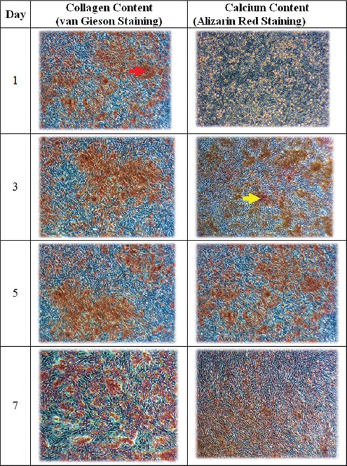
Collagen was detected on day 1 and calcium on day 3 (figure 1) after the OB culture reached 90% confluency at a cell concentration of 6000/cm2. This implies that the PBMNCs could be added to the OB culture on day 3 as the OBs were fully differentiated.
Figure 1.

Characterization of the collagen content according to the van Gieson staining and the calcium content using Alizarin Red staining on days 1, 3, 5, and 7. Collagen appeared as early as day 1 and was seen abundantly present until day 7. Calcium emerged on day 3 and was seen until day 7. Collagen and calcium are shown with red and yellow arrows, respectively (40X magnification).
Co-culture Ratio Optimization
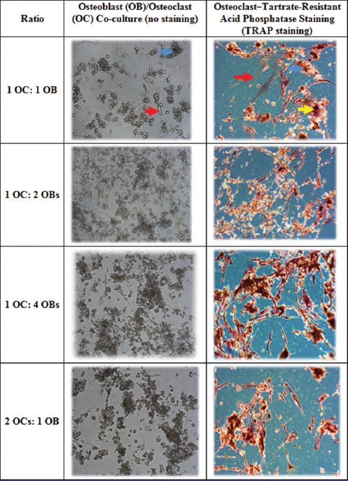
The PBMNCs were co-cultured with primary hFOB 1.19 at the ratios of 1 OC: 1 OB, 1 OC: 2 OBs, 1 OC: 4 OBs, and 2 OCs: 1 OB, respectively. The ratio of 1 OC: 2 OBs was chosen as the best ratio because the TRAP-positive cells were evenly distributed as compared to the other experimental groups, which were aggregated visually (figure 2). Aggregation of TRAP-positive cells at a certain area leads to the destruction of cells.13
Figure 2.

Characterization of the co-culture of peripheral blood mononuclear cells with hFOB 1.19 at the ratios of 1 OC: 1 OB, 1 OC: 2 OBs, 1 OC: 4 OBs, and 2 OCs: 1 OB, respectively. The ratio of 1 OC: 2 OBs was chosen as the best ratio because both OBs and OCs were viable and TRAP was moderately expressed. OBs, OCs, and TRAP are shown with red, blue, and yellow arrows, correspondingly (40X magnification).
Discussion
Optimization of the human OB/OC co-culture system in the present study involved 2 steps. In the first step, we performed time-point optimization of OB differentiation and identified a suitable ratio for the co-culture of PBMNCs and differentiated hFOB 1.19. We detected collagen on day 1 and calcium on day 3, indicating that the OBs were fully differentiated on day 3 (figure 1). In the second step, we optimized the OB/OC co-culture ratio. We chose the ratio of 1 OC: 2 OBs as the best ratio because the TRAP-positive cells were evenly distributed as compared to the other experimental groups, which were aggregated visually (figure 2).12 Aggregation of TRAP-positive cells at a certain area results in the destruction of cells.13
In the current study, we obtained hFOB 1.19 (ATCC® CRL-11372™) via biopsy from the limb tissue of a fetus from a spontaneous miscarriage. Not only were the cells homogenous, well established, and able to differentiate into mature OBs expressing common OB phenotypes, but also they were rapidly proliferating. Under induction by certain stimuli, osteoprogenitors differentiate into premature OBs and further into mature OBs.3 This process of maturity is marked by an increase in ALP and the formation of a mineralized calcium layer.3 In addition, a mature OB also secretes high levels of osteopontin, osteonectin, bone sialoprotein, and type I collagen. Sequential expression of type I collagen, ALP, and calcium deposition can be used as markers of OB maturation. As reported by previous researchers, hFOB 1.19 takes 14 days to fully differentiate. In our case, however, we managed to fully differentiate the OBs on day 3 as the last sequential expression of the molecular marker of calcium was formed. The time for OBs to fully differentiate might differ as the cell concentration differs with respect to the size of the flask culture. In our case, we used a cell concentration of 1×10⁶ in a culture flask of T175 (equivalent to a density of 6000/cm2). It took Yen et al.14 in 2007 a period of 14 days to fully differentiate premature OBs seeded at 3×10⁴ per cm2. Furthermore, Shaminea et al.9 in 2014 cultured OBs seeded at 5000 cells for 14 days before maturation. Liang et al.15 in 2012 also reported that it took hFOB cells seeded in 48-well plates at a density of 2×10⁵ cells/well 14 days to fully differentiate. Thus, the concentration of hFOB cells influences the time of differentiation.
OBs together with the other bone cells are responsible to ensure the continuity of bone remodeling. Bone cell/cell interaction is crucial to ensure that they function as an organized unit.3 OBs secrete the macrophage colony-stimulating factor (MCSF), which is required for macrophage/OC lineage survival, OC migration, and cytoskeleton reorganization. OBs also secrete the receptor activator of the nuclear factor-κB ligand (RANKL) to stimulate the differentiation of OCs.3 Therefore, interaction between OC precursors and OBs is vital to initiate osteoclastogenesis. In our experiment, the presence of the OBs stimulated the formation of the OCs, probably by providing the stimuli required such as MCSF and RANKL. TRAP is a unique biomarker for bone-resorbing OCs. Consequently, the expression of TRAP is important to mark OC maturity and ability to resorb bone. Additionally, TRAP also plays a critical role in important biological mechanisms such as the development of the skeleton, synthesis of collagen, and bone mineralization. The OCs in our co-culture system expressed TRAP, implying that they were fully differentiated and functional.
Our co-culture system is efficient because no additional inducers such as RANKL and MCSF were required as those key regulators were naturally released by the mature OBs. This is an advantage over other systems that require MCSF and RANKL, such as those developed by Kleinhans et al.16 in 2015. Our system shares similarities with the system employed by Penolazzi et al.17 in 2016 insofar as MCSF and RANKL were naturally produced by the bone-lining cells and the OBs in the system. This makes our optimized static co-culture system more reliable and brings it a step closer to mimicking the human bone-remodeling process. We plan to apply this co-culture system in a 3D scaffold in future experiments.
In the current study, we chose the ratio of 1 OC (5×10⁵): 2 OBs (1×10⁶) as a suitable ratio for co-culturing PBMNCs and differentiated OBs (6000/cm2) as the TRAP-positive cells were evenly distributed in comparison with the other experimental groups, which were aggregated visually. Aggregation of TRAP-positive cells at a certain area brings about the destruction of cells.13 The ratio of 1 OC (5×10⁵): 2 OBs (1×10⁶), evenly distributed in the TRAP-positive cells, enabled the co-cultured cells to survive healthily as compared to the other experimental groups, which were aggregated visually.
The current study has some limitations. Firstly, we were not able to provide images at finer resolutions due to technical difficulties. Still, at 40X magnification, we succeeded in illustrating the deposition of collagen and calcium and the formation of TRAP-positive cells. Secondly, we did not implement a scoring system and a definite cutoff for collagen, calcium, and TRAP-positive cells. Be that as it may, our qualitative evaluations chime in with the results reported by Jones et al.12 in 2009. Thirdly, we were unable to quantify the TRAP-positive cells. Nevertheless, our qualitative evaluations of the TRAP-positive cells are in concordance with the results reported by Jones et al.12 in 2009.
Conclusion
We developed a 2-step OB/OC co-culturing system. The first step involved the optimization of the OB culture to determine the time of differentiation, 3 days after confluency at a cell concentration of 6000/cm2. The second step involved co-culturing PBMNCs and differentiated OBs at a suitable ratio (1 OC [5×10⁵]: 2 OBs [1×10⁶], where the TRAP-positive cells were evenly distributed as compared to the other experimental groups, which were aggregated visually). This system did not require exogenous MCSF and RANKL. Moreover, this co-culture ratio optimization ensured a good interaction between the bone cells, thus providing us with a reliable bone cell co-culture system capable of mimicking the bone-remodeling process in humans.
Acknowledgement
We gratefully acknowledge the financial support from Laureate Research and Novelist Mindset Project Grant (Laureate-003-2013) and FF-2017-270. We thank Universiti Kebangsaan Malaysia for providing us facilities to perform the study. Me authors thank to technical support by Nur Sabariah Adnan and Madam Nurul Hafizah Abas.
Conflict of Interest: None declared.
References
- 1.Chang CY, Rosenthal DI, Mitchell DM, Handa A, Kattapuram SV, Huang AJ. Imaging Findings of Metabolic Bone Disease. Radiographics. 2016;36:1871–87. doi: 10.1148/rg.2016160004. [DOI] [PubMed] [Google Scholar]
- 2.An J, Yang H, Zhang Q, Liu C, Zhao J, Zhang L, et al. Natural products for treatment of osteoporosis: The effects and mechanisms on promoting osteoblast-mediated bone formation. Life Sci. 2016;147:46–58. doi: 10.1016/j.lfs.2016.01.024. [DOI] [PubMed] [Google Scholar]
- 3.Heinemann C, Heinemann S, Worch H, Hanke T. Development of an osteoblast/osteoclast co-culture derived by human bone marrow stromal cells and human monocytes for biomaterials testing. Eur Cell Mater. 2011;21:80–93. doi: 10.22203/ecm.v021a07. [DOI] [PubMed] [Google Scholar]
- 4.Nakagawa K, Abukawa H, Shin MY, Terai H, Troulis MJ, Vacanti JP. Osteoclastogenesis on tissue-engineered bone. Tissue Eng. 2004;10:93–100. doi: 10.1089/107632704322791736. [DOI] [PubMed] [Google Scholar]
- 5.Mbalaviele G, Jaiswal N, Meng A, Cheng L, Van Den Bos C, Thiede M. Human mesenchymal stem cells promote human osteoclast differentiation from CD34+bone marrow hematopoietic progenitors. Endocrinology. 1999;140:3736–43. doi: 10.1210/endo.140.8.6880. [DOI] [PubMed] [Google Scholar]
- 6.Heinemann S, Heinemann C, Wenisch S, Alt V, Worch H, Hanke T. Calcium phosphate phases integrated in silica/collagen nanocomposite xerogels enhance the bioactivity and ultimately manipulate the osteoblast/osteoclast ratio in a human co-culture model. Acta Biomater. 2013;9:4878–88. doi: 10.1016/j.actbio.2012.10.010. [DOI] [PubMed] [Google Scholar]
- 7.Clarke MS, Sundaresan A, Vanderburg CR, Banigan MG, Pellis NR. A three-dimensional tissue culture model of bone formation utilizing rotational co-culture of human adult osteoblasts and osteoclasts. Acta Biomater. 2013;9:7908–16. doi: 10.1016/j.actbio.2013.04.051. [DOI] [PubMed] [Google Scholar]
- 8.Hayden RS, Quinn KP, Alonzo CA, Georgakoudi I, Kaplan DL. Quantitative characterization of mineralized silk film remodeling during long-term osteoblast-osteoclast co-culture. Biomaterials. 2014;35:3794–802. doi: 10.1016/j.biomaterials.2014.01.034. [ PMC Free Article] [DOI] [PMC free article] [PubMed] [Google Scholar]
- 9.Shaminea S, Kannan T, Norazmi M, Abdullah NA. Interleukin 6 and Interleukin 17a enhance proliferation and differentiation of murine osteoblast and human foetal osteoblast cell lines. The International Medical Journal of Malaysia. 2014;13:35–40. [Google Scholar]
- 10.Beşkardeş IG, Hayden RS, Glettig DL, Kaplan DL, Gümüşderelioğlu M. Bone tissue engineering with scaffold-supported perfusion co-cultures of human stem cell-derived osteoblasts and cell line-derived osteoclasts. Process Biochemistry. 2017;59:303–11. doi: 10.1016/j.procbio.2016.05.008. [DOI] [Google Scholar]
- 11.Somanchi SS, Senyukov VV, Denman CJ, Lee DA. Expansion, purification, and functional assessment of human peripheral blood NK cells. J Vis Exp. 2011 doi: 10.3791/2540. [ PMC Free Article] [DOI] [PMC free article] [PubMed] [Google Scholar]
- 12.Jones GL, Motta A, Marshall MJ, El Haj AJ, Cartmell SH. Osteoblast: osteoclast co-cultures on silk fibroin, chitosan and PLLA films. Biomaterials. 2009;30:5376–84. doi: 10.1016/j.biomaterials.2009.07.028. [DOI] [PubMed] [Google Scholar]
- 13.Tsuboi H, Matsui Y, Hayashida K, Yamane S, Maeda-Tanimura M, Nampei A, et al. Tartrate resistant acid phosphatase (TRAP) positive cells in rheumatoid synovium may induce the destruction of articular cartilage. Ann Rheum Dis. 2003;62:196–203. doi: 10.1136/ard.62.3.196. [ PMC Free Article] [DOI] [PMC free article] [PubMed] [Google Scholar]
- 14.Yen PH, Kiem PV, Nhiem NX, Tung NH, Quang TH, Minh CV, et al. A new monoterpene glycoside from the roots of Paeonia lactiflora increases the differentiation of osteoblastic MC3T3-E1 cells. Arch Pharm Res. 2007;30:1179–85. doi: 10.1007/BF02980258. [DOI] [PubMed] [Google Scholar]
- 15.Liang W, Lin M, Li X, Li C, Gao B, Gan H, et al. Icariin promotes bone formation via the BMP-2/Smad4 signal transduction pathway in the hFOB 1.19 human osteoblastic cell line. Int J Mol Med. 2012;30:889–95. doi: 10.3892/ijmm.2012.1079. [DOI] [PubMed] [Google Scholar]
- 16.Kleinhans C, Schmid FF, Schmid FV, Kluger PJ. Comparison of osteoclastogenesis and resorption activity of human osteoclasts on tissue culture polystyrene and on natural extracellular bone matrix in 2D and 3D. J Biotechnol. 2015;205:101–10. doi: 10.1016/j.jbiotec.2014.11.039. [DOI] [PubMed] [Google Scholar]
- 17.Penolazzi L, Lolli A, Sardelli L, Angelozzi M, Lambertini E, Trombelli L, et al. Establishment of a 3D-dynamic osteoblasts-osteoclasts co-culture model to simulate the jawbone microenvironment in vitro. Life Sci. 2016;152:82–93. doi: 10.1016/j.lfs.2016.03.035. [DOI] [PubMed] [Google Scholar]


