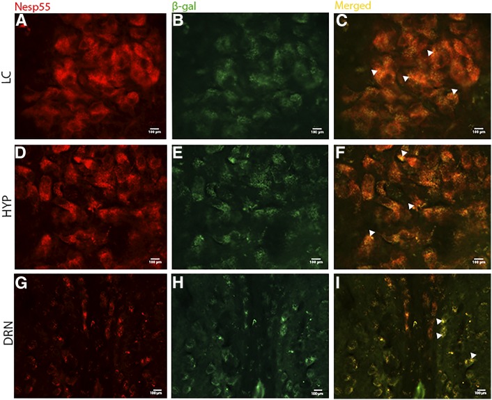
Figure 3.
Dual-labeling immunofluorescence histochemistry of Nesp55 and Grb10 in coronal sections of adult brain. Sections were dual-labeled with antibodies against Nesp55 and β-gal, where the reporter gene LacZ is expressed in place of Grb10 in tissue from Grb10+/p mice, and can be used to identify Grb10-positive cells. Images were viewed at different light intensities (568 and 488 nm for Nesp55 and β-gal, respectively), and were then merged to gauge cellular colocalization of the two target proteins, depicted by white arrows in the merged figures. The majority of cells showed evidence of colocalization: within the locus coeruleus (LC) (A–C), the hypothalamus (HYP) (D–F), and the dorsal raphe nuclei (DRN) (G–I). LC and HYP images at ×40 magnification, DRN at ×20.

