Inherent strain in lattice mismatched lateral heterostructures can help to create quantum-well superlattices in the 2D limit.
Abstract
The advent of two-dimensional (2D) materials has led to extensive studies of heterostructures for novel applications. 2D lateral multiheterojunctions and superlattices have been recently demonstrated, but the available growth methods can only produce features with widths in the micrometer or, at best, 100-nm scale and usually result in rough and defective interfaces with extensive chemical intermixing. Widths smaller than 5 nm, which are needed for quantum confinement effects and quantum-well applications, have not been achieved. We demonstrate the growth of sub–2-nm quantum-well arrays in semiconductor monolayers, driven by the climb of misfit dislocations in a lattice-mismatched sulfide/selenide heterointerface. Density functional theory calculations provide an atom-by-atom description of the growth mechanism. The calculated energy bands reveal type II alignment suitable for quantum wells, suggesting that the structure could, in principle, be turned into a “conduit” of conductive nanoribbons for interconnects in future 2D integrated circuits via n-type modulation doping. This misfit dislocation–driven growth can be applied to different combinations of 2D monolayers with lattice mismatch, paving the way to a wide range of 2D quantum-well superlattices with controllable band alignment and nanoscale width.
INTRODUCTION
Two-dimensional (2D) materials have attracted extensive research efforts in recent years due to their unique properties and promise in a wide range of applications (1–4). By stitching different 2D materials side by side or stacking them vertically layer by layer, heterostructures can be created with interesting optical and electronic properties emerging from the heterointerface due to the coupling between the different 2D components (4–16). In particular, the structural similarity among different semiconducting transition metal dichalcogenides has made it feasible to grow high-quality 2D lateral semiconductor heterostructures with atomically sharp interfaces via lateral epitaxy growth (7–14). Further combining lateral epitaxy growth with lithography patterning or multistep sequential growth, 2D multiheterojunctions or superlattices with featured width in the micrometer scale have also been demonstrated (5, 12–14). However, these methods are limited in spatial resolution and usually produce rough or defective interfaces with extensive chemical intermixing (5, 12–14). 2D lateral multiheterostructures or superlattices with width smaller than 5 nm, a regime where quantum size effect would come into play, have never been reported and remain a major challenge for 2D materials research.
Quantum-well structures in the 2D limit can, in principle, be created by laterally sandwiching a nanoscale strip of a 2D semiconductor between two strips of another 2D semiconductor with a different band gap. Because of their unique electronic structure and quantum confinement, quantum wells in conventional semiconductors have found important applications in quantum cascade lasers, infrared photodetectors, high–electron mobility transistors, high-efficiency thermoelectrics, and solar cells (17–21). In order for quantum size effects to take place, it is necessary to control the width of the 2D quantum well to be comparable to the de Broglie wavelength of the carriers (that is, in the sub–10-nm regime and ideally less than 5 nm).
Here, we report the growth of high-quality sub–2-nm-wide quantum wells within semiconductor monolayers, making use of the lattice mismatch between two semiconducting materials in the 2D lateral heterostructures. The growth is controlled by individual misfit dislocations formed at the lateral heterointerface between a selenide monolayer (WSe2 or MoSe2) and a sulfide monolayer (WS2 or MoS2). Atomic-resolution scanning transmission electron microscopy (STEM) images reveal that these sulfide quantum wells are less than 2 nm in width and form fully coherent lateral interfaces with the selenide monolayer matrix without extended defects. Density functional theory (DFT) calculations demonstrate that the strain field around the misfit dislocations makes them highly reactive during the heterostructure growth. We show that insertion of metal and S atoms into the dislocation cores induces dislocation climb, whereas concomitant selective substitution of Se atoms around the dislocation core by S atoms, driven by the local strain field, leads to growth of WS2 and MoS2 quantum-well arrays embedded in the WSe2 and MoSe2 monolayers. The regular dislocation spacing in the misfit array initially present at the lattice-mismatched interface results in nanoscale quantum-well arrays with periodic spacing. Because of the 2D nature of these structures, the quantum wells can also be thought of as ultranarrow nanoribbons. If doped to be metallic, these nanoribbons could serve as interconnects in future 2D integrated circuits (that is, the 2D material can serve as a “conduit” of conductive nanoribbons). Because misfit dislocations could form in periodic arrays at heterointerfaces, we expect that similar growth mechanisms can be used to fabricate 2D semiconductor quantum wells using different combinations of 2D monolayers with lattice mismatch and create a wide range of 2D quantum-well superlattices or nanoribbon conduits with controllable width and separation.
RESULTS
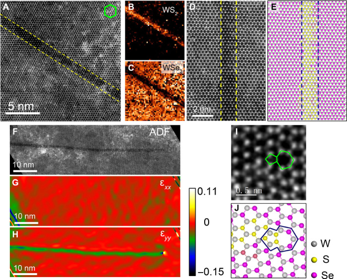
Figure 1A shows a WS2 quantum well formed inside the WSe2 monolayer in a lateral WSe2/WS2 heterostructure. Under our experimental setup for STEM annular dark-field (ADF) imaging, the STEM-ADF image intensity for Se and S sites roughly follows Z1.54, where Z is the atomic number, showing distinguishable atomic number contrast (22, 23). As shown in Fig. 1A, the WS2 monolayer strip displays lower image intensity than the surrounding WSe2 matrix, where the lateral interfaces are highlighted by the yellow dashed lines. Chemical mapping using electron energy loss spectroscopy (EELS) imaging (see Materials and Methods and fig. S1), acquired simultaneously with the ADF image from the same region, further confirms that the darker strip is a WS2 quantum well embedded within the WSe2 monolayer, as shown in Fig. 1 (B and C). The bright background features in the ADF images are contaminates, mainly carbon and Si from the sample growth and TEM sample transfer, as confirmed by EELS mapping. Measuring from the atomic-resolution image, this WS2 quantum well has a width of 1.2 nm (that is, four WS2 unit cells in width).
Fig. 1. Structure and strain analysis of a WS2 quantum well embedded in a WSe2 matrix.

(A) Atomic-resolution STEM-ADF image of the embedded WS2 quantum well with a width of 1.2 nm. The coherent interfaces between the WS2 quantum well and the WSe2 matrix are highlighted by the yellow dashed line. The hexagon highlights the orientation of the lattice. (B and C) Chemical mapping from the same region as in (A) showing the spatial distribution of WS2 and WSe2, respectively. (D and E) High-resolution STEM-ADF image of a WS2 quantum well and the corresponding atomic structural model. The dashed lines highlight the coherent lateral interface along the armchair direction. (F to H) STEM-ADF of the entire 65-nm-long WS2 quantum well and the corresponding strain distribution (see Materials and Methods) around the quantum well. (I and J) STEM-ADF image showing the atomic arrangement of the dislocation core at the tip of the WS2 quantum well in (F) and the corresponding atomic model.
Careful inspection of the ADF image reveals that the WS2 quantum well grows along the armchair direction of the hexagonal lattice and forms fully coherent lateral interfaces with the WSe2 matrix, free of any misfit dislocations or extended defects (Fig. 1, D and E). This is further confirmed by the strain analysis (see Fig. 1, F to H; fig. S2; and Materials and Methods) over the entire length of the WS2 quantum well (65 nm in length, representing an aspect ratio over 50). Here, the perfect lattice from the WSe2 monolayer is used as reference for calculating the strain. As shown in Fig. 1G, in parallel to the growth direction (x direction), the quantum well shows the same lattice spacing as the surrounding WSe2 monolayer. The fact that the WSe2 lattice constant is ~4% larger than WS2 in perfect monolayers indicates a high and uniform tensile strain accommodated in the WS2 quantum well along its growth direction, which leads to the observed dislocation-free lateral interface. In contrast, considerable difference (~4.3 ± 0.5%) in lattice spacing is observed between the WS2 quantum well and the WSe2 monolayer along the y direction (Fig. 1H), arising from the inherent lattice mismatch between the two materials. This lattice spacing difference is accommodated in a misfit dislocation core, composed of a pentagon-heptagon (5|7) pair (24–26), at the tip of the WS2 quantum well, with the heptagon pointing away from the WS2 (Fig. 1, I and J).
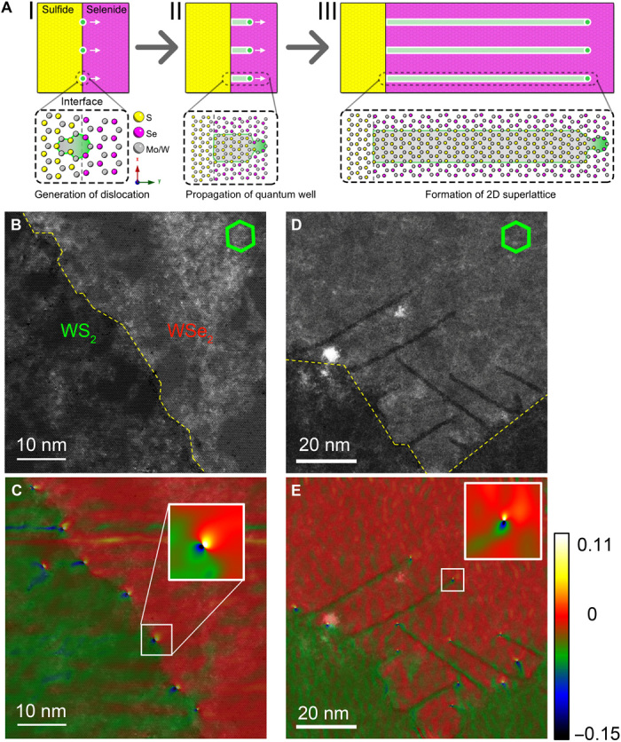
These ultranarrow WS2 quantum wells are frequently observed in almost all the lateral WSe2/WS2 heterostructure samples (fig. S3) we have studied, growing in arrays from the heterointerface into the WSe2 monolayer. As is well established in thin-film growth, strain relaxation at epitaxial heterointerfaces with inherent lattice mismatch can generate misfit dislocations once the film passes a certain critical thickness (27, 28). For the lateral WSe2/WS2 heterostructure, because of the ~4% lattice mismatch, misfit dislocation arrays are expected to form at the lateral interface, with an average separation of ~8 nm along the zigzag direction (fig. S4), to relieve the lattice strain, as schematically shown in Fig. 2A (I). This is observed experimentally in the heterostructure samples. Figure 2 (B and C) shows such an epitaxial heterointerface with quasi-periodic misfit dislocation arrays, where the heptagons at the dislocation cores all point into the WSe2 lattice (fig. S5). However, in most of the samples, we observe these misfit dislocations propagating (or penetrating) into the 2D matrix, leaving a trace of arrays of the sub–2-nm-wide WS2 quantum wells, as illustrated in Fig. 2A (II and III) and evidenced by Fig. 2 (D and E) and fig. S6. Statistical analysis reveals that these WS2 quantum wells have an average width of 1.19 ± 0.09 nm (fig. S6C) (that is, four WS2 unit cells in width). The fact that the WS2 quantum wells are always growing from the lateral WSe2/WS2 heterointerface, with a single dislocation core at the growth front, indicates that the growth of the WS2 quantum wells and climb of the misfit dislocations are intimately connected and that the growth is controlled by the dislocation.
Fig. 2. Formation of periodic dislocation arrays and dislocation-driven growth of WS2 quantum wells at the WSe2/WS2 lateral interface.

(A) Schematic showing (I) the formation of periodic dislocation array, (II) dislocation-driven growth of WS2 quantum wells, and (III) formation of 2D quantum-well superlattice in the WSe2/WS2 lateral heterostructure. (B) STEM-ADF image of a WSe2/WS2 lateral interface without the formation of WS2 quantum wells. The epitaxial interface is highlighted by the yellow dashed line. (C) Corresponding strain distribution, overlaid onto the ADF image, showing the formation of periodic dislocation array at the heterointerface. (D) STEM-ADF image of a WSe2/WS2 lateral interface with the formation of WS2 quantum wells. The WS2 quantum wells appear as darker stripes with the same width. (E) Corresponding strain map, overlaid onto the ADF image, showing the presence of a dislocation core at the tip of each WS2 quantum well. Insets in (C) and (E) are magnified views of the strain maps at the highlighted dislocation cores.
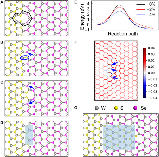
To explore the atom-by-atom mechanism of the misfit dislocation–driven growth of quantum wells, we posited a scenario and then performed DFT calculations to validate it. A misfit dislocation, modeled as a 5|7 pair as shown in Fig. 3A, is created at the WSe2/WS2 interface for every 24 WSe2 or 25 WS2 unit cells to release lattice strain (fig. S4). Because the climb of the misfit dislocation from the interface into the WSe2 lattice involves the insertion of an extra line of atoms, we consider the insertion of one W atom and two S atoms (highlighted by the blue circle in Fig. 3B, denoted as W-S2 hereafter) from the gas source into the 5|7 rings. This insertion pushes the 5|7 dislocation one unit cell forward into WSe2 (that is, dislocation climb). If this step is followed by substitution of Se atoms by S atoms (denoted as SSe) selectively in the pentagon and then right next to the 5|7 dislocation core (blue arrows in Fig. 3, B and C) to relieve local strain, a WS2 nanoseed that is four unit cells wide and one unit cell long would penetrate into the WSe2 monolayer (shaded in light blue in Fig. 3D). Repeating the above processes of W-S2 insertion and subsequent three SSe substitutions would lead to the growth of the WS2 nanoseed into the WSe2 lattice and ultimately form a WS2 quantum well. An atomic model with six steps of dislocation climb and SSe substitution is shown in Fig. 3G. It should be noted that based on this growth mechanism, the WS2 quantum well should, on average, grow along the armchair direction, which is in excellent agreement with our experimental observations.
Fig. 3. Growth mechanism of the WS2 quantum well in WSe2.

Atomic models of (A) 5|7 dislocation at the WS2/WSe2 interface due to lattice mismatch, (B) dislocation climbs one unit cell into WSe2 by inserting a W atom and an S2 pair, (C) substitution of Se by S in pentagon of the 5|7 dislocation, and (D) subsequent substitution of Se atoms next to the 5|7 dislocation by S resulting in a four–unit cell–wide WS2 nanoseed. (E) Energy barrier for SSe substitution under different levels of compressive strain. (F) Strain mapping based on bond length analysis for the atomic model in (B). See Materials and Methods for details. The green dashed line indicates the WS2/WSe2 interface. (G) Structural model showing a short WS2 strip four unit cells wide and six unit cells long after repeating the insertion-substitution process six times.
DFT calculations verified that the above scenario is energetically possible. We constructed a very large supercell accommodating 24 WSe2 and 25 WS2 unit cells (fig. S4), accommodating the lattice misfit by a 5|7 dislocation. For the insertion of a W-S2 unit, we compared the competition between insertion into the dislocation core (that is, dislocation climb) and attachment to a straight or a stepped WS2 edge (that is, growth at the fresh WS2 edge) (fig. S7). The energy gain for edge growth is 2.45 eV at a step edge (fig. S7, C and D) and 3.53 eV at a straight edge (fig. S7, E and F), respectively. For comparison, insertion into a 5|7 dislocation core leads to an energy gain of 2.81 eV. These results suggest that the climb of misfit dislocations is energetically feasible and should occur in parallel with the heterostructure growth, given the availability of W and S atoms during the WS2 growth.
We also evaluated the energetics of SSe substitution. Using the DFT-based climbing image nudged elastic band (DFT-CINEB) method, we first calculated the substitution barrier in a perfect WSe2 lattice (see Materials and Methods and fig. S8). As shown in Fig. 3E, the barrier for SSe substitution in a perfect WSe2 lattice is 3.8 eV, which indicates that SSe substitution is unlikely to happen under the growth temperature of 700°C. This result is consistent with the experimental fact that we do not obtain alloying in the WSe2 monolayer. However, this barrier can be significantly lowered by strain. We found that the SSe substitution energy barrier drastically decreases to 2.6 eV (blue curve in Fig. 3E) under 4% compressive strain and lowers by 0.5 to 3.3 eV for 2% strain (see Materials and Methods). Bond length analysis in Fig. 3F, based on the DFT-relaxed interface structural model in Fig. 3B, shows that compressive strain is mainly distributed in and next to the pentagon of the 5|7 dislocation and quickly fades out as one goes further away from the dislocation core. Therefore, the calculations support the notion that the localized compressive strain at the misfit dislocation cores reduces the substitution energy barrier by a sufficient amount to make the SSe substitution feasible and highly selective under typical growth conditions of the heterostructures (700°C).
Because the SSe substitution process is governed by the strain field around the dislocation core at the growth front, we can expect that the WS2 quantum wells have uniform width and a sharp coherent interface with the WSe2 matrix over the entire length if a thermodynamically stable growth condition is maintained. Experimental results suggest that this is very promising. As demonstrated in Fig. 1 (E and D), this WS2 quantum well shows a uniform width of 1.2 nm and an atomically sharp interface over a length of 12 nm. A small amount (~8%) of Se atoms remains inside the WS2 quantum well due to incomplete SSe substitution during the growth, which may well be eliminated if the growth conditions can be better controlled.
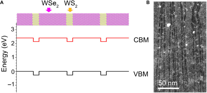
From the mechanism discussed above, we can expect these quantum wells to grow into equally spaced parallel arrays over macroscopic length scales (that is, forming a 2D quantum-well superlattice) starting with periodic misfit dislocations at the heterointerface, if mild growth conditions can be precisely controlled over a long period of time. The WS2 quantum wells grown via this mechanism have a type II band alignment with the WSe2 monolayer matrix as shown in Fig. 4A. Charge separation is a possible function of the WS2/WSe2 superlattice. In addition, the WS2 quantum wells can also be thought of as ultranarrow nanoribbons. Because of the type II band alignment in the present system, if the material is doped n-type (for example, by Re atoms substituting W atoms in either WSe2 or WS2) (29), all the electrons would drop into the WS2 pockets in the conduction bands, making the WS2 nanoribbons conductive (fig. S9). This kind of “modulation doping” leads to high carrier mobilities because the ionized donors lie largely in the WSe2 regions, which reduces the Coulomb scattering in the nanoribbons (30).
Fig. 4. Toward 2D quantum-well superlattice with atomically sharp lateral interfaces.

(A) Atomic structural model and band alignment for a WSe2/WS2 superlattice calculated with the HSE06 functional. Valence band maximum (VBM) and conduction band minimum (CBM) are plotted by black and red lines, respectively. Yellow, S; purple, Se; gray, W. (B) Low-magnification STEM-ADF image showing the formation of parallel MoS2 quantum wells of a few hundred nanometers long in a MoSe2 monolayer toward the formation of quantum-well superlattice.
DISCUSSION
On the basis of the results from the lateral WSe2/WS2 heterostructure system, we can expect that similar quantum-well superlattice structures can also form in lateral MoSe2/MoS2 heterostructures (fig. S10). Figure 4B shows such an example. Appearing as quasi-parallel narrow dark stripes across the whole image, these MoS2 quantum wells are seen to extend more than a few hundred nanometers (although we were not able to see the entire length of these quantum wells due to the fracture and folding of the atomic film during TEM specimen transfer and the 1-μm visible window on the carbon support film) while still maintaining their general armchair growth direction and a uniform width of 1.8 ± 0.2 nm (that is, six unit cells). As can be noticed from the atomic mechanism illustrated in Fig. 3 (A and B), each dislocation climb step actually has a small component along the zigzag direction due to atomic reconstruction after the insertion of the W-S2 units (fig. S11A). That is, the dislocation wiggles slightly sidewise around the armchair direction while climbing forward. Ideally, this small wiggling has equal probability toward both sides of the armchair direction (fig. S11A); therefore, the overall consequence is that the dislocation climbs and the corresponding quantum well grows along the armchair direction (movie S1). In the case when a few steps of dislocation climbing have the same sidewise component, a nanosized kink (see fig. S11B for an example) would develop, and as a consequence, the quantum wells would not look so straight and parallel at a larger scale. Nevertheless, each segment of the quantum wells still follows the general armchair growth direction.
Although the quantum wells shown in Fig. 4B are not evenly spaced, presumably because the misfit dislocations were not initially formed in a periodic array as those shown in Fig. 2C, we believe that this is an important demonstration toward the formation of 2D quantum-well superlattice at a length scale where quantum confinement would have a strong effect. With better control over the growth parameters of chemical vapor deposition (CVD) systems, or alternatively by using molecular beam epitaxy or metal-organic CVD techniques that are commonly used for the growth of bulk semiconductor superlattices, it is very promising that high-quality 2D semiconductor quantum-well superlattices can be grown via this dislocation-driven mechanism.
The successful growth of quantum-well arrays in both WSe2/WS2 and MoSe2/MoS2 lateral heterostructures suggests that this misfit dislocation–driven growth mechanism should apply in a much broader combination of 2D monolayers with lattice mismatch given their structural similarities. For example, NbSe2 and MoSe2 monolayers share the same 2H structure but with ~4.5% lattice mismatch (a = 3.44 Å for NbSe2 and 3.29 Å for MoSe2), which should lead to misfit dislocation arrays with average spacing of 7.5 nm at the lateral heterointerface and, consequently, quantum-well superlattices with average separation of 7.5 nm. Considering the wide spectrum of electronic and optical properties in various 2D monolayers, including topological insulators and superconductors, this opens exciting opportunities to create a large family of 2D quantum wells and their superlattices with novel properties.
MATERIALS AND METHODS
Growth of the WSe2/WS2 heterostructures
The WSe2/WS2 heterojunctions were grown on SiO2/Si substrates using a two-step ambient pressure CVD method (12). WO3 powder (about 5.0 mg) was placed on SiO2/Si growth substrates and located in the heating zone center of the furnace. Se powder (1.2 g) was placed in a quartz test tube at the upper stream side as the source for the selenization of WO3. H2 [2.2 standard cubic centimeter per minute (sccm)] and Ar (20 sccm) were used as the carrier gas. The center heating zone was heated to 700°C at a ramping rate of 20°C/min. As the temperature approached 700°C, the temperature of the Se powder was maintained at ~280°C. After a 30-min growth at 700°C, the furnace was naturally cooled down to room temperature in a gas flow of 5 sccm H2 and 20 sccm Ar. The growth substrate with WSe2 crystals and WO3 powder (as W precursor) on the surface was transferred to another CVD system for WS2 growth. Similarly, a smaller quartz test tube containing 0.8 g of sulfur powder was located upstream as S precursor. Notably, the growth substrate was kept out of the furnace during the heating stage. As the temperature approached 700°C, the heating zone in the furnace was adjusted by moving the furnace to set the temperature of the S powders at ~180°C and the temperature of the substrates at ~700°C. After a 15-min growth at 700°C, the furnace was cooled down to room temperature quickly.
Growth of the MoSe2/MoS2 heterostructures
A two-step CVD method was used for the growth of the MoSe2/MoS2 lateral heterostructures. Briefly, the MoSe2 monolayer was first grown by a CVD method in a 5.08-cm tube. A mixed Ar/H2 flow of 80:5 sccm was used as the carrier gas, and a silicon boat containing 10 mg of MoO3 was put in the center of the tube. The SiO2/Si substrate was placed on the boat with surface downside. Another silicon boat containing 0.5 g of Se powder was located on the upstream. The temperature ramped up to 750°C in 15 min, where it was kept for about 10 min. The as-grown MoSe2 was then transferred into another CVD setup for subsequent MoS2 growth. For the growth of MoS2, Ar flow of 60 sccm was used as the carrier gas, and a silicon boat containing 10 mg of MoO3 was put in the center of a 2.54-cm tube. The MoSe2/SiO2/Si substrate was placed on the boat with surface downside. Another silicon boat containing 0.5 g of S powder was located on the upstream. The temperature was ramped up to 650°C in 13 min and was kept there for about 5 min.
Electron microscopy experiments
The TEM samples were prepared with a poly(methyl methacrylate) (PMMA)–assisted method. A layer of PMMA about 1 μm thick was spin-coated on the wafer with heterostructure samples deposited and then baked at 180°C for 3 min. Afterward, the wafer was immersed in NaOH solution (1 M) to etch the SiO2 layer overnight. After liftoff, the sample was transferred into deionized water for several cycles to wash away the residual contaminants, and then it was fished by a TEM grid. For the WSe2/WS2 lateral heterostructure samples, conventional lacey carbon film TEM grids, with random holes of 1 to 5 μm in diameter, were used. Quantifoil grids with regular 1-μm holes were used for the MoSe2/MoS2 lateral heterostructure samples. The as-transferred specimen was dried naturally in ambient environment and then dropped into acetone overnight to wash away the PMMA coating layers.
To avoid hydrocarbon contamination, all the TEM samples were baked at 160°C for 8 hours under vacuum before the microscopy experiment. STEM imaging and EELS analysis were performed on a Nion UltraSTEM 100 equipped with a cold field-emission gun and a fifth-order aberration corrector operating at 60 kV. The convergence semiangle for the incident probe was 31 mrad. The ADF images were collected for a half-angle range of ~86 to 200 mrad. The collection semiangle for EELS was set to 48 mrad. The WSe2 and WS2 maps were obtained by multiple linear least-squares fitting of the experimental EELS spectrum image with the reference spectra from pure WSe2 and WS2 monolayers (fig. S1), acquired under the same experimental conditions. Strain analysis was performed on the basis of the geometric phase analysis method (31) using the FRWRtools plugin (www.physics.hu-berlin.de/en/sem/software/software_frwrtools) for DigitalMicrograph. The strain was calculated using the perfect WSe2 lattice as reference. All STEM experiments were performed at room temperature.
Theoretical calculations
Quantum mechanical calculations based on DFT were performed using the Vienna Ab initio Simulation Package (VASP) (32, 33). The projector augmented wave method was used to describe the core electrons (34). The Perdew-Becke-Ernzerhof (PBE) functional was used for exchange and correlation (35). The theoretical calculated lattice constants of WSe2 and WS2 were 3.315 and 3.181 Å, respectively, which are in agreement with experimental values (3.282 Å for WSe2 and 3.153 Å for WS2) (36). The atomic model we used is shown in fig. S3. In the y direction, we used 24 units of WSe2 (79.56 Å), which matched 25 units of WS2 (79.53 Å). In the x direction, the supercell contained four WS2 hexagons, six WSe2 hexagons, and a vacuum layer larger than 15 Å. In the z direction, a vacuum layer larger than 15 Å was used. Because of the large size of the supercell, a 259-eV energy cutoff and Γ-only k-point sampling were used for structure relaxation and energy band calculations. Total energies were converged to 10−4 eV, and forces were converged to 0.02 eV/Å.
Energy barriers for the SSe substitution were calculated using the CINEB method (37, 38). A (5 × 5) WSe2 supercell was used to model the WSe2 basal plane. A 3 × 3 Γ-center k-grid mesh was used to sample the first Brillouin zone (39). Four images were inserted between initial and final states. Calculation of the substitution energy barrier in the big supercell containing a heterostructure with a dislocation (fig. S3) was not practical. Instead, we applied different levels of compressive strain to the otherwise perfect WSe2 monolayer to model the effect of compressive strain on the energy barrier for the SSe substitution.
The strain mapping shown in Fig. 3E in the main text was obtained by comparing the bond length in the DFT-optimized atomic model of the 5|7 dislocation at the WS2/WSe2 interface (Fig. 3B) to the standard bond lengths in pristine WS2 and WSe2 (that is, 2.39 Å for W-S bonds and 2.52 Å for W-Se bonds) (40). Specifically, we used the following formulae to calculate the bond strain, for W-S bonds and for W-Se bonds
where dw-s and dW-Se are measured bond lengths for the W-S and W-Se bonds, respectively, in the structural model.
The energy band alignment of a lateral WSe2/WS2 superlattice was examined by calculations at different theoretical levels. A 20 WSe2/5 WS2 superlattice was set up. Atomic structures were fully relaxed at the PBE level with a fixed 79.56 Å lattice constant in the y direction (24 WSe2 units) and 3.38 Å lattice constant in the x direction (1 WSe2 unit). Energy band alignment was calculated at both PBE and HSE06 levels, in which a portion α = 25% of exact nonlocal Hartree-Fock exchange was mixed (41). Both calculations showed a type II band alignment.
Supplementary Material
Acknowledgments
Dedicated to the 80th birthday of Prof. Jing Zhu. We thank X. Zhao for help with the x-ray photoelectron spectroscopy measurement. Funding: This research was supported by the National Natural Science Foundation of China (51622211); the Key Research Program of the Chinese Academy of Sciences (CAS) (XDPB08-1); the CAS Pioneer Hundred Talents Program; the CAS Key Research Program of Frontier Sciences; the U.S. Department of Energy, Office of Science, Basic Energy Sciences, Materials Science and Engineering Division; and through a user project supported by Oak Ridge National Laboratory’s Center for Nanophase Materials Sciences, which is sponsored by the Scientific User Facilities Division of U.S. Department of Energy (DOE). A portion of the research was performed at the CAS Key Laboratory of Vacuum Physics. Work at Vanderbilt was supported by the DOE (grant DE-FG02-09ER46554) and by the McMinn Endowment. This research is also supported by the Singapore National Research Foundation under NRF RF award no. NRF-RF2013-08, Tier 2 MOE2016-T2-2-153, and MOE2015-T2-2-007. Computations were carried out at the National Energy Research Scientific Computing Center, a DOE Office of Science User Facility supported by the Office of Science of the DOE under contract no. DE-AC02-05CH11231. Author contributions: W.Z. conceived the idea, performed the electron microscopy experiments, and wrote the paper. D.L. and M.F.C. participated in the electron microscopy data analysis. Y.-Y.Z. and S.T.P. performed the theoretical calculations. J.C. and K.P.L. provided the WSe2/WS2 heterostructure sample. J.Z. and Z.L. provided the MoSe2/MoS2 heterostructure sample. All authors discussed the results and commented on the manuscript. Competing interests: The authors declare that they have no competing interests. Data and materials availability: All data needed to evaluate the conclusions in the paper are present in the paper and/or the Supplementary Materials. Additional data related to this paper may be requested from the authors.
SUPPLEMENTARY MATERIALS
Supplementary material for this article is available at http://advances.sciencemag.org/cgi/content/full/4/3/eaap9096/DC1
fig. S1. Reference EELS spectra from pure WSe2 and WS2 monolayers.
fig. S2. STEM-ADF of the entire 65-nm-long WS2 quantum well and the corresponding strain distribution around the quantum well.
fig. S3. Optical images and spectroscopy measurements of the WSe2/WS2 lateral heterostructure.
fig. S4. Atomic model of WSe2/WS2 heterostructure.
fig. S5. Additional structural characterization data from the lateral WSe2/WS2 heterointerface.
fig. S6. Additional low-magnification STEM-ADF images showing the formation of arrays of WS2 quantum wells at the WSe2/WS2 lateral interface, driven by dislocations.
fig. S7. Comparison between dislocation climb and extension of a WS2 edge during the sample growth.
fig. S8. Atomic models for the SSe substitution barrier calculations.
fig. S9. Band structure of lateral WSe2/WS2 superlattice.
fig. S10. Optical images and spectroscopy measurements of the MoSe2/MoS2 lateral heterostructure.
fig. S11. Dislocation climb and formation of nanosize kinks during the growth of quantum well.
movie S1. Scheme showing the dislocation climb and the corresponding quantum well growth process.
REFERENCES AND NOTES
- 1.Novoselov K. S., Geim A. K., Morozov S. V., Jiang D., Zhang Y., Dubonos S. V., Grigorieva I. V., Firsov A. A., Electric field effect in atomically thin carbon films. Science 306, 666–669 (2004). [DOI] [PubMed] [Google Scholar]
- 2.Novoselov K. S., Fal’ko V. I., Colombo L., Gellert P. R., Schwab M. G., Kim K., A roadmap for graphene. Nature 490, 192–200 (2012). [DOI] [PubMed] [Google Scholar]
- 3.Bhimanapati G. R., Lin Z., Meunier V., Jung Y., Cha J., Das S., Xiao D., Son Y., Strano M. S., Cooper V. R., Liang L., Louie S. G., Ringe E., Zhou W., Kim S. S., Naik R. R., Sumpter B. G., Terrones H., Xia F., Wang Y., Zhu J., Akinwande D., Alem N., Schuller J. A., Schaak R. E., Terrones M., Robinson J. A., Recent advances in two-dimensional materials beyond graphene. ACS Nano 9, 11509–11539 (2015). [DOI] [PubMed] [Google Scholar]
- 4.Geim A. K., Grigorieva I. V., Van der Waals heterostructures. Nature 499, 419–425 (2013). [DOI] [PubMed] [Google Scholar]
- 5.Liu Z., Ma L., Shi G., Zhou W., Gong Y., Lei S., Yang X., Zhang J., Yu J., Hackenberg K. P., Babakhani A., Idrobo J.-C., Vajtai R., Lou J., Ajayan P. M., In-plane heterostructures of graphene and hexagonal boron nitride with controlled domain sizes. Nat. Nanotechnol. 8, 119–124 (2013). [DOI] [PubMed] [Google Scholar]
- 6.Liu L., Park J., Siegel D. A., McCarty K. F., Clark K. W., Deng W., Basile L., Idrobo J. C., Li A.-P., Gu G., Heteroepitaxial growth of two-dimensional hexagonal boron nitride templated by graphene edges. Science 343, 163–167 (2014). [DOI] [PubMed] [Google Scholar]
- 7.Duan X., Wang C., Shaw J. C., Cheng R., Chen Y., Li H., Wu X., Tang Y., Zhang Q., Pan A., Jiang J., Yu R., Huang Y., Duan X., Lateral epitaxial growth of two-dimensional layered semiconductor heterojunctions. Nat. Nanotechnol. 9, 1024–1030 (2014). [DOI] [PMC free article] [PubMed] [Google Scholar]
- 8.Gong Y., Lin J., Wang X., Shi G., Lei S., Lin Z., Zou X., Ye G., Vajtai R., Yakobson B. I., Terrones H., Terrones M., Tay B. K., Lou J., Pantelides S. T., Liu Z., Zhou W., Ajayan P. M., Vertical and in-plane heterostructures from WS2/MoS2 monolayers. Nat. Mater. 13, 1135–1142 (2014). [DOI] [PubMed] [Google Scholar]
- 9.Huang C., Wu S., Sanchez A. M., Peters J. J. P., Beanland R., Ross J. S., Rivera P., Yao W., Cobden D. H., Xu X., Lateral heterojunctions within monolayer MoSe2–WSe2 semiconductors. Nat. Mater. 13, 1096–1101 (2014). [DOI] [PubMed] [Google Scholar]
- 10.Gong Y., Lei S., Ye G., Li B., He Y., Keyshar K., Zhang X., Wang Q., Lou J., Liu Z., Vajtai R., Zhou W., Ajayan P. M., Two-step growth of two-dimensional WSe2/MoSe2 heterostructures. Nano Lett. 15, 6135–6141 (2015). [DOI] [PubMed] [Google Scholar]
- 11.Li M.-Y., Shi Y., Cheng C.-C., Lu L.-S., Lin Y.-C., Tang H.-L., Tsai M.-L., Chu C.-W., Wei K.-H., He J.-H., Chang W.-H., Suenaga K., Li L.-J., Epitaxial growth of a monolayer WSe2-MoS2 lateral p-n junction with an atomically sharp interface. Science 349, 524–528 (2015). [DOI] [PubMed] [Google Scholar]
- 12.Chen J., Zhou W., Tang W., Tian B., Zhao X., Xu H., Liu Y., Geng D., Tan S. J. R., Fu W., Loh K. P., Lateral epitaxy of atomically sharp WSe2/WS2 heterojunctions on silicon dioxide substrates. Chem. Mater. 28, 7194–7197 (2016). [Google Scholar]
- 13.Mahjouri-Samani M., Lin M.-W., Wang K., Lupini A. R., Lee J., Basile L., Boulesbaa A., Rouleau C. M., Puretzky A. A., Ivanov I. N., Xiao K., Yoon M., Geohegan D. B., Patterned arrays of lateral heterojunctions within monolayer two-dimensional semiconductors. Nat. Commun. 6, 7749 (2015). [DOI] [PMC free article] [PubMed] [Google Scholar]
- 14.Zhang Z., Chen P., Duan X., Zang K., Luo J., Duan X., Robust epitaxial growth of two-dimensional heterostructures, multiheterostructures, and superlattices. Science 357, 788–792 (2017). [DOI] [PubMed] [Google Scholar]
- 15.Ling X., Lin Y., Ma Q., Wang Z., Song Y., Yu L., Huang S., Fang W., Zhang X., Hsu A. L., Bie Y., Lee Y.-H., Zhu Y., Wu L., Li J., Jarillo-Herrero P., Dresselhaus M., Palacios T., Kong J., Parallel stitching of 2D materials. Adv. Mater. 28, 2322–2329 (2016). [DOI] [PubMed] [Google Scholar]
- 16.Chen L., He L., Wang H. S., Wang H., Tang S., Cong C., Xie H., Li L., Xia H., Li T., Wu T., Zhang D., Deng L., Yu T., Xie X., Jiang M., Oriented graphene nanoribbons embedded in hexagonal boron nitride trenches. Nat. Commun. 8, 14703 (2017). [DOI] [PMC free article] [PubMed] [Google Scholar]
- 17.Esaki L., Tsu R., Superlattice and negative differential conductivity in semiconductors. IBM J. Res. Dev. 14, 61–65 (1970). [Google Scholar]
- 18.Tsu R., Esaki L., Tunneling in a finite superlattice. Appl. Phys. Lett. 22, 562–564 (1973). [Google Scholar]
- 19.Hicks L. D., Harman T. C., Dresselhaus M. S., Use of quantum-well superlattice to obtain a high figure of merit from nonconvential thermoelectric materials. Appl. Phys. Lett. 63, 3230–3232 (1993). [Google Scholar]
- 20.Faist J., Capasso F., Sivco D. L., Sirtori C., Hutchinson A. L., Cho A. Y., Quantum cascade laser. Science 264, 553–556 (1994). [DOI] [PubMed] [Google Scholar]
- 21.Gudiksen M. S., Lauhon L. J., Wang J., Smith D. C., Lieber C. M., Growth of nanowire superlattice structures for nanoscale photonics and electronics. Nature 415, 617–620 (2002). [DOI] [PubMed] [Google Scholar]
- 22.Krivanek O. L., Chisholm M. F., Nicolosi V., Pennycook T. J., Corbin G. J., Dellby N., Murfitt M. F., Own C. S., Szilagyi Z. S., Oxley M. P., Pantelides S. T., Pennycook S. J., Atom-by-atom structural and chemical analysis by annular dark-field electron microscopy. Nature 464, 571–574 (2010). [DOI] [PubMed] [Google Scholar]
- 23.Zhou W., Oxley M. P., Lupini A. R., Krivanek O. L., Pennycook S. J., Idrobo J.-C., Single atom microscopy. Microsc. Microanal. 18, 1342–1354 (2012). [DOI] [PubMed] [Google Scholar]
- 24.Zou X., Liu Y., Yakobson B. I., Predicting dislocations and grain boundaries in two-dimensional metal-disulfides from the first principles. Nano Lett. 13, 253–258 (2013). [DOI] [PubMed] [Google Scholar]
- 25.Zhou W., Zou X., Najmaei S., Liu Z., Shi Y., Kong J., Lou J., Ajayan P. M., Yakobson B. I., Idrobo J.-C., Intrinsic structural defects in monolayer molybdenum disulfide. Nano Lett. 13, 2615–2622 (2013). [DOI] [PubMed] [Google Scholar]
- 26.Najmaei S., Liu Z., Zhou W., Zou X., Shi G., Lei S., Yakobson B. I., Idrobo J.-C., Ajayan P. M., Lou J., Vapour phase growth and grain boundary structure of molybdenum disulphide atomic layers. Nat. Mater. 12, 754–759 (2013). [DOI] [PubMed] [Google Scholar]
- 27.Matthews J. W., Blakeslee A. E., Defects in epitaxial multilayers. 1. Misfit dislocations. J. Cryst. Growth 27, 118–125 (1974). [Google Scholar]
- 28.Jain S. C., Harker A. H., Cowley R. A., Misfit strain and misfit dislocations in lattice mismatched epitaxial layers and other systems. Philos. Mag. A 75, 1461–1515 (1997). [Google Scholar]
- 29.Lin Y.-C., Dumcenco D. O., Komsa H.-P., Niimi Y., Krasheninnikov A. V., Huang Y.-S., Suenaga K., Properties of individual dopant atoms in single-layer MoS2: Atomic structure, migration, and enhanced reactivity. Adv. Mater. 26, 2857–2861 (2014). [DOI] [PubMed] [Google Scholar]
- 30.Boland J. L., Conesa-Boj S., Parkinson P., Tütüncüoglu G., Matteini F., Rüffer D., Casadei A., Amaduzzi F., Jabeen F., Davies C. L., Joyce H. J., Herz L. M., Fontcuberta i Morral A., Johnston M. B., Modulation doping of GaAs/AlGaAs core-shell nanowires with effective defect passivation and high electron mobility. Nano Lett. 15, 1336–1342 (2015). [DOI] [PubMed] [Google Scholar]
- 31.Hytch M. J., Snoeck E., Kilaas R., Quantitative measurement of displacement and strain fields from HREM micrographs. Ultramicroscopy 74, 131–146 (1998). [Google Scholar]
- 32.Kresse G., Furthmüller J., Efficient iterative schemes for ab initio total-energy calculations using a plane-wave basis set. Phys. Rev. B 54, 11169–11186 (1996). [DOI] [PubMed] [Google Scholar]
- 33.Kresse G., Furthmuller J., Efficiency of ab-initio total energy calculations for metals and semiconductors using a plane-wave basis set. Comput. Mater. Sci. 6, 15–50 (1996). [DOI] [PubMed] [Google Scholar]
- 34.Blöchl P. E., Projector augmented-wave method. Phys. Rev. B 50, 17953–17979 (1994). [DOI] [PubMed] [Google Scholar]
- 35.Perdew J. P., Burke K., Ernzerhof M., Generalized gradient approximation made simple. Phys. Rev. Lett. 77, 3865–3868 (1996). [DOI] [PubMed] [Google Scholar]
- 36.Kang J., Tongay S., Zhou J., Li J., Wu J., Band offsets and heterostructures of two-dimensional semiconductors. Appl. Phys. Lett. 102, 012111 (2013). [Google Scholar]
- 37.Henkelman G., Uberuaga B. P., Jónsson H., A climbing image nudged elastic band method for finding saddle points and minimum energy paths. J. Chem. Phys. 113, 9901–9904 (2000). [Google Scholar]
- 38.Henkelman G., Jónsson H., Improved tangent estimate in the nudged elastic band method for finding minimum energy paths and saddle points. J. Chem. Phys. 113, 9978–9985 (2000). [Google Scholar]
- 39.Methfessel M., Paxton A. T., High-precision sampling for Brillouin-zone integration in metals. Phys. Rev. B 40, 3616–3621 (1989). [DOI] [PubMed] [Google Scholar]
- 40.Kaloni T. P., Kou L., Frauenheim T., Schwingenschlögl U., Quantum spin Hall states in graphene interacting with WS2 or WSe2. Appl. Phys. Lett. 105, 233112 (2014). [Google Scholar]
- 41.Heyd J., Scuseria G. E., Ernzerhof M., Hybrid functionals based on a screened coulomb potential. J. Chem. Phys. 118, 8207–8215 (2003). [Google Scholar]
Associated Data
This section collects any data citations, data availability statements, or supplementary materials included in this article.
Supplementary Materials
Supplementary material for this article is available at http://advances.sciencemag.org/cgi/content/full/4/3/eaap9096/DC1
fig. S1. Reference EELS spectra from pure WSe2 and WS2 monolayers.
fig. S2. STEM-ADF of the entire 65-nm-long WS2 quantum well and the corresponding strain distribution around the quantum well.
fig. S3. Optical images and spectroscopy measurements of the WSe2/WS2 lateral heterostructure.
fig. S4. Atomic model of WSe2/WS2 heterostructure.
fig. S5. Additional structural characterization data from the lateral WSe2/WS2 heterointerface.
fig. S6. Additional low-magnification STEM-ADF images showing the formation of arrays of WS2 quantum wells at the WSe2/WS2 lateral interface, driven by dislocations.
fig. S7. Comparison between dislocation climb and extension of a WS2 edge during the sample growth.
fig. S8. Atomic models for the SSe substitution barrier calculations.
fig. S9. Band structure of lateral WSe2/WS2 superlattice.
fig. S10. Optical images and spectroscopy measurements of the MoSe2/MoS2 lateral heterostructure.
fig. S11. Dislocation climb and formation of nanosize kinks during the growth of quantum well.
movie S1. Scheme showing the dislocation climb and the corresponding quantum well growth process.


