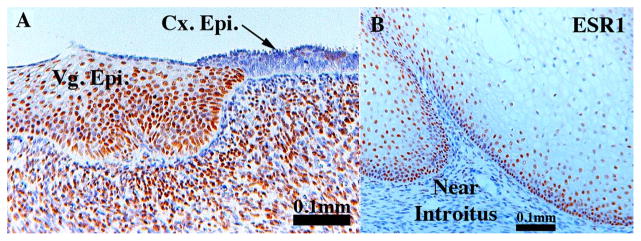
Fig. 13.
Sections of vaginal epithelium immunostained for ESR1. Section (A) is at the junction of vaginal/cervical border. Note ESR1 expression throughout the vaginal epithelium and the high percentage of ESR1-positive mesenchymal cells. Section (B) is vaginal epithelium near the introitus stained for ESR1. Note the paucity of ESR1-positive mesenchymal cells. Vg. Epi. = vaginal epithelium, Cx. Epi. = cervical epithelium.

