Fig. 15.
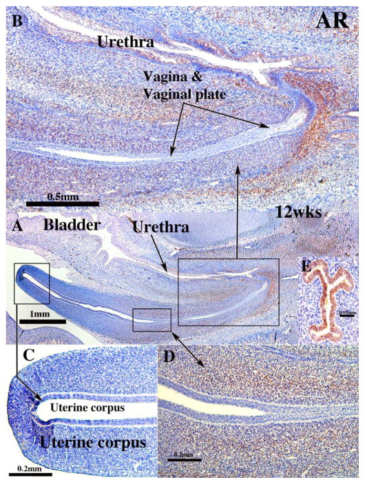
Androgen receptor immunohistochemistry of human fetal female reproductive tracts. (A) low power overview sagittal section. Note AR staining in mesenchyme associated with the vaginal plate and vagina. (B) Higher magnification view of vaginal region showing mesenchymal AR staining. (C) Absence of AR staining in uterine corpus A & C). Intense mesenchymal AR staining associated with the junction of the cervix and vagina (D). (E) Uterine tube of a 16-week fetus showing strong AR immuno-reactivity.

