

Abstract
The lactose operon (lacTEGF) from Lactobacillus casei strain BL23 has been previously studied. The lacT gene codes for a transcriptional antiterminator, lacE and lacF for the lactose-specific phosphoenolpyruvate: phosphotransferase system (PTSLac) EIICB and EIIA domains, respectively, and lacG for the phospho-β-galactosidase. In this work, we have shown that L. casei is able to metabolize N-acetyllactosamine (LacNAc), a disaccharide present at human milk and intestinal mucosa. The mutant strains BL153 (lacE) and BL155 (lacF) were defective in LacNAc utilization, indicating that the EIICB and EIIA of the PTSLac are involved in the uptake of LacNAc in addition to lactose. Inactivation of lacG abolishes the growth of L. casei in both disaccharides and analysis of LacG activity showed a high selectivity toward phosphorylated compounds, suggesting that LacG is necessary for the hydrolysis of the intracellular phosphorylated lactose and LacNAc. L. casei (lacAB) strain deficient in galactose-6P isomerase showed a growth rate in lactose (0.0293 ± 0.0014 h−1) and in LacNAc (0.0307 ± 0.0009 h−1) significantly lower than the wild-type (0.1010 ± 0.0006 h−1 and 0.0522 ± 0.0005 h−1, respectively), indicating that their galactose moiety is catabolized through the tagatose-6P pathway. Transcriptional analysis showed induction levels of the lac genes ranged from 130 to 320–fold in LacNAc and from 100 to 200–fold in lactose, compared to cells growing in glucose.
Introduction
All human milk oligosaccharides (HMOs) contain lactose (Galβ1-4Glc) at the reducing end, which can be elongated with lacto-N-biose (LNB, type-1 core; Galβ1-3GlcNAc) by a β-1,3 bond and/or with N-acetyllactosamine (LacNAc, type-2 core; Galβ1-4GlcNAc) by a β-1,3 or β-1,6-bond1,2. Recently, LacNAc has also been found as a free disaccharide in human milk, with concentrations that decrease from 310 μg/ml on colostrum to 6.7 μg/m after the first week of lactation3. LacNAc is also a common structure of human glycans present at mucosal surfaces and other specific tissues and cells4. The LacNAc residues usually constitute the terminal sugars that form part of the variable portions of the glycan epitopes, including O-glycans, N-glycans and glycolipids. Sometimes these proteins and lipids carry poly-N-acetyllactosamines chains, which can be acceptors for subsequent glycosylations and serve as specific arms to present other functional terminal glycans4. Additionally, LacNAc is a constituent of the ABO and Lewis blood group antigens, which are expressed on the membrane of blood red cells and other tissues, including the gastrointestinal epithelium5.
The gastrointestinal tract of breast-fed infants is rapidly colonized by Bifidobacterium species6, which are well adapted to metabolize HMOs7. This capacity has been associated with a complete array of enzymes that includes various types of intra- and extracellular glycosidases8–10. LacNAc and other LacNAc-containing oligosaccharides, such as lacto-N-neotetraose and lacto-N-hexaose, are digested in vitro by the extracellular membrane-bound β-galactosidase BbgIII cloned from Bifidobacterium bifidum11,12. This species also contains three intracellular β-glycosidases (BbgI, BbgII and BbgIV) able to hydrolyze LacNAc in vitro, although different from BgbIII, they have higher affinity for lactose than for LacNAc11,12. Galactose is also liberated from the LacNAc contained in the lacto-N-neotetraose structure by β-galactosidases described in Bifidobacterium longum and Bifidobacterium breve strains13. The genus Lactobacillus has many characterized probiotic strains14,15 and some species have also been isolated from the gastrointestinal tract of infants16,17. However, contrarily to Bifidobacterium species, genome analysis of lactobacilli revealed that they have a limited capability to ferment HMOs or mucosa-derived glycans18–20. One exception to this are the members of the Lactobacillus casei/paracasei/rhamnosus phylogenetically close group. We have previously characterized in L. casei three genes encoding α-L-fucosidases and purified the corresponding enzymes, which hydrolyze in vitro fucosylated HMOs21. Specifically, the operon alf, encoding the α-L-fucosidase AlfB, the transcriptional repressor AlfR and the EII components of a mannose-class phosphoenolpyruvate: phosphotransferase system (PTS), is involved in these species in the metabolism of fucosyl-α1,3-GlcNAc disaccharide, an structure present in HMOs and mucins22,23. The type-1 HMOs core LNB and the type-1 core O-glycosylation galacto-N-biose (GNB) are also metabolized by L. casei24. Both disaccharides are transported and phosphorylated by the PTSGnb and then are hydrolyzed by the phospho-β-galactosidase GnbG, which are encoded by the gnb operon24. Recently, we have characterized the cell-wall anchored N-acetylglucosaminidase BnaG necessary for the utilization of the trisaccharide lacto-N-triose by L. casei25. BnaG is the only extracellular glycosidase described until now for the metabolism of HMOs in lactobacilli. L. casei also metabolizes lactose and the lac operon (Fig. 1a) from L. casei strain BL23 has been extensively studied in our laboratory26–29. The lacT gene codes for a transcriptional antiterminator, lacE and lacF genes for the lactose-specific PTS, EIICB and EIIA domains, respectively, and lacG for the phospho-β-galactosidase27. The lac operon is induced by lactose through transcription antitermination mediated by LacT28. Additionally, the expression of this operon is subject to carbon catabolite repression mediated by the general regulatory protein CcpA (catabolite control protein A) and also by PTS elements via LacT26,28. Despite the relevance of the LacNAc disaccharide, its metabolism has not been studied in the genus Lactobacillus. Here we report that L. casei strain BL23 is able to grow in the presence of LacNAc and that the lac operon is involved in its metabolism. Additionally, we demonstrated that the tagatose-6P pathway and the N-acetylglucosamine-6P deacetylase NagA are involved in the catabolism of this disaccharide.
Figure 1.
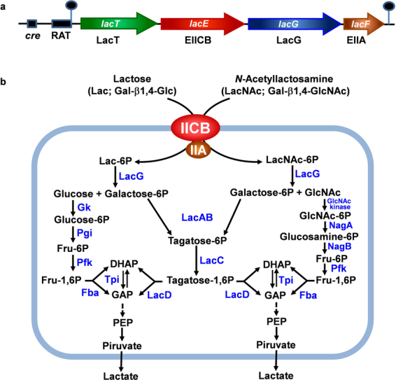
(a) Genetic organization of the lac operon in Lactobacillus casei strain BL23. Hairpin loops indicate rho-independent transcriptional terminators. cre, catabolite responsive element; RAT, ribonucleic antiterminator; LacT is a transcriptional antiterminator; EIICB and EIIA, domains of the lactose-specific phosphoenolpyruvate: phosphotransferase system (PTSLac); LacG, phospho-β-galactosidase. (b) Schematic presentation of the pathways for N-acetyllactosamine (LacNAc) and lactose (Lac) transport and metabolism in L. casei BL23. GlcNAc: N-acetylglucosamine; Fru, fructose; DHAP: dyhidroxyacetone phosphate; GAP: glyceraldehyde 3-phosphate; PEP: phosphoenolpyruvate; NagA, N-acetylglucosamine-6P deacetylase; NagB: glucosamine-6P deaminase; Pfk: 6-phosphofructo-1-kinase; Gk, glucokinase; Pgi, phosphoglucose isomerase; LacAB: galactose-6P isomerase; LacC: tagatose-6P kinase; LacD: tagatose-1,6 P aldolase; Fba: fructose-1,6 P aldolase; Tpi: triose phosphate isomerase.
Results
L. casei BL23 can be cultured in the presence of LacNAc and transports it by the PTSLac
We have previously shown that L. casei is able to transport and ferment LNB, the type-1 disaccharide building block of HMOs24. In order to determine the ability of this species to metabolize the type-2 core structure of HMOs, we cultured L. casei BL23 in sugar-free MRS supplemented with 4 mM LacNAc as carbon source (Fig. 2). The results showed that L. casei can grow in the presence of this disaccharide and that the maximum cell density reached is similar to that obtained in lactose, which was used as a positive control.
Figure 2.
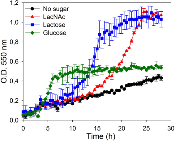
Growth curves of Lactobacillus casei wild type strain BL23 on sugar-free MRS without carbon source (black), with N-acetyllactosamine (LacNAc) (red), lactose (blue) or glucose (green). Data presented are mean values based on at least three replicates. Error bars indicate standard deviations.
Most of the characterized PTS are specific for one carbohydrate, however some PTS can transport two or more structurally related sugars30–32. Therefore, we analyzed if LacNAc, which is structurally similar to LNB and lactose, is transported in L. casei BL23 by the PTSGnb or by the PTSLac, which are involved in LNB and lactose uptake, respectively24,28. The mutant strains BL385 (gnbC)24, that is disrupted in the gene encoding the EIIC domain of the PTSGnb, BL153 (lacE) and BL155 (lacF)28, which are impaired in the EIICB and EIIA, respectively, of the PTSLac, were tested for their capacity to ferment LacNAc. BL385 (gnbC) was able to grow in the presence of LacNAc as carbon source (Fig. 3a). Contrarily, strains BL153 (lacE) and BL155 (lacF) showed a poor growth with LacNAc which was similar to that of the negative controls (lactose supplemented and non-supplemented sugar-free MRS) (Fig. 3b,c). The growth pattern of strains BL153 (lacE) and BL155 (lacF) in the presence of glucose as a positive control is also shown (Fig. 3b,c). Analysis for sugar content in the supernatants demonstrated that LacNAc was consumed by strain BL385 (gnbC) but not by strains BL153 (lacE) and BL155 (lacF) (data not shown), indicating that the domains EIICB and EIIA encoded by lacE and lacF, respectively, are involved in the uptake of LacNAc (Fig. 1). It has been previously shown that the PTS from L. casei can accomplish sugar transport not coupled to phosphorylation23. Then, to further confirm the involvement of the PTSLac in the transport of LacNAc and to test if transport though the EII permease was coupled to phosphorylation, the growth pattern of BL126 (ptsI), a mutant lacking the PTS-general component Enzyme I33, was tested in LacNAc as carbon source. BL126 (ptsI) did not grow in the presence of LacNAc (Fig. 3d), confirming that its utilization needs a functional complete PTS. These results suggest that LacNAc is internalized as a phosphorylated derivative. BL126 (ptsI) was grown with glucose and lactose as positive and negative controls, respectively. The growth pattern of strain BL126 in the presence of glucose differs from that of strains BL153 (lacE) and BL155 (lacF) (Fig. 3). This might be due to the fact that the latest strains contain a functional PTS for glucose uptake while strain BL126 (ptsI) can only transport this sugar by the proton-driven permease34.
Figure 3.

Growth curves of Lactobacillus casei mutant strain BL385 (gnbG) (a) on sugar-free MRS without carbon source (black) or with N-acetyllactosamine (LacNAc) (red). Data presented are mean values based on at least three replicates. Error bars indicate standard deviations. Growth curves of L. casei mutant strain BL153 (lacE) (b), L. casei mutant strain BL155 (lacF) (c) and L. casei mutant strain BL126 (pstI) (d) on sugar-free MRS without carbon source (black), with N-acetyllactosamine (LacNAc) (red), glucose (green) or lactose (blue). Data presented are mean values based on at least three replicates. Error bars indicate standard deviations.
LacG is involved in the metabolism of LacNAc and lactose
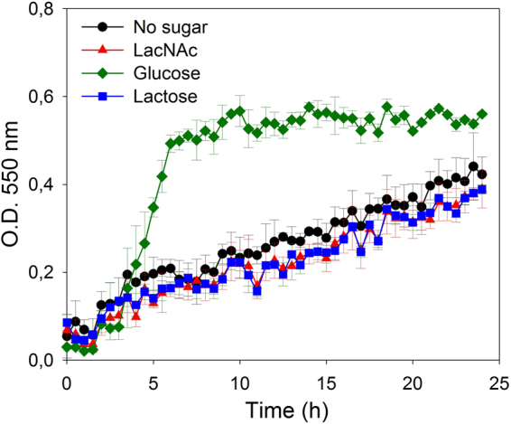
To determine if the phospho-β-galactosidase LacG was involved in the utilization of LacNAc in L. casei BL23, a mutant in lacG was constructed (strain BL400). This mutant was cultured in sugar-free MRS supplemented with 4 mM LacNAc as carbon source (Fig. 4). The growth pattern showed that BL400 (lacG) strain did not ferment LacNAc and neither did lactose, the only substrate described until now for the lac operon26–28. These results indicated that LacG is necessary for the utilization of both disaccharides (Fig. 1b). Sugar content analysis of the culture supernatants detected LacNAc and lactose, respectively, in the supernatants from BL400 (lacG), while they were completely consumed by the wild-type BL23 strain (data not shown).
Figure 4.

Growth curves of Lactobacillus casei mutant strain BL400 (lacG) on sugar-free MRS without carbon source (black), with N-acetyllactosamine (LacNAc) (red), glucose (green) or lactose (blue). Data presented are mean values based on at least three replicates. Error bars indicate standard deviations.
LacG (EC 3.2.1.85) belongs to the glycosyl hydrolase family 1 (GH 1; www.cazy.org), which includes β-glycosidases as well as phospho-β-glycosidases. In order to characterize that enzyme, the lacG gene was expressed in E. coli as a His-tagged protein and purified to homogeneity (data not shown). The purified protein displayed a molecular weight of 55 kDa, in agreement with the calculated mass of the 6x(His)-tagged protein (55,305 Da). 6x(His)LacG did not hydrolyze o-NP-β-D-galactopyranoside but it does when this substrate is phosphorylated (Table 1), suggesting a high selectivity toward phosphorylated compounds. The kinetic analysis showed a high Km and low Vmax for o-NP-β-D-galactopyranoside-6P, and it displayed an optimal pH of 7.0 and an optimal temperature of 41 °C (Table 1). 6x(His)LacG was unable to hydrolyze any of the natural oligosaccharides tested (Table 1), including lactose and LacNAc, possibly because these need to be phosphorylated before turned into substrates for this glycosidase.
Table 1.
Activity and characterization of the phospho-β-galactosidase LacG.
| Substratea (Structure) | Activityb |
|---|---|
| o-NP-β-D-galactopyranoside | − |
| p-NP-β-D-glucopyranoside | − |
| o-NP-1-thio-β-D-galactopyranoside | − |
| p-NP-β-D-glucuronide | − |
| p-NP-N-acetyl-β-D-glucosaminide | − |
| p-NP-α-D-glucopyranoside | − |
| p-NP-α-D- galactopyranoside | − |
| p-NP-α-L- fucopyranoside | − |
| o-NP-β-D-galactopyranoside-6-phosphate | + |
| Lacto-N-biose (Galβ1-3GlcNAc) | − |
| Galacto-N-biose (Galβ1-3 GalNAc) | − |
| Lacto-N-tetraose (Galβ1-3GlcNAcβ1-3 Galβ1-4Glc) | − |
| Lacto-N-neotetraosa (Galβ1-4GlcNAcβ1-3 Galβ1-4Glc) | − |
| 3′-N-Acetilgalactosaminil-Gal (GalNAcβ1-3Gal) | − |
| 3′-N-Acetilglucosaminil-Man (GlcNAcβ1-3Man) | − |
| 4′-Galactofuranosil-GlcNAc (Galβ1-4GlcNAc) | − |
| Fucosyl-α1-3GlcNAc | − |
| Lactose (Galβ1-4Glc) | − |
| N-acetyl-lactosamine (Galβ1-4GlcNAc) | − |
| Lactulose (Galβ1-4Fru) | − |
| Maltose (Glcα1-4Glc) | − |
| Maltotriose (Glcα1-4Glcα1-4Glc) | − |
| Characterizationc | |
| Vmax (μmol (mg protein−1 min−1) | 1.4 |
| Km (mM) | 3.0 |
| Optimal pH | 7.0 |
| Optimal temperature (°C) | 41 |
aCarbohydrates used as substrates. NP, nitrophenyl; Glc, glucose; Gal, Galactose; GlcNAc, N-acetylglucosamine; GalNAc, N-acetylgalactosamine; Man, mannose; Fru, fructose.
b+, substrate is totally hydrolyzed after 16 h reaction in the conditions described in the “Materials and methods” section; −, no activity detected.
cThe enzyme activity was determined with o-nitrophenyl-β-D-galactopyranoside-6-P as the substrate.
The tagatose-6P pathway and the N-acetylglucosamine-6P deacetylase NagA are involved in the metabolism of LacNAc
The results described above suggest that, as occurs with lactose, LacNAc is transported and phosphorylated by the PTSLac, and then hydrolyzed by the phospho-β-galactosidase LacG into GlcNAc and Gal-6P. It has been assumed that this phosphorylated sugar resulting from the lactose metabolism is catabolized through the Tag-6P pathway in L. casei35,36. To analyze this at the genetic level, the mutant strain L. casei BL393 (lacAB), deficient in the heteromeric Gal-6P isomerase of the Tag-6P route24, was cultured on lactose or LacNAc (Fig. 5). This strain showed a growth rate in lactose (0.0293 ± 0.0014 h−1) and LacNAc (0.0307 ± 0.0009 h−1) significantly lower than the wild-type on these disaccharides (0.1010 ± 0.0006 h−1 and 0.0522 ± 0.0005 h−1, respectively) (wild-type versus lacAB mutant, P = 0.0004 (lactose); P = 0.0012 (LacNAc)). These results supported that lactose and LacNAc metabolism in L. casei utilizes the Tag-6P pathway for the catabolism of the galactose moiety (Fig. 1b). Additionally, these results suggest that the residual growth showed by BL393 (lacAB) strain on those carbohydrates would be maintain by the catabolism of the glucose and GlcNAc moieties, resulting from the lactose and LacNAc hydrolysis, respectively.
Figure 5.

Growth curves of Lactobacillus casei wild type strain BL23 (blue) and mutant strain BL393 (lacAB) (cyan) on sugar-free MRS with lactose. BL23 (red) and mutants BL393 (lacAB) (pink) and BL388 (nagA) (green) on sugar-free MRS supplemented with N-acetyllactosamine (LacNAc). Data presented are mean values based on at least three replicates. Error bars indicate standard deviations.
We have previously shown that in L. casei the nagA gene, encoding an N-acetylglucosamine-6P deacetylase, is involved in the metabolism of GlcNAc either free o derived from LNB24. Here, we tested the growth of the mutant strain L. casei BL388 disrupted in nagA on LacNAc as carbon source (Fig. 5). The results showed a growth rate for this mutant (0.0436 ± 0.0008 h−1) significantly lower (P = 0.004) than the wild-type (0.0522 ± 0.0005 h−1), suggesting that nagA is required for the catabolism of the GlcNAc moiety resulting from the hydrolysis of this disaccharide.
Transcriptional analyses of the lac operon
Northern blot analyses have previously shown that the lacTEGF operon from L. casei is induced by lactose and subjected to carbon catabolite repression by glucose28. In order to determine if the transcription of the lac genes are also regulated by LacNAc, RNA was isolated from L. casei wild-type strain BL23 grown in sugar-free MRS containing galactose, GlcNAc, glucose, LacNAc or LacNAc plus glucose and used for RT-qPCR analyses. Additionally, RNA obtained from cultures grown on lactose or lactose plus glucose were also included in these analyses to quantify the expression levels of the lac genes in these carbon sources (Fig. 6). Taking as a reference the transcript levels in cells growing in glucose, the lacT, lacE, lacG and lacF were induced by LacNAc and lactose. The induction levels ranged from 130 to 320–fold and from 100 to 200–fold in LacNAc and lactose, respectively. The expression levels were highly reduced when these carbohydrates were mixed with glucose (Fig. 6). These results indicate that the lac operon in addition to lactose is also induced by LacNAc and confirmed that it is repressed by glucose. The lac genes were barely expressed in the presence of galactose and GlcNAc, denoting that their induction relies on the presence of the disaccharide and not on the monosaccharides resulting from the hydrolysis.
Figure 6.
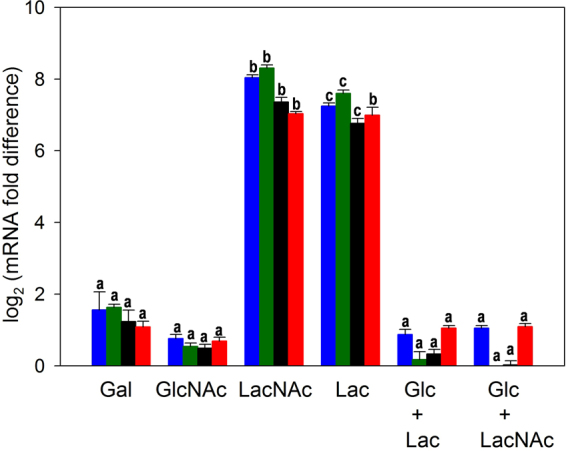
Transcriptional analysis by RT-qPCR of the expression of lacT (blue bars), lacE (green bars), lacG (black bars) and lacF (red bars) in Lactobacillus casei wild type strain BL23 grown in sugar-free MRS containing galactose (Gal), N-acetylglucosamine (GlcNAc), N-acetyllactosamine (LacNAc), lactose (Lac), a mix of glucose and lactose (Glc + Lac) or a mix of glucose and N-acetyllactosamine (Glc + LacNAc). Cells grown in sugar-free MRS supplemented with glucose were used as reference condition. Data presented are mean values based on three replicates of at least two biological independent samples. Bars indicate standard errors. For each lac gene, significantly different values (P < 0.05) among culture conditions are marked by different lower-case letters.
The transcriptional antiterminator LacT is required for LacNAc metabolism
The L. casei LacT protein prevents transcription termination of the lac operon in response to the presence of lactose in the culture medium26. In order to determine the involvement of this transcriptional antiterminator in the metabolism of LacNAc, the mutant strain L. casei BL195 (lacT), deficient in LacT26, was cultured on LacNAc as carbon source (Fig. 7). BL195 (lacT) strain was also grown with glucose and lactose as positive and negative controls, respectively. The results show that this mutant strain exhibited a growth in the presence of LacNAc similar to that of the negative controls (lactose supplemented and non-supplemented sugar-free MRS), indicating that the transcriptional antitermiator LacT is also involved on LacNAc metabolism. Additionally, the results suggest that LacT antiterminates transcription of lac operon not only depending on the presence of lactose if not also on the presence of LacNAc in the growth medium.
Figure 7.
Growth curves of Lactobacillus casei mutant strain BL195 (lacT) on sugar-free MRS without carbon source (black), with N-acetyllactosamine (LacNAc) (red), glucose (green) or lactose (blue). Data presented are mean values based on at least three replicates. Error bars indicate standard deviations.
Discussion
The disaccharide N-acetyllactosamine (LacNac) has an important role in many cell recognition processes such as parasite-host cell interaction37, autoimmune38 and inflammatory39 diseases, and also in cancer40. Additionally, LacNAc is a key structure present at human milk oligosaccharides and also at the glycan domains of glycoproteins and glycolipids present in the gastrointestinal tract41–43. We have demonstrated that L. casei is able to metabolize LacNAc. Curiously, the lac operon, which has been widely studied in this strain26–28, is also the responsible of the transport and catabolism of LacNAc. As previously described for the PTSGnb, that is involved in the transport of N-acetyl-galactosamine, LNB and GNB in L. casei24, the PTSLac represents a new example of a PTS able to transport two structurally related substrates, lactose and LacNAc. L. casei mutants deficient in either EIICBLac or EIIALac 28, were unable to grow in the presence of LacNAc. Due to the great biotechnological and economic importance of lactose fermentation in the dairy industry there are a great number of studies directed to analyze lactose fermentation by lactic acid bacteria44–47. Lactose in these bacteria can be transported through proton symport permeases, lactose/galactose antiport systems44,48 or via PTS28,49–51. Analysis of the genome sequence of lactobacilli (http://www.ncbi.nlm.nih.gov/genomes) showed that genes encoding PTSs homologous to the PTSLac from L. casei BL23, are present in the Lactobacillus casei/paracasei/rhamnosus/zeae group of phylogenetically related lactobacilli, and also in a few strains of Lactobacillus heilongjiangensis, Lactobacillus futsaii, Lactobacillus farciminis, Lactobacillus perolens, Lactobacillus fermentum, Lactobacillus sharpeae, Lactobacillus crustorum, Lactobacillus pobuzihii, Lactobacillus kimchiensis, Lactobacillus ruminis, Lactobacillus gasseri and Lactobacillus johnsonii, suggesting that lactose-specific PTSs are widely distributed among lactobacilli. For L. gasseri strain ATCC 33323, which is autochthonous of the gut, it has already been demonstrated that it contains two different PTSs involved in the transport of lactose and that the expression of both was induced by this carbohydrate51. Another strain indigenous of the gut is L. rhamnosous TCELL-1, for which it has also been demonstrated that the lac operon is induced by lactose50. Whether those PTS are functional for lactose and LacNAc remains to be investigated. Transcriptional analysis demonstrated here that the lac operon from L. casei is induced by lactose as well as by LacNAc and that induction levels are higher on LacNAc than on lactose. This might suggest that LacNAc is the substrate to which the lac operon had been adapted first. Indeed, many L. casei strains have been isolated from the human gastrointestinal tract52,53, which is very rich in LacNAc5,41. In E. coli, the lac operon is not regulated directly by lactose if not by allolactose, a transient product synthesized by the β-galactosidase, and this has generated controversy about the true physiological role of the lac operon in this bacterium54.
L. casei transport LacNAc or lactose through the PTSLac resulting in the formation of LacNAc-P or lactose-P, which are further hydrolyzed inside the cell by the phospho-β-galactosidase LacG into Gal-6P and GlcNAc or glucose, respectively (Fig. 1b). We have biochemically characterized this enzyme and confirmed that can only hydrolyze phosphorylated substrates. This also occurs with all the proteins homologs to LacG that have been characterized until now, which have been isolated from Streptococcus mutants55, Saphylococcus aureus, Lactococcus lactis, L. casei strain 64H56 and L. gasseri57. Contrarily, β-galactosidases isolated from Bifidobacterium bifidum are able to hydrolyze non-phosphorylated lactose and LacNAc11,12, showing two different mechanisms to metabolize these disaccharides in species that would compete for them in environmental niches such as the gastrointestinal tract. The Gal-6P generated after the hydrolysis of LacNAc-P or lactose-P by LacG is channeled through the Tag-6P pathway. We have showed that the mutant strain L. casei BL393 (lacAB) is impaired in the growth on lactose and LacNAc. The genes encoding the Tag-6P route in L. casei are present in the operon lacR1ABD2C, which includes a transcriptional regulator LacR1, the two subunits of the heteromeric Gal-6P isomerase (lacAB), a Tag-6P kinase (lacC) and a Tag-1,6-bisP aldolase (lacD2). Unlike L. casei, in Lactococcus lactis58 and Streptococcus mutans59, the lac operon contains the genes lacRABCDFEG encoding the Tag-6P catabolic proteins in addition to the lactose-specific PTS and the phospho-β-galactosidase LacG.
L. casei species have been isolated from dairy products, plant material and reproductive and gastrointestinal tracts of humans and animals52,53, which reveals their great adaptability to different environments. Genome analyses have showed that gene loss and acquisition are the main events resulting in niche adaptation60. Additionally, lactobacilli also contain genes involved in sugar uptake, metabolism and regulation grouped in genomic islands61. Lactose metabolism is well known in the dairy industry, but few data is found about its metabolism by the gastrointestinal microbiota. Lactose and LacNAc are constituents of HMO molecules that reach the breastfeeding infant gut microbiota, and LacNAc and poly-LacNAc molecules are also present in high amounts in the newborn gut62,63. Here we showed that L. casei metabolizes both disaccharides by using the same transport system and catabolic enzymes, which could be another niche adaptation mechanism of this bacterium to optimize the metabolic machinery minimizing energy consumption in a very competitive environment such as the gastrointestinal tract. Furthermore, the present work evidences that a catabolic pathway designed for survival of lactobacilli in the children gut has become an important tool for the development of dairy fermented products.
Materials and Methods
Strains, growth conditions and plasmids
The strains and plasmids used in this work are enumerated in Table 2. The L. casei strains were grown at 37 °C in MRS broth (Difco). E. coli was utilized as a cloning host and was grown in Luria-Bertani medium (Pronadisa) at 37 °C. E. coli DH10B transformants were selected with ampicillin (100 μg ml−1), E. coli BE50 with ampicillin (100 μg ml−1) and kanamycin (25 μg ml−1) and L. casei with erythromycin (5 μg ml−1). The vectors pRV30064 and pQE80 (Qiagen) were used for disruption of genes in L. casei and overproduction of proteins, respectively. E. coli strains were transformed by electroporation with a Gene Pulser apparatus (Bio-Rad Laboratories) as indicated by the manufacturer, and L. casei strains were transformed as described previously65.
Table 2.
Strains and plasmids used in this studya.
| Strain or plasmid | Relevant genotype or properties | Source or reference |
|---|---|---|
| Strains | ||
| Lactobacillus casei | ||
| BL23 | Wild type | CECT 5275 (Acedo-Félix et al.70) |
| BL126 | BL23 ptsI | Viana et al.33 |
| BL153 | BL23 lacE | Gosalbes et al.28 |
| BL155 | BL23 lacF | Gosalbes et al.28 |
| BL195 | BL23 lacT | Gosalbes et al.26 |
| BL385 | BL23 gnbC::pRV300 ErmR | Bidart et al.24 |
| BL388 | BL23 nagA::pRV300 ErmR | Bidart et al.24 |
| BL393 | BL23 lacAB | Bidart et al.24 |
| BL400 | BL23 lacG (frameshift at SphI site) | This work |
| Escherichia coli | ||
| DH10B | F− endA1 recA1 galE15 galK16 nupG rpsL ΔlacX74 Φ80lacZΔM15 araD139 Δ(ara,leu)7697 mcrA Δ(mrr) sdRMS-mcrBC) λ− | Invitrogen |
| BE50 | BL21(DE3) containing pREPGroES/GroEL | Dale et al.71 |
| PE172 | BE50 containing pQElacG | This work |
| Plasmids | ||
| pRV300 | Suicide vector carrying ErmR from pAMβ1 | Leloup et al.64 |
| pRVlacG | pRV300 with a frameshift at SphI site in lacG fragment | This work |
| pQE80 | E. coli expression vector; AmpR | Qiagen |
| pQElacG | pQE80 containing lacG-coding region | This work |
aCECT, Colección Española de Cultivos Tipo; ErmR, erythromycin resistance; AmpR, ampicillin resistant.
Culture of L. casei strains with lactose and LacNAc
The L. casei strains were cultured as previously described24 on sugar-free MRS containing: bactopeptone (Difco), 10 g l−1; yeast extract (Pronadisa), 4 g l−1; sodium acetate, 5 g l−1; tri-ammonium citrate, 2 g l−1; magnesium sulphate 7-hydrate, 0.2 g l−1; manganese sulphate monohydrate, 0.05 g l−1; and Tween 80, 1 ml l−1. LacNAc (Carbosynth, Compton, Berkshire, UK), lactose (Sigma-Aldrich, St. Louis, MO, USA), N-acetylglucosamine, galactose or glucose were added to the sugar-free MRS medium at a concentration of 4 mM. Bacterial growth was assayed in microtiter plates (100 μl culture broth per well) at 37 °C in a POLARstar Omega plate reader (BMG Labtech, Offenburg, Germany). The Gompertz model (GraphPad Software, San Diego, CA) was used for the analysis of the growth rates (μ).
DNA manipulation and sequencing
DNA was obtained from L. casei BL23 as previously described65. Plasmid DNA was isolated from E. coli by using the kit illustra plasmidPrep Mini Spin (GE Healthcare, UK). Standard methods were used for recombinant DNA techniques66 and PCR reactions were carried out with the Expand High Fidelity PCR System (Roche). The DNA sequencing reactions were performed by the Central Service of Research Support of the University of Valencia (Spain). Specific primers hybridizing within the proper DNA fragments and universal primers were used for sequencing. The analysis of DNA sequences was performed with the aid of the DNAMAN 4.03 software package (Lynnon BioSoft) and sequence similarities were analyzed with the BLAST program67.
Construction of a lacG mutant strain
A DNA fragment containing part of lacG was obtained by PCR using L. casei BL23 chromosomal DNA and the oligonucleotides lacGfor (5′-CAAGGAAGACGGTAAAGG) and lacGrev (5′-CCAACGGATAGTCATTATG). The PCR product was cloned into pRV300 digested with EcoRV. The resulting plasmid pRVlacG was cleaved at the unique SphI restriction site, made blunt with the Klenow fragment, ligated and transformed to select a plasmid with a frameshift at the SphI site in lacG. L. casei was transformed with this plasmid and one integrant where single recombination occurred was selected and cultured at 37 °C without antibiotic selection for at least 200 generations. Cells were grown on MRS-agar plates and screened for erythromycin-sensitive phenotype by replica plating on MRS-agar plates with erythromycin. Antibiotic-sensitive clones were selected and, among them, one was chosen (BL400 strain) in which a double-crossover event conducted to the excision of the plasmid resulting in a mutated lacG copy, as was confirmed by sequencing of PCR-amplified fragment spanning the mutated region.
Disaccharide and monosaccharide analysis in culture supernatants
The cells from the L. casei cultures were eliminated by centrifugation and the supernatants were collected for sugar determination. The analyses were carried out by high-performance liquid chromatography (HPLC) with a Jasco PU2080Plus system. A Rezex RCM-Monosaccharide column (Phenomenex) (at 80 °C) was used and the samples elution was performed in isocratic mode using a flow rate of 0.6 ml min−1. The mobile phase was water and a refractive index detector (Jasco RI-2031 Plus) was used for carbohydrate detection. Peaks in the chromatograms were identified by comparing the retention times with those of the standards (LacNAc, lactose, glucose, galactose and GlcNAc).
Expression and purification of His-tagged LacG
lacG coding region was amplified by PCR using genomic DNA of wild-type L. casei strain BL23 as template and the primers LacGBamHIFW (5′-TTTTGGATCCATGAGTAAACAGCTACCTCAAG) and LacGPstIRV (5′-TTTTCTGCAGTTAATCCGGAATGATGTGGG) with added BamHI and PstI restriction sites to the 5′- and 3′-ends (underlined). The obtained PCR product was digested with BamHI and PstI, cloned into pQE80 and the resulting plasmid pQElacG was used to transform E. coli BE50. DNA sequencing was carried out to confirm the correct construction. One clone, PE172 (pQElacG) was selected, grown in Luria-Bertani medium and induced with IPTG (1 mM) as described before25. The recombinant protein was purified and analyzed as described previously24.
His-tagged LacG enzyme activity
100 μl reaction mixtures containing different o/p-nitrophenyl(NP)-sugars (Table 1) at 5 mM were performed in 96-well microtiter plates. The LacG activity was measured at 37 °C in 100 mM Tris-HCl buffer, pH 7.0, and the reaction started by adding 1 μg of enzyme. The amount of released o/p-nitrophenol was tested by tracking the absorbance change with time at 404 nm using a microplate reader (POLARstar Omega, BMG Labtech, Offenburg, Germany). The optimal pH and temperature reaction, and kinetic analysis were determined with o-NP-β-D-galactopyranoside-6-phosphate as previously described24.
LacG was tested for its capacity to hydrolyse different natural oligosaccharides (Table 1). 100 μl reactions containing substrate (4 mM) in 100 mM Tris-HCl buffer, pH7.0 were performed at 37 °C for 16 h. The reaction mixtures were analysed by HPLC as described above. Peaks in the chromatograms were identified by comparing the retention times with those of the standards (LNB, GNB, lacto-N-tetraose, lacto-N-neotetraose, 3′-N-acetilgalactosaminil-Gal, 3′-N-acetilglucosaminil-Man, 4′-galactofuranosil-GlcNAc, fucosyl-α1–3GlcNAc, LacNAc, lactose, lactulose, maltose, maltotriose, glucose, galactose, GlcNAc, mannose, GalNAc, L-fucose and fructose). All the oligo- and disaccharides were obtained from Carbosynth (Compton, Berkshire, UK), except lactose, lactulose, maltose and maltotriose that were obtained from Sigma-Aldrich (St. Louis, MO, USA).
RNA isolation and Reverse Transcriptase quantitative PCR (RT-qPCR)
Total RNA was isolated from L. casei strain BL23 grown in sugar-free MRS supplemented with 4 mM of different sugars as previously described24. Cells were harvested at mid-exponential phase of growth (OD550 of 0.3 for cultures on glucose, galactose or GlcNAc and 0.5 for cultures on LacNAc, lactose, a mix of LacNAc plus glucose or lactose plus glucose). The isolated RNA was digested with DNaseI and retro-transcribed using the Maxima First strand cDNA Synthesis Kit (Fermentas)24. The resulted cDNA was subjected to quantitative PCR for the genes lacT, lacE, lacG and lacF. RT-qPCR was performed using the Lightcycler 2.0 system (Roche), LC Fast Start DNA Master SYBR green I (Roche) and primers pairs that produce amplicons ranging from 70 to 200 bp in size. RT-qPCR was performed for each cDNA sample in triplicate and using the primers pairs: qPCRlacTfor (5′-TTGTAAGGGGACGTGGCATC)/qPCRlacTrev (5′-TTGTCGGGAAGTCTCGTTCG) (lacT), qPCRlacEfor (5′-TTGGCCATGAACACGATGGA)/qPCRlacErev (5′-CCGAAAGTGCATGGCACAAA) (lacE), qPCRlacGfor (5′-AAGTCGAAGGAGCCACCAAG)/qPCRlacGrev (5′-GAACCGCCCCTGTTTATCCA) (lacG) and qPCRlacFfor (5′-GGTTTTGCACTTGTGGCGTA)/qPCRlacFrev (5′-CTGTTGGGCCTTCTCAACCA) (lacF). The reaction mixtures and cycling conditions were performed as previously described24. The pyrG, lepA and IleS genes were chosen as reference genes68. Cells growing in sugar-free MRS supplemented with glucose were used as reference condition. The relative expression based on the expression ratio between the target genes and reference genes was calculated using the software tool REST (relative expression software tool)69. The efficiency of all the primer pairs was between 1.9 and 2 (close to 100%). RT-qPCR reactions were performed in triplicate of two biological independent samples.
Statistical analysis
Statistical analysis was performed using the Statgraphics Plus, ver. 2.1 (Statistical Graphics Corp., USA). One way analysis of variance (ANOVA) was used to assess the effects of the carbon source (galactose, GlcNAc, LacNAc, lactose, a mix of glucose and lactose, and a mix of glucose and LacNAc) on the expression levels of the lac genes. Student’s t-test was used to detect statistically significant differences between growth rate from L. casei wild-type BL23 strain versus each mutant BL393 (lacAB) and BL388 (nagA) strains. Statistical significance was accepted at P < 0.05.
Data availability statement
All data generated or analyzed during this study are included in this published article.
Acknowledgements
This work was financed by funds of the Spanish Ministry for Economy and Competitiveness (MINECO)/FEDER through the Project AGL2014-52996-C2 (1-R and 2-R). G.N.B. was supported by a predoctoral fellowship from the Carolina Foundation and Argentinian Ministry of Education. J.R.-D. was supported by a Ramon y Cajal Contract by the Spanish Ministry for Economy and Competitiveness.
Author Contributions
G.N.B. performed the experimental work. J.R.-D., G.P.-M. and M.J.Y. designed the study and supervised it. M.J.Y. drafted the manuscript. G.N.B., J.R.-D. and G.P.-M. helped improving the draft.
Competing Interests
The authors declare no competing interests.
Footnotes
Publisher's note: Springer Nature remains neutral with regard to jurisdictional claims in published maps and institutional affiliations.
References
- 1.Kunz C, Rudloff S, Baier W, Klein N, Strobel S. Oligosaccharides in human milk: structural, functional, and metabolic aspects. Annu Rev Nutr. 2000;20:699–722. doi: 10.1146/annurev.nutr.20.1.699. [DOI] [PubMed] [Google Scholar]
- 2.Kobata A. Structures and application of oligosaccharides in human milk. Proc Jpn Acad Ser B Phys Biol Sci. 2010;86:731–747. doi: 10.2183/pjab.86.731. [DOI] [PMC free article] [PubMed] [Google Scholar]
- 3.Balogh R, Jankovics P, Beni S. Qualitative and quantitative analysis of N-acetyllactosamine and lacto-N-biose, the two major building blocks of human milk oligosaccharides in human milk samples by high-performance liquid chromatography-tandem mass spectrometry using a porous graphitic carbon column. J Chromatogr A. 2015;1422:140–146. doi: 10.1016/j.chroma.2015.10.006. [DOI] [PubMed] [Google Scholar]
- 4.Stanley, P. & Cummings, R. D. In Essentials of Glycobiology (eds Varki, A. et al.) (2009). [PubMed]
- 5.Marionneau S, et al. ABH and Lewis histo-blood group antigens, a model for the meaning of oligosaccharide diversity in the face of a changing world. Biochimie. 2001;83:565–573. doi: 10.1016/S0300-9084(01)01321-9. [DOI] [PubMed] [Google Scholar]
- 6.Klaassens ES, et al. Mixed-species genomic microarray analysis of fecal samples reveals differential transcriptional responses of bifidobacteria in breast- and formula-fed infants. Appl Environ Microbiol. 2009;75:2668–2676. doi: 10.1128/AEM.02492-08. [DOI] [PMC free article] [PubMed] [Google Scholar]
- 7.Sela DA, Mills DA. Nursing our microbiota: molecular linkages between bifidobacteria and milk oligosaccharides. Trends Microbiol. 2010;18:298–307. doi: 10.1016/j.tim.2010.03.008. [DOI] [PMC free article] [PubMed] [Google Scholar]
- 8.Fushinobu S. Unique sugar metabolic pathways of bifidobacteria. Biosci Biotechnol Biochem. 2010;74:2374–2384. doi: 10.1271/bbb.100494. [DOI] [PubMed] [Google Scholar]
- 9.LoCascio RG, Desai P, Sela DA, Weimer B, Mills DA. Broad conservation of milk utilization genes in Bifidobacterium longum subsp. infantis as revealed by comparative genomic hybridization. Appl Environ Microbiol. 2010;76:7373–7381. doi: 10.1128/AEM.00675-10. [DOI] [PMC free article] [PubMed] [Google Scholar]
- 10.Locascio RG, et al. A versatile and scalable strategy for glycoprofiling bifidobacterial consumption of human milk oligosaccharides. Microbial Biotechnol. 2009;2:333–342. doi: 10.1111/j.1751-7915.2008.00072.x. [DOI] [PMC free article] [PubMed] [Google Scholar]
- 11.Goulas T, Goulas A, Tzortzis G, Gibson GR. Expression of four beta-galactosidases from Bifidobacterium bifidum NCIMB41171 and their contribution on the hydrolysis and synthesis of galactooligosaccharides. Appl Microbiol Biotechnol. 2009;84:899–907. doi: 10.1007/s00253-009-2009-5. [DOI] [PubMed] [Google Scholar]
- 12.Miwa M, et al. Cooperation of beta-galactosidase and beta-N-acetylhexosaminidase from bifidobacteria in assimilation of human milk oligosaccharides with type 2 structure. Glycobiology. 2010;20:1402–1409. doi: 10.1093/glycob/cwq101. [DOI] [PubMed] [Google Scholar]
- 13.Asakuma S, et al. Physiology of consumption of human milk oligosaccharides by infant gut-associated bifidobacteria. J Biol Chem. 2011;286:34583–34592. doi: 10.1074/jbc.M111.248138. [DOI] [PMC free article] [PubMed] [Google Scholar]
- 14.Turroni F, et al. Molecular dialogue between the human gut microbiota and the host: a Lactobacillus and Bifidobacterium perspective. Cell Mol Life Sci. 2014;71:183–203. doi: 10.1007/s00018-013-1318-0. [DOI] [PMC free article] [PubMed] [Google Scholar]
- 15.Hill C, et al. Expert consensus document. The International Scientific Association for Probiotics and Prebiotics consensus statement on the scope and appropriate use of the term probiotic. Nat Rev. Gastroenterol Hepatol. 2014;11:506–514. doi: 10.1038/nrgastro.2014.66. [DOI] [PubMed] [Google Scholar]
- 16.Martin R, Heilig GH, Zoetendal EG, Smidt H, Rodriguez JM. Diversity of the Lactobacillus group in breast milk and vagina of healthy women and potential role in the colonization of the infant gut. J Appl Microbiol. 2007;103:2638–2644. doi: 10.1111/j.1365-2672.2007.03497.x. [DOI] [PubMed] [Google Scholar]
- 17.Albesharat R, Ehrmann MA, Korakli M, Yazaji S, Vogel RF. Phenotypic and genotypic analyses of lactic acid bacteria in local fermented food, breast milk and faeces of mothers and their babies. Syst Appl Microbiol. 2011;34:148–155. doi: 10.1016/j.syapm.2010.12.001. [DOI] [PubMed] [Google Scholar]
- 18.Pridmore RD, et al. The genome sequence of the probiotic intestinal bacterium Lactobacillus johnsonii NCC 533. Proc Natl Acad Sci USA. 2004;101:2512–2517. doi: 10.1073/pnas.0307327101. [DOI] [PMC free article] [PubMed] [Google Scholar]
- 19.Altermann E, et al. Complete genome sequence of the probiotic lactic acid bacterium Lactobacillus acidophilus NCFM. Proc Natl Acad Sci USA. 2005;102:3906–3912. doi: 10.1073/pnas.0409188102. [DOI] [PMC free article] [PubMed] [Google Scholar]
- 20.Azcarate-Peril MA, et al. Analysis of the genome sequence of Lactobacillus gasseri ATCC 33323 reveals the molecular basis of an autochthonous intestinal organism. Appl Environ Microbiol. 2008;74:4610–4625. doi: 10.1128/AEM.00054-08. [DOI] [PMC free article] [PubMed] [Google Scholar]
- 21.Rodriguez-Diaz J, Monedero V, Yebra MJ. Utilization of natural fucosylated oligosaccharides by three novel alpha-L-fucosidases from a probiotic Lactobacillus casei strain. Appl Environ Microbiol. 2011;77:703–705. doi: 10.1128/AEM.01906-10. [DOI] [PMC free article] [PubMed] [Google Scholar]
- 22.Becerra JE, Coll-Marques JM, Rodriguez-Diaz J, Monedero V, Yebra MJ. Preparative scale purification of fucosyl-N-acetylglucosamine disaccharides and their evaluation as potential prebiotics and antiadhesins. Appl Microbiol Biotechnol. 2015;99:7165–7176. doi: 10.1007/s00253-015-6666-2. [DOI] [PubMed] [Google Scholar]
- 23.Rodriguez-Diaz J, Rubio-del-Campo A, Yebra MJ. Lactobacillus casei ferments the N-Acetylglucosamine moiety of fucosyl-alpha-1,3-N-acetylglucosamine and excretes L-fucose. Appl Environ Microbiol. 2012;78:4613–4619. doi: 10.1128/AEM.00474-12. [DOI] [PMC free article] [PubMed] [Google Scholar]
- 24.Bidart GN, Rodriguez-Diaz J, Monedero V, Yebra MJ. A unique gene cluster for the utilization of the mucosal and human milk-associated glycans galacto-N-biose and lacto-N-biose in Lactobacillus casei. Mol Microbiol. 2014;93:521–538. doi: 10.1111/mmi.12678. [DOI] [PubMed] [Google Scholar]
- 25.Bidart GN, Rodriguez-Diaz J, Yebra MJ. The Extracellular Wall-Bound beta-N-Acetylglucosaminidase from Lactobacillus casei Is Involved in the Metabolism of the Human Milk Oligosaccharide Lacto-N-Triose. Appl Environ Microbiol. 2015;82:570–577. doi: 10.1128/AEM.02888-15. [DOI] [PMC free article] [PubMed] [Google Scholar]
- 26.Gosalbes MJ, Esteban CD, Perez-Martinez G. In vivo effect of mutations in the antiterminator LacT in Lactobacillus casei. Microbiology. 2002;148:695–702. doi: 10.1099/00221287-148-3-695. [DOI] [PubMed] [Google Scholar]
- 27.Gosalbes MJ, Monedero V, Alpert CA, Perez-Martinez G. Establishing a model to study the regulation of the lactose operon in Lactobacillus casei. FEMS Microbiol Lett. 1997;148:83–89. doi: 10.1111/j.1574-6968.1997.tb10271.x. [DOI] [PubMed] [Google Scholar]
- 28.Gosalbes MJ, Monedero V, Perez-Martinez G. Elements involved in catabolite repression and substrate induction of the lactose operon in Lactobacillus casei. J Bacteriol. 1999;181:3928–3934. doi: 10.1128/jb.181.13.3928-3934.1999. [DOI] [PMC free article] [PubMed] [Google Scholar]
- 29.Monedero V, Gosalbes MJ, Perez-Martinez G. Catabolite repression in Lactobacillus casei ATCC 393 is mediated by CcpA. J Bacteriol. 1997;179:6657–6664. doi: 10.1128/jb.179.21.6657-6664.1997. [DOI] [PMC free article] [PubMed] [Google Scholar]
- 30.Le Coq D, Lindner C, Kruger S, Steinmetz M, Stulke J. New beta-glucoside (bgl) genes in Bacillus subtilis: the bglP gene product has both transport and regulatory functions similar to those of BglF, its Escherichia coli homolog. J Bacteriol. 1995;177:1527–1535. doi: 10.1128/jb.177.6.1527-1535.1995. [DOI] [PMC free article] [PubMed] [Google Scholar]
- 31.Deutscher J, Francke C, Postma PW. How phosphotransferase system-related protein phosphorylation regulates carbohydrate metabolism in bacteria. Microbiol Mol Biol Rev. 2006;70:939–1031. doi: 10.1128/MMBR.00024-06. [DOI] [PMC free article] [PubMed] [Google Scholar]
- 32.Tanaka Y, Teramoto H, Inui M, Yukawa H. Translation efficiency of antiterminator proteins is a determinant for the difference in glucose repression of two beta-glucoside phosphotransferase system gene clusters in Corynebacterium glutamicum R. J Bacteriol. 2011;193:349–357. doi: 10.1128/JB.01123-10. [DOI] [PMC free article] [PubMed] [Google Scholar]
- 33.Viana R, et al. Enzyme I and HPr from Lactobacillus casei: their role in sugar transport, carbon catabolite repression and inducer exclusion. Mol Microbiol. 2000;36:570–584. doi: 10.1046/j.1365-2958.2000.01862.x. [DOI] [PubMed] [Google Scholar]
- 34.Veyrat A, Monedero V, Perez-Martinez G. Glucose transport by the phosphoenolpyruvate:mannose phosphotransferase system in Lactobacillus casei ATCC 393 and its role in carbon catabolite repression. Microbiology. 1994;140(Pt 5):1141–1149. doi: 10.1099/13500872-140-5-1141. [DOI] [PubMed] [Google Scholar]
- 35.Alpert CA, Siebers U. The lac operon of Lactobacillus casei contains lacT, a gene coding for a protein of the Bg1G family of transcriptional antiterminators. J Bacteriol. 1997;179:1555–1562. doi: 10.1128/jb.179.5.1555-1562.1997. [DOI] [PMC free article] [PubMed] [Google Scholar]
- 36.Bettenbrock K, Siebers U, Ehrenreich P, Alpert CA. Lactobacillus casei 64H contains a phosphoenolpyruvate-dependent phosphotransferase system for uptake of galactose, as confirmed by analysis of ptsH and different gal mutants. J Bacteriol. 1999;181:225–230. doi: 10.1128/jb.181.1.225-230.1999. [DOI] [PMC free article] [PubMed] [Google Scholar]
- 37.Ryan CM, Mehlert A, Richardson JM, Ferguson MA, Johnson PJ. Chemical structure of Trichomonas vaginalis surface lipoglycan: a role for short galactose (beta1-4/3) N-acetylglucosamine repeats in host cell interaction. J Biol Chem. 2011;286:40494–40508. doi: 10.1074/jbc.M111.280578. [DOI] [PMC free article] [PubMed] [Google Scholar]
- 38.Cappione AJ, Pugh-Bernard AE, Anolik JH, Sanz I. Lupus IgG VH4.34 antibodies bind to a 220-kDa glycoform of CD45/B220 on the surface of human B lymphocytes. J Immunol. 2004;172:4298–4307. doi: 10.4049/jimmunol.172.7.4298. [DOI] [PubMed] [Google Scholar]
- 39.Liu FT, Yang RY, Hsu DK. Galectins in acute and chronic inflammation. Annals New York Acad Sci. 2012;1253:80–91. doi: 10.1111/j.1749-6632.2011.06386.x. [DOI] [PubMed] [Google Scholar]
- 40.Holst S, Wuhrer M, Rombouts Y. Glycosylation characteristics of colorectal cancer. Adv Cancer Res. 2015;126:203–256. doi: 10.1016/bs.acr.2014.11.004. [DOI] [PubMed] [Google Scholar]
- 41.Moran AP, Gupta A, Joshi L. Sweet-talk: role of host glycosylation in bacterial pathogenesis of the gastrointestinal tract. Gut. 2011;60:1412–1425. doi: 10.1136/gut.2010.212704. [DOI] [PubMed] [Google Scholar]
- 42.Bode L. Human milk oligosaccharides: every baby needs a sugar mama. Glycobiology. 2012;22:1147–1162. doi: 10.1093/glycob/cws074. [DOI] [PMC free article] [PubMed] [Google Scholar]
- 43.Garrido D, Ruiz-Moyano S, Mills DA. Release and utilization of N-acetyl-D-glucosamine from human milk oligosaccharides by Bifidobacterium longum subsp. infantis. Anaerobe. 2012;18:430–435. doi: 10.1016/j.anaerobe.2012.04.012. [DOI] [PMC free article] [PubMed] [Google Scholar]
- 44.de Vos WM, Vaughan EE. Genetics of lactose utilization in lactic acid bacteria. FEMS Microbiol Rev. 1994;15:217–237. doi: 10.1111/j.1574-6976.1994.tb00136.x. [DOI] [PubMed] [Google Scholar]
- 45.Lapierre L, Mollet B, Germond JE. Regulation and adaptive evolution of lactose operon expression in Lactobacillus delbrueckii. J Bacteriol. 2002;184:928–935. doi: 10.1128/jb.184.4.928-935.2002. [DOI] [PMC free article] [PubMed] [Google Scholar]
- 46.Cavanagh D, Fitzgerald GF, McAuliffe O. From field to fermentation: the origins of Lactococcus lactis and its domestication to the dairy environment. Food Microbiol. 2015;47:45–61. doi: 10.1016/j.fm.2014.11.001. [DOI] [PubMed] [Google Scholar]
- 47.Stefanovic E, Fitzgerald G, McAuliffe O. Advances in the genomics and metabolomics of dairy lactobacilli: A review. Food Microbiol. 2017;61:33–49. doi: 10.1016/j.fm.2016.08.009. [DOI] [PubMed] [Google Scholar]
- 48.Leong-Morgenthaler P, Zwahlen MC, Hottinger H. Lactose metabolism in Lactobacillus bulgaricus: analysis of the primary structure and expression of the genes involved. J Bacteriol. 1991;173:1951–1957. doi: 10.1128/jb.173.6.1951-1957.1991. [DOI] [PMC free article] [PubMed] [Google Scholar]
- 49.Alpert CA, Chassy BM. Molecular cloning and DNA sequence of lacE, the gene encoding the lactose-specific enzyme II of the phosphotransferase system of Lactobacillus casei. Evidence that a cysteine residue is essential for sugar phosphorylation. J Biological Chem. 1990;265:22561–22568. [PubMed] [Google Scholar]
- 50.Tsai YK, Lin TH. Sequence, organization, transcription and regulation of lactose and galactose operons in Lactobacillus rhamnosus TCELL-1. J Appl Microbiol. 2006;100:446–459. doi: 10.1111/j.1365-2672.2005.02790.x. [DOI] [PubMed] [Google Scholar]
- 51.Francl AL, Hoeflinger JL, Miller MJ. Identification of lactose phosphotransferase systems in Lactobacillus gasseri ATCC 33323 required for lactose utilization. Microbiology. 2012;158:944–952. doi: 10.1099/mic.0.052928-0. [DOI] [PubMed] [Google Scholar]
- 52.Toh H, et al. Genomic adaptation of the Lactobacillus casei group. PloS one. 2013;8:e75073. doi: 10.1371/journal.pone.0075073. [DOI] [PMC free article] [PubMed] [Google Scholar]
- 53.Cai H, Thompson R, Budinich MF, Broadbent JR, Steele JL. Genome sequence and comparative genome analysis of Lactobacillus casei: insights into their niche-associated evolution. Genome Biol Evol. 2009;1:239–257. doi: 10.1093/gbe/evp019. [DOI] [PMC free article] [PubMed] [Google Scholar]
- 54.Wheatley RW, Lo S, Jancewicz LJ, Dugdale ML, Huber RE. Structural explanation for allolactose (lac operon inducer) synthesis by lacZ beta-galactosidase and the evolutionary relationship between allolactose synthesis and the lac repressor. J Biol Chem. 2013;288:12993–13005. doi: 10.1074/jbc.M113.455436. [DOI] [PMC free article] [PubMed] [Google Scholar]
- 55.Honeyman AL, Curtiss R., 3rd Isolation, characterization and nucleotide sequence of the Streptococcus mutans lactose-specific enzyme II (lacE) gene of the PTS and the phospho-beta-galactosidase (lacG) gene. J Gen Microbiol. 1993;139:2685–2694. doi: 10.1099/00221287-139-11-2685. [DOI] [PubMed] [Google Scholar]
- 56.Witt E, Frank R, Hengstenberg W. 6-Phospho-beta-galactosidases of gram-positive and 6-phospho-beta-glucosidase B of gram-negative bacteria: comparison of structure and function by kinetic and immunological methods and mutagenesis of the lacG gene of Staphylococcus aureus. Protein Eng. 1993;6:913–920. doi: 10.1093/protein/6.8.913. [DOI] [PubMed] [Google Scholar]
- 57.Honda H, et al. Purification and characterization of two phospho-beta-galactosidases, LacG1 and LacG2, from Lactobacillus gasseri ATCC33323(T) J Gen Appl Microbiol. 2012;58:11–17. doi: 10.2323/jgam.58.11. [DOI] [PubMed] [Google Scholar]
- 58.van Rooijen RJ, van Schalkwijk S, de Vos WM. Molecular cloning, characterization, and nucleotide sequence of the tagatose 6-phosphate pathway gene cluster of the lactose operon of Lactococcus lactis. J Biol Chem. 1991;266:7176–7181. [PubMed] [Google Scholar]
- 59.Jagusztyn-Krynicka EK, et al. Streptococcus mutans serotype c tagatose 6-phosphate pathway gene cluster. J Bacteriol. 1992;174:6152–6158. doi: 10.1128/jb.174.19.6152-6158.1992. [DOI] [PMC free article] [PubMed] [Google Scholar]
- 60.Douglas GL, Klaenhammer TR. Genomic evolution of domesticated microorganisms. Ann Rev Food Sci Tech. 2010;1:397–414. doi: 10.1146/annurev.food.102308.124134. [DOI] [PubMed] [Google Scholar]
- 61.Bellanger X, Payot S, Leblond-Bourget N, Guedon G. Conjugative and mobilizable genomic islands in bacteria: evolution and diversity. FEMS Microbiol Rev. 2014;38:720–760. doi: 10.1111/1574-6976.12058. [DOI] [PubMed] [Google Scholar]
- 62.Liu Y, et al. Poly-LacNAc as an age-specific ligand for rotavirus P[11] in neonates and infants. PloS one. 2013;8:e78113. doi: 10.1371/journal.pone.0078113. [DOI] [PMC free article] [PubMed] [Google Scholar]
- 63.Haltiwanger RS, Lowe JB. Role of glycosylation in development. Ann Rev Biochem. 2004;73:491–537. doi: 10.1146/annurev.biochem.73.011303.074043. [DOI] [PubMed] [Google Scholar]
- 64.Leloup L, Ehrlich SD, Zagorec M, Morel-Deville F. Single-crossover integration in the Lactobacillus sake chromosome and insertional inactivation of the ptsI and lacL genes. App Environ Microbiol. 1997;63:2117–2123. doi: 10.1128/aem.63.6.2117-2123.1997. [DOI] [PMC free article] [PubMed] [Google Scholar]
- 65.Posno M, et al. Incompatibility of Lactobacillus Vectors with Replicons Derived from Small Cryptic Lactobacillus Plasmids and Segregational Instability of the Introduced Vectors. App Environ Microbiol. 1991;57:1822–1828. doi: 10.1128/aem.57.6.1822-1828.1991. [DOI] [PMC free article] [PubMed] [Google Scholar]
- 66.Sambrook, J., E. F. Fritsch &.T. M. Molecular cloning: a laboratory manual, 2nd ed. Cold Spring Harbor Laboratory, Cold Spring Harbor, N.Y. (1989).
- 67.Altschul SF, Gish W, Miller W, Myers EW, Lipman DJ. Basic local alignment search tool. J Mol Biol. 1990;215:403–410. doi: 10.1016/S0022-2836(05)80360-2. [DOI] [PubMed] [Google Scholar]
- 68.Landete JM, et al. Requirement of the Lactobacillus casei MaeKR two-component system for L-malic acid utilization via a malic enzyme pathway. App Environ Microbiol. 2010;76:84–95. doi: 10.1128/AEM.02145-09. [DOI] [PMC free article] [PubMed] [Google Scholar]
- 69.Pfaffl MW, Horgan GW, Dempfle L. Relative expression software tool (REST) for group-wise comparison and statistical analysis of relative expression results in real-time PCR. Nucleic Acids Res. 2002;30:e36. doi: 10.1093/nar/30.9.e36. [DOI] [PMC free article] [PubMed] [Google Scholar]
- 70.Acedo-Felix E, Perez-Martinez G. Significant differences between Lactobacillus casei subsp. casei ATCC 393T and a commonly used plasmid-cured derivative revealed by a polyphasic study. Int J Syst Evol Microbiol. 2003;53:67–75. doi: 10.1099/ijs.0.02325-0. [DOI] [PubMed] [Google Scholar]
- 71.Dale, G. E., Schonfeld, H. J., Langen, H. & Stieger, M. Increased solubility of trimethoprim-resistant type S1 DHFR from Staphylococcus aureus In Escherichia coli cells overproducing the chaperonins GroEL and GroES. Protein Eng7, 925–931 (1994). [DOI] [PubMed]
Associated Data
This section collects any data citations, data availability statements, or supplementary materials included in this article.
Data Availability Statement
All data generated or analyzed during this study are included in this published article.