Abstract
Purpose
To investigate the association between aortic arch width on frontal chest radiography and systemic hypertension.
Methods
A total of 200 consecutive patients were included. Relationships between aortic arch width measurement on chest radiography and blood pressure measurement were investigated using Student’s t-tests and Fisher’s exact tests.
Results
Twenty-five patients were normotensive (< 130/90 mmHg), and 175 were hypertensive. Using cut-off values, 136 patients had an aortic arch width ≥ 3.5 cm, and 65 had an aortic arch width ≥ 4 cm. We found a significant relationship between aortic arch width and hypertension (p < 0.001) as well between aortic arch width cut-off values of 3.5 cm and 4 cm and hypertension (p < 0.001 and p < 0.005, respectively). An aortic arch width ≥ 3.5 cm was associated with a positive likelihood ratio (LR) of 2.3, negative LR of 0.39, sensitivity of 73, specificity of 68, positive predictive value of 94, negative predictive value of 26.6, pretest odds of 7, posttest odds of 16, and posttest probability of 94%. An aortic arch width ≥ 4 cm was associated with a positive LR of 4.50, negative LR of 0.70, sensitivity of 36, specificity of 92, positive predictive value of 97, negative predictive value of 17, pretest odds of 7, posttest odds of 31.5, and posttest probability of 97%.
Conclusions
Aortic arch width measurement on chest radiography can be used to predict the presence of long-standing systemic arterial hypertension.
Keywords: Aortic arch width, Chest radiography, Hypertension
Introduction
Systemic hypertension affects 25% of the adult population, including 78 million persons in the United States and more than 1 billion persons worldwide. Its prevalence increases with age. Hypertension is the most common cause for an outpatient visit to a physician and the most important preventable risk factor for stroke, myocardial infarction, heart failure, peripheral vascular disease, aortic dissection, atrial fibrillation, end-stage renal disease, and premature death worldwide. In the United States, it has surpassed smoking as a cause of preventable death. At least 20% of affected persons are initially unaware of their condition. Chest radiography is arguably the most commonly performed radiographic examination, with at least 70 million radiographs performed each year in the United States. Aortic arch width is a landmark identified on every chest radiograph and can easily be used for screening purposes. To our knowledge, no scientific study has been performed in a United States population to determine the predictive value of aortic arch width on frontal chest radiography for the diagnosis of systemic hypertension. Therefore, we investigated the association between aortic arch width and systemic hypertension.
Materials and Methods
Study Population
A total of 200 consecutive patients of Veterans Health Administration San Diego Health Care System who underwent posteroanterior chest radiography were included in this study, which was approved by the Institutional Review Board. The requirement for informed consent was waived. Due to the nature of the veteran patient population, the study population consisted of predominantly male and high-risk subjects for cardiovascular disease processes.
Aortic Arch Measurement
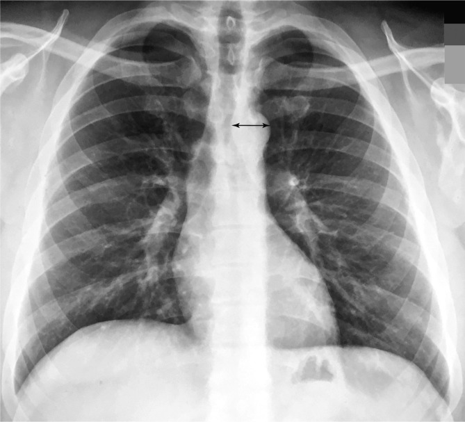
The width of the aortic arch was measured prospectively. Aortic arch width was measured from the point of the left lateral edge of the trachea to the left lateral wall of the aortic arch (Figure 1).
Figure 1.
Aortic arch measurement technique on frontal chest radiography. Transverse measurement from the left margin of the trachea to the left margin of the aortic arch.
Blood Pressure Measurement
Blood pressure measurements were obtained retrospectively through medical record review. Measurements were performed using standard medical office sphygmomanometers and recorded in electronic medical records as part of routine physician visit procedures. A blood pressure measurement of < 130/90 mmHg was considered normal.
Statistical Analysis
The relationship between aortic arch width measurement on chest radiography and blood pressure measurement was investigated using Student’s t-tests. Aortic arch width measurements were also categorized using cut-off values of 3.5 cm and 4 cm, and their correlations with blood pressure were investigated using Fisher’s exact tests (two-sided). A p < 0.05 was considered statistically significant. Likelihood ratios (LRs) were calculated to determine the predictive value of aortic arch width measurement for long-standing arterial systolic hypertension.
Results
Of the included patients, 25 (12.5%) were normotensive (< 130/90 mmHg), and 175 (87.5%) were hypertensive (≥ 130/90 mmHg) (Table 1.) Mean systolic blood pressure was 146 mmHg (standard deviation (SD): 14.7; range: 96-178; median: 145). Mean diastolic blood pressure was 85 mmHg (SD: 11.5; range: 48-117; median: 84.5).
Table 1.
Demographic data of the study population. A clear majority of the patients were male due to nature of the patient population where the study was conducted.
| Age (Mean, Range) | Obesity (%) | Hyperlipidemia (%) | Tobacco (%) | DM (%) | |
|---|---|---|---|---|---|
| Main population | 57.7 (23-96) | 77 (38.5) | 102 (51) | 10 (5) | 50 (25) |
| Hypertensive | 60.4 (24-95) | 70 (35) | 100 (50) | 9 (4.5) | 49 (23) |
| Normotensive | 39.4 (23-80) | 7 (3.5) | 2 (1) | 1 (0.5) | 1 (2) |
DM = diabetes mellitus
One hundred thirty-six (68%) patients had an aortic arch width ≥ 3.5 cm, and 65 (33%) patients had an aortic arch width ≥ 4 cm. Mean aortic arch width was 38 mm (SD: 5.8; range: 24.9-57.8 mm; median: 36.8).
We found a significant relationship between aortic arch width on chest radiography and blood pressure measurement (p < 0.0001) as well as also significant relationships between aortic arch width cut-off values of 3.5 cm and 4 cm and the presence of hypertension (p < 0.0001 and p < 0.005, respectively).
An aortic arch width ≥ 3.5 cm was associated with a positive LR of 2.3 (95% confidence interval (CI): 1.28, 4.08), negative LR of 0.39 (95% CI: 0.27, 0.57), sensitivity of 73, specificity of 68, positive predictive value of 94, negative predictive value of 26.6, pretest odds of 7, posttest odds of 16, and posttest probability of 94%.
An aortic arch width ≥ 4 cm was associated with a positive LR of 4.50 (95% CI: 1.17, 17), negative LR of 0.70 (95% CI: 0.59, 0.82), sensitivity of 36, specificity of 92, positive predictive value of 97, negative predictive value of 17, pretest odds of 7, posttest odds of 31.5, and posttest probability of 97%.
Discussion
Hypertension affects more than 1 billion persons worldwide. In the United States alone, 78 million individuals have hypertension. High blood pressure may not cause symptoms until end organ damage manifests clinically. For this reason, hypertension has been called the “silent killer” [1]. Subclinical organ damage has been considered an intermediate stage in the spectrum of vascular disease and is a strong determinant of total cardiovascular risk [2]. Chest radiography has been shown to predict associated target organ damage in hypertensive patients [3]. Specifically, aortic arch width is positively correlated with both systolic and diastolic blood pressure and was found to be a reliable marker of target organ damage due to its correlation with blood creatinine values, retinal fundoscopic changes, cardiothoracic ratio, and left ventricular mass on echocardiography [3]. Left ventricular hypertrophy is considered the most powerful determinant of cardiovascular outcome in hypertensive patients [4], and an aortic arch width of > 3.6 cm is suggested as a marker of target organ damage [3]. Subsequent studies demonstrated a significant and independent association between aortic arch width and cardio-ankle vascular index [5], which is considered to reflect arteriosclerosis of the aorta and is a novel parameter of arterial stiffness and surrogate marker of subclinical atherosclerosis [6]. Based on this association, an aortic arch width of 4.1 cm has been suggested as a cut-off value for the presence of subclinical atherosclerosis with modest sensitivity and specificity [5]. In our study, an aortic arch width ≥ 3.5 cm demonstrated a positive LR of 2.3 (95% CI: 1.28, 4.08) with modest sensitivity and specificity but a very high positive predictive value of 94 for hypertension. On the other hand, an aortic arch width ≥ 4 cm demonstrated a positive LR of 4.50 (95% CI: 1.17, 17.00) with relatively low sensitivity of 36, very high specificity of 92, and very high positive predictive value of 97 for hypertension.
Aortic arch width does not exceed 3 cm in most adult chest radiographs in the United States [7], whereas a recent study among an Asian-Indian population reports a width of 3.04 cm in healthy adults [8]. Felson [7] analyzed the width of the aortic “knob” in 500 normal persons from the left border of the trachea transversely to the lateral margin of the aorta and found an age-related increase in the width of the aortic “knob”. That is, 95% of persons under 30 years of age had a width < 3 cm, 91% of persons between 30 and 40 years of age had a width < 3 cm, and 69% of persons over 40 years of age had a width < 3 cm. In no case did the aortic “knob” width measure > 4 cm.
However, these studies were not focused on the “size and pressure” association. Permanent changes in the size of the aorta happen gradually and progressively as part of the aging process and are probably exacerbated by systemic arterial hypertension [3]. Dilation is most likely related to degradation of elastic fibers in the aortic wall. When monitored over years, especially in high-risk populations, systemic arterial blood pressure often demonstrates dramatic variability, with short-term episodic spikes reaching or exceeding 200 mmHg and sustained undulations of longer duration between 100 to 200 mmHg triggered by emotional states, exercise, medications, life style, habits, diet, or other disease processes. The literature about aortic arch width on chest radiographs in relation to systemic arterial blood pressure primarily consists of cross-sectional studies evaluating populations at one point in time without taking into account the wall-damaging impact of short-term and long-term fluctuations in blood pressure over time. This can explain some inconsistent results among studies attempting to assess the correlation of “size and pressure”, which yield a variety of suggested cut-off values for the width of the aortic arch as a predictor of systemic hypertension. This challenge is also reflected in variable guidelines for protective target blood pressures.
Our study was conducted in a high-risk patient population for cardiovascular disease including systemic hypertension. Most patients were monitored over a long period of time, which allowed us to more accurately assess their blood pressure status. Most of our patients had recurrent visits to our healthcare system over several months to several years, which allowed for multiple blood pressure measurements. We decided to select a representative blood pressure measurement among multiple blood pressure measurements from the time period preceding the date of the optimal posteroanterior chest radiograph used for measurement of the aortic arch width.
Higaki et al. [9] reported a strong correlation between aortic arch width and age, central systolic blood pressure, and central pulse pressure in a study population with known or suspected coronary artery disease. They showed that larger aortic arch widths were associated with not only dilation but also stiffening of the aorta. Rayner et al. [3] demonstrated a significant difference in aortic arch width between normotensive and hypertensive populations (3.28 vs. 3.69 cm, respectively) using standard mercury sphygmomanometer measurements in a hypertension clinic. These results are comparable to our findings (3.31 vs. 3.87 cm, respectively).
One limitation of our study is that we did not perform multivariate linear regression analysis of variables to determine independent predictors of progressive aortic arch widening, such as aging. However, studies show that in a healthy aging population, cardiovascular dimensions in frontal chest radiographs show only modest age-related changes [10]. The other limitation of our study is the relatively high frequency of multiple confounding cardiovascular disease processes in our patient population, which made isolation of systemic hypertension as the only factor determining the aortic arch width more difficult.
In conclusion, aortic arch width measurements on chest radiography can be used to predict the presence of long-standing systemic arterial hypertension.
Conflict of Interest
The authors have no conflicts of interest relevant to this publication.
References
- 1.Torpy JM, Lynm C, Glass RM. Hypertension. JAMA.2010;303:2098 DOI: 10.1001/jama.303.20.2098 [DOI] [PubMed] [Google Scholar]
- 2.Mancia G, De Backer G, Dominiczak A, Cifkova R, Fagard R, Germano G, et al. Guidelines for the management of arterial hypertension: the task force for the management of arterial hypertension of the European Society of Hypertension (ESH) and of the European Society of Cardiology (ESC). J Hypertens.2007;25:1105-1187. DOI: 10.1097/HJH.0b013e3281fc975a [DOI] [PubMed] [Google Scholar]
- 3.Rayner BL, Goodman H, Opie LH. The chest radiograph. A useful investigation in the evaluation of hypertensive patients. Am J Hypertens.2004;17:507-510. DOI: 10.1016/j.amjhyper.2004.02.012 [DOI] [PubMed] [Google Scholar]
- 4.Kannel WB, Cobb J. Left ventricular hypertrophy and mortality – results from the Framingham Study. Cardiology.1992;81:291-298. DOI: 10.1159/000175819 [DOI] [PubMed] [Google Scholar]
- 5.Korkmaz L, Erkan H, Korkmaz AA, Acar Z, Ağaç MT, Bektaş H, et al. Relationship of aortic knob width with cardio-ankle vascular stiffness index and its value in diagnosis of subclinical atherosclerosis in hypertensive patients: a study on diagnostic accuracy. Anadolu Kardiyol Derg.2012;12:102-106. DOI: 10.5152/akd.2012.034 [DOI] [PubMed] [Google Scholar]
- 6.Takaki A, Ogawa H, Wakeyama T, Iwami T, Kimura M, Hadano Y, et al. Cardio-ankle vascular index is a new noninvasive parameter of arterial stiffness. Circ J.2007;71:1710-1714. DOI: 10.1253/circj.71.1710 [DOI] [PubMed] [Google Scholar]
- 7.Felson B. A review of over 30,000 normal chest roentgenograms. In: Chest Roentgenology. Philadelphia: WB Saunders; 1973, p. 494-495. [Google Scholar]
- 8.Ray A, Mandal D, Kundu P, Manna S, Mandal S. Aortic knob diameter in chest X-ray and its relation with age, heart diameter and transverse diameter of thorax in a population of Bankura District of West Bengal, India: a cross sectional study. J Evol Med Dent Sci.2014;3:8595-8600. DOI: 10.14260/jemds/2014/3092 [DOI] [Google Scholar]
- 9.Higaki T, Kurisu S, Watanabe N, Ikenaga H, Shimonaga T, Iwasaki T, et al. Usefulness of aortic knob width on chest radiography to predict central hemodynamics in patients with known or suspected coronary artery disease. Clin Exp Hypertens.2015;37:440-444. DOI: 10.3109/10641963.2015.1057834 [DOI] [PubMed] [Google Scholar]
- 10.Simon G, Gamsu G. Cardiopulmonary dimensions: stability over a long period. AJR Am J Roentgenol.1977;128:239-241. DOI: 10.2214/ajr.128.2.239 [DOI] [PubMed] [Google Scholar]


