Abstract
Rationale:
Stevens–Johnson syndrome (SJS) is an acute blistering disease of the skin and mucous membranes. SJS in children is not common but potentially serious disease. But the epidemiology of SJS in China is not well defined.
Patient concerns:
A 6-year-old boy was initially diagnosed as pneumonia admitted to hospital after admission, and the body appears red rash with blisters, skin damage, lip debaucjed, repeated high fever, and rapid progression.
Diagnoses:
SJS often results from an allergy reaction response to a range of drugs. It is a clinical diagnosis suggested by fever and malaise followed by an extensive painful, nonblanching, macular rash that commonly progresses to blistering or sloughing, and mucositis.
Interventions:
The boy was treated with continuous renal replacement therapy, anti-infection therapy, high-dose glucocorticoid treatment, and symptomatic treatment.
Outcomes:
The patient was recovered after 33 days of treatment.
Lessons:
The current treatment is mainly symptomatic treatment, and for the patient, it is important to make skin care related well, included early out blisters at effusion, reducing skin ulceration of the mucosa area, keeping skin clean, removing mucosa secretion and blood clots, doing eye care related, preventing the complications, ensuring adequate intake of nutrition and warm and so on.
Keywords: case study research, infection control, nursing care
1. Introduction
Stevens–Johnson syndrome (SJS) was first reported in 1922 by Stevens and Johnson,[1] Bastuj-Garin et al,[2] classified SJS, SJS/toxic epidermal necrolysis overlap, and toxic epidermal necrolysis according to the percentage of detachment of the body surface area and widespread purpuric macules or flat atypical targets. SJS is a life-threatening blistering disease. It often results from an allergy reaction response to a range of drugs. A lot of drugs, such as antibiotics, sulfonamides, pyrazolones, corticosteroids, barbiturates, and antiepileptics, are associated with SJS.[3] It is characterized by a prodromal illness followed by severe mucocutaneous symptoms.[1] The incidence, mortality, and cost of SJS in Chinese pediatrics are unknown.
Elzagallaai et al[4] suggested more than one mechanistic pathway for different carbamazepine-induced hypersensitivity reactions in patients with different ethnic backgrounds. The study by Wang et al[5] showed that bronchiolitis obliterans occurred in 9.5% of cases after SJS. Azithromycin is a commonly prescribed antibiotic but is not considered a common etiologic agent for SJS.[6] SJS has been rarely reported in patients on azithromycin therapy. Destruction of the skin and mucosa, especially in the body and limbs, with significant blisters, occurs in this disease. This is a reaction for the body to protect the underlying skin from infection. Patient's contact with the outside world should be reduced as much as possible before the underlying skin regenerates completely. When the skin scab is formed, the black scab should be removed to improve wound healing and treatment.[7] Murphy and Amblum[8] suggested that blisters that were bigger in size than the patient's own nail should be cleared, but small blisters should be left to shrink and heal on their own. Severe delayed drug-induced skin reactions in children are not common,[9] but this study aimed to present aspects concerning the clinical manifestations, treatment, and nursing care in a pediatric patient with SJS.
2. Case report
The case report was approved by the Ethics Committee of the Xin Hua Hospital Affiliated to Shanghai Jiao Tong University School of Medicine (approval no. XHEC-D-2017-065). The written informed consent was obtained from patient's parents.
A 6-year-old Chinese boy who was born prematurely and grew well was diagnosed with pneumonia because of fever and cough for 7 days without obvious predisposing causes. Before admission, the child was treated for 4 days in a local hospital and given oral medication and “Cefotiam” infusion therapy, following which the temperature returned to normal, but his cough did not improve obviously. After admission, his physical examination was normal except double lung breath sounds with moist crackles. The systemic skin mucous membrane was not yellow, no subcutaneous petechia and rashes were present, and the oral mucosa was complete. The child had one healthy sister. Parents denied consanguineous marriage, any similar family history, or a familial history of genetic and metabolic diseases. He lived with his parents and sister. He was in a hospital and was treated with azithromycin. The daily dosage of azithromycin given to him was 0.25 g as intravenous drips, with cefmetazole sodium used in conjunction for anti-infection treatment. He still had hyperpyrexia on the second day. In the whole process, inducing physical hypothermia using drug and ice pillow was the main nursing measure. On the second day in the hospital, his oral mucosa and tarsal conjunctiva yielded a copious purulent material. Red maculopapules and blisters were scattered over his whole body. On the second day after admission, exudate in the eyelids and erythema scattered on the trunk, with no obvious blisters, appeared due to facial erythema and erosion of lips; the right finger had a blister with no obvious exudation; and the palmoplantar had erythema with no obvious visible blisters or a pustule. On consulting the dermatologist, he was diagnosed as having developed erythema multiforme on account of the aforementioned physical signs and the possibility of an adverse drug reaction due to azithromycin. The dermatologist suggested discontinuing azithromycin and external application of zinc oxide powder on the affected part of the skin (Fig. 1).
Figure 1.
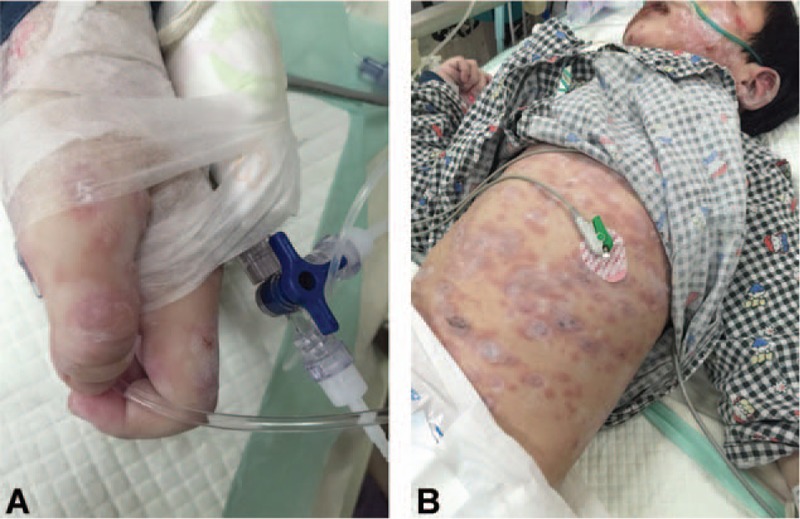
Condition after using zinc oxide powder on consultation with a dermatologist.
His symptoms were increasingly severe with pyohemia. He had a fever with chills, polypnea, blisters, ulceration from face to body, conjunctival congestion, and visible purulent secretion. Involvement of the oral, nasal, ocular, genital, and anal mucosa was seen. According to the clinical manifestation, he had SJS (Fig. 2).
Figure 2.
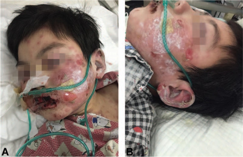
Treatment with indwelling gastric tube and oxygen absorbing by nasal trachea.
The child had eating difficulty; therefore, he was on indwelling gastric tube support to supplement enough energy to guarantee enteral nutrition intake. He was fed compound amino yogurt. He was given oxygen uptake with a nasal catheter of 2 L/min to ensure compliance with oxygenation.
Laboratory examination revealed the following: white blood count 13.84 × 10⁹/L, aspartate aminotransferase 143 U/L, C-reactive protein 7.60 mg/L, mycoplasma pneumonia antibody (+), cold agglutination test 1:256, neutrophil 84.4%, lymphocyte 29.8%, monocyte 14.9%, amylase 1718 U/L, creatine kinase 890 U/L, creatine kinase–MB 94.00 U/L, and procalcitonin 2.80 ng/mL.
The child's inflammatory factors increased; he was in a critical condition, and the internal environment was not stable. His liver and kidney functions were damaged. After multidisciplinary consultation, he was treated with continuous renal replacement (CRRT) and other anti-infective therapies to control and remove the inflammation mediator effectively. He was treated with individualized CRRT in which the blood pump speed was 70 mL/min, the displacement fluid speed was 750 mL/h, the dialysate speed was 750 mL/h, the anticoagulant fluid was 3.7 mL/h, and every 4 hours, the blood electrolytes, blood gas analysis, blood routine, and disseminated intravascular coagulation were reviewed. At the time of removing inflammatory mediators, he was given Imipenem for anti-infection therapy and acyclovir for antiviral treatment. Fixing the central venous catheters (CVCs) properly was crucial because of the large areas of skin erosion. The charge nurse regularly evaluated the puncture site, position, and pressure of the catheters to ensure successful intravenous infusion and CRRT treatment (Fig. 3).
Figure 3.
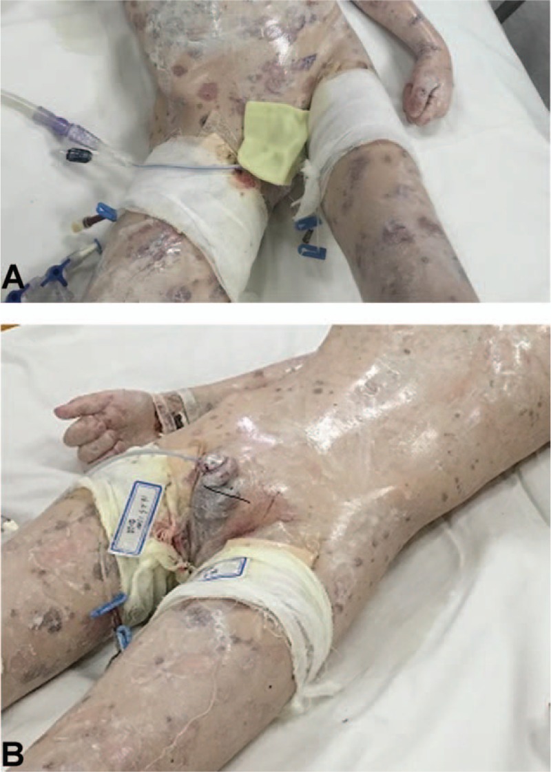
It showed how to fixed CVCs with the sterile gauze. CVCs = central venous catheters.
Because of the prolonged systemic skin blisters with a burst in the child, a transparent dressing paste could not be used to fix the CVCs. According to the nursing measures for acute patients, the physician used sutures to fix the CVCs. The sterile gauze was replaced with a transparent dressing, followed by a bandage wrapped around the puncture site.[10] His charge nurse disinfected the puncture site with chlorhexidine disinfectant and changed the sterile gauze every day.
Because of the acuteness of the blistering disease of the skin and mucous membranes, the pediatricians used a lot of medicines to treat the disease. They used ofloxacin eye drops every 2 hours and ofloxacin every 4 hours for the eyes, and b-FGF spray and stomatitis spray for mouth twice a day. The stomatologist suggested using dexamethasone injection 5 mg, gentamicin injection 16,000 U, lidocaine injection 5 mL, and sterile water 200 mL to clean the mouth 3 times a day; and self-made zinc oxide oil solution (made in Xinhua Hospital, Shanghai, China) and other medicines to cure his infection (Fig. 4).
Figure 4.
Nursing has affected part of skin, especially his eyes and oral mucosa.
The charge nurse cleaned the affected part of his skin with a sterile saline cotton ball. The blisters were aspirated using a 1-mL injector according to the aseptic operation. Before each medication, the skin was cleaned with a sterile saline lint cotton ball; a 1-mL syringe was used to aspirate the blisters according to sterile operation norms, without tearing the scarred skin. Then the whole body was daubed with a zinc oxide powder oil solution made by Xin Hua Hospital Affiliated to Shanghai Jiao Tong Unieversity School of Medicine to achieve the effects, such as cleaning, protection, anti-inflammation, analgesic, and so on. This application was used 3 times a day, and the body was kept warm.
As scabs formed easily due to secretions, damage, errhysis, and so on, the wound of the scab was cleaned daily to protect the eyes and oral mucosa; eye drops and ointments were used according to his symptoms to prevent conjunctival lesions, and the oral mucosa was daubed with olive oil to prevent the damage of skin and mucous membrane caused by the dry skin.
Even after clearing the blood scabs, lips and eyes were still exudated forming new blood scabs because of serious mucosal involvement. Therefore, when removing the blood scabs, first an oil agent was used gently to soften the scabs, preventing forced removal of the scab and avoiding any new injury to the mucous membrane of the skin. As friction material can easily cause the skin to burst, the skin was daubed with zinc oxide to protect it before removing the scab. In the whole process of the treatment, the child showed an extremely strong adherence and will power both physiologically and psychologically. He endured the pain that was almost unbearable for an adult.
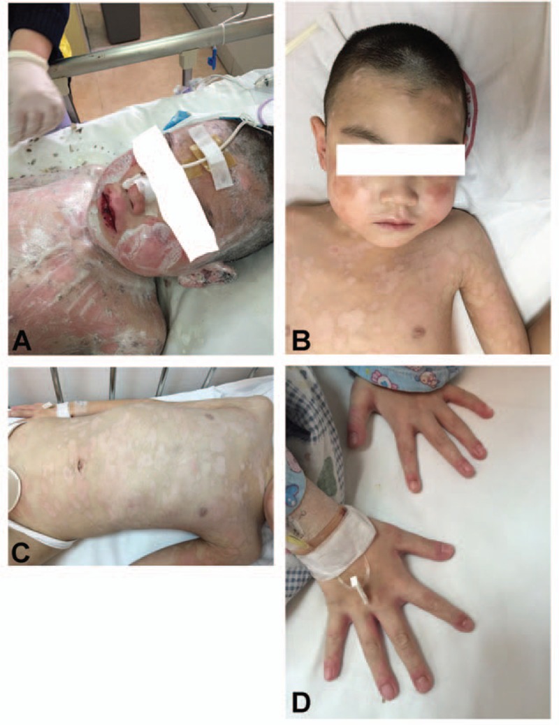
In addition to the symptomatic care, strategies to strengthen the management of the airway and gastrointestinal tract, maintain homeostasis, and improve nutrition were adopted. After the treatment, a new red skin grew, and the skin around the mouth and eyes and other mucosa lesions got better (Fig. 5).
Figure 5.
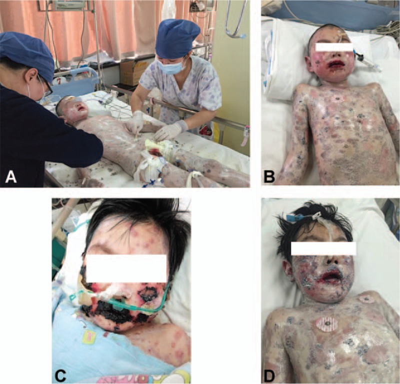
After treatment, the old skin rashes fell off, and he gradually recovered.
After 18 days of active treatment and nursing, his double eyelids were a bit red, the eye scabs had fallen off, eyes opened, and the face was slightly swollen. The oral mucosa still had dark red scabs with a little hemorrhagic fluid. The child had systemic visible old dark skin rashes, with pulmonary purple spots.
After treatment, the old skin rashes fell off, with no new rash or pigmentation.
After 33 days of careful treatment and care, he gradually recovered and was then discharged with continued follow-up.
3. Discussion
This study aimed to emphasize the skin care effectively, timely, and accurately to prevent complications in children with SJS and promote the rehabilitation of this disease. SJS is a group of serious diseases, involving the whole-body skin mucous membrane, usually caused by drugs. However, SJS has been rarely reported in patients on azithromycin therapy.[11] In several studies, the initial symptoms were high fever, discomfort, or respiratory symptoms, and rashes with blisters or lesions causing mucosal inflammation.[12] Lesions cause corneal ulceration or perforation, eventually leading to blindness. The urethral mucosa gets damaged, causing urethritis and leading to dysuria. The ventilatory function is affected by the peeling off of the respiratory tract mucous membrane. After cure, the scars would be formed on the skin due to mucosal damage and inflammation.[13] The oral and systemic skin in the process of the disease can be characterized by serious mucosal lesions and can cause severe pain at every treatment and nursing, which impacts both physiologically and psychologically. SJS has been considered a late-onset allergic reaction and can cause serious long-term sequelae and high mortality rate. Studies have reported a recurrence of the disease to a certain extent in children, and in adolescents, the recurrence rate may be higher than that in smaller children.[14]
A few studies have reported on SJS; it lacks a clinical control test and does not have a systematic treatment and prevention program. Immunoglobulin or hormone therapy has been recommended for treating SJS.
4. Conclusion
SJS should be actively treated symptomatically, and inflammation factors should be controlled to further protect the body. For this kind of disease, taking good care of the skin is extremely important. The internal fluid should be drawn before the bubble bursts as far as possible to reduce the area of the skin burst and to prevent further infection. At the same time, it can prevent hypothermia caused by excessive exposure to wounds and other adverse reactions. When giving medicine, nurses must promptly remove eye and oral mucosal secretion, blood scab, and so forth to prevent related complications.
Footnotes
Abbreviations: CRRT = continuous renal replacement therapy, CVCs = central venous catheters, IVIG = intravenous immunoglobulin, SJS = Stevens–Johnson syndrome.
The authors have no conflicts of interest to disclose.
References
- [1].Stevens AM, Johnson FC. A new eruptive fever associated with stomatitis and ophthalmia: report of two cases in children. Am J Dis Child 1922;24:526–33. [Google Scholar]
- [2].Bastuji-Garin S, Rzany B, Stern RS, et al. Clinical classification of cases of toxic epidermal necrolysis, stevens-johnson syndrome, and erythema multiforme. Arch Dermatol 1993;129:92–6. [PubMed] [Google Scholar]
- [3].Horner ME, Abramson AK, Warren RB, et al. The spectrum of oculocutaneous disease: Part I. Infectious, inflammatory, and genetic causes of oculocutaneous disease. J Am Acad Dermatol 2014;70:795.e1–25. [DOI] [PubMed] [Google Scholar]
- [4].Elzagallaai AA, Garcia-Bournissen F, Finkelstein Y, et al. Severe bullous hypersensitivity reactions after exposure to carbamazepine in a Han-Chinese child with a positive HLA-B∗1502 and negative in vitro toxicity assays: evidence for different pathophysiological mechanisms. J Popul Ther Clin Pharmacol 2011;18:e1–9. [PubMed] [Google Scholar]
- [5].Wang W, Shen KL, Zeng JJ. Clinical studies of children with bronchiolitis obliterans. Zhonghua Er Ke Za Zhi 2008;46:732–8. [PubMed] [Google Scholar]
- [6].Hur J, Zhao C, Bai JP. Systems pharmacological analysis of drugs inducing Stevens–Johnson syndrome and toxic epidermal necrolysis. Chem Res Toxicol 2015;28:927–34. [DOI] [PubMed] [Google Scholar]
- [7].Sun YH, Yu DN, Chen X, et al. Preliminary study on the improvement of wound microcirculation and retrospection on several methods of the management of deep partial thickness burn wound. Zhonghua Shao Shang Za Zhi 2005;21:17–20. [PubMed] [Google Scholar]
- [8].Murphy F, Amblum J. Treatment for burn blisters: debride or leave intact? Emerg Nurse 2014;22:24–7. [DOI] [PubMed] [Google Scholar]
- [9].Bellon T, Alvarez L, Mayorga C, et al. Differential gene expression in drug hypersensitivity reactions: induction of alarmins in severe bullous diseases. Br J Dermatol 2010;162:1014–22. [DOI] [PubMed] [Google Scholar]
- [10].Sheridan RL, Neely AN, Castillo MA, et al. A survey of invasive catheter practices in U.S. burn centers. J Burn Care Res 2012;33:741–6. [DOI] [PubMed] [Google Scholar]
- [11].Nappe TM, Goren-Garcia SL, Jacoby JL. Stevens–Johnson syndrome after treatment with azithromycin: an uncommon culprit. Am J Emerg Med 2016;34:676.e1–3. [DOI] [PubMed] [Google Scholar]
- [12].Treat J. Stevens–Johnson syndrome and toxic epidermal necrolysis. Pediatr Ann 2010;39:667–72. [DOI] [PubMed] [Google Scholar]
- [13].Di Marco N, Schaink JM, Cranswick NE, et al. Stevens–Johnson syndrome: old and new opportunities for prevention. J Paediatr Child Health 2015;51:924–6. quiz 926. [DOI] [PubMed] [Google Scholar]
- [14].Finkelstein Y, Soon GS, Acuna P, et al. Recurrence and outcomes of Stevens–Johnson syndrome and toxic epidermal necrolysis in children. Pediatrics 2011;128:723–8. [DOI] [PubMed] [Google Scholar]


