


Abstract
Background:
Exposure to crystalline silica results in silicosis with initiation and progression of pulmonary fibrosis. The impaired lung parenchyma leads to pulmonary arterial hypertension and increased pressure in the right ventricle of the heart. Usually, the silicosis may be followed by enlargement of hilar lymphnodes, but silicosis-induced pulmonary artery stenosis with severe pulmonary hypertension is rare. Percutaneous pulmonary artery stenting and balloon angioplasty were performed to relieve stenosis and pulmonary hypertension.
Methods:
We report the case of a 52-year-old man who was admitted for persistent dyspnea for 2 years and progressive dyspnea for half a month. He had been a stonemason for 20 years. The computer tomography pulmonary angiography scan images showed partially fibrotic lungs with a disseminated nodular pattern and enlarged bilateral hilar and mediastinal lymphnodes. The echocardiogram and right heart catheterization confirmed the diagnosis of severe pulmonary arterial hypertension.
Results:
Pulmonary angiograms showed severe stenosis of the proximal upper right and lower left pulmonary artery. Moderate stenosis occured in a branch of the lower right pulmonary artery and a branch of the upper left pulmonary artery. A total of 2 stents and 4 balloons were used to relieve lesions. The final angiograms showed a significantly increased pulmonary artery caliber. The clinical symptom and 6-minute walk distance of the patient were much improved.
Conclusion:
To our knowledge, this is the first reported case of percutaneous treatment for silicosis-induced pulmonary artery stenosis and pulmonary hypertension. The clinical symptom, 6-minute walking test, and vessel caliber at areas of stenosis improved significantly following stent implantation and balloon dilatation. However, the patient was followed up for a short period and long-term outcomes have not yet been sufficiently evaluated.
Keywords: percutaneous treatment, pulmonary artery stenosis, pulmonary hypertension, silicosis
1. Introduction
Exposure to crystalline silica results in silicosis with initiation and progression of pulmonary fibrosis. The clinical presentation is nonspecific, including dyspnea, cough, and fatigue. Silica-exposed patients could be asymptomatic for decades when eventually diagnosed by regular screening chest radiographs.[1] Silicosis is typically diagnosed by patient history, chest radiography, computed tomography(CT), and functional parameters including spirometry. Exposure to crystalline silica may occur in a variety of working during sandblasting, surface drilling, stone cutting, construction, pottery making, silica flour mill operations, and other occupations in which silica dust exposures occur. Environmental exposure to silica may also result in silicosis during inhalation of very fine particles of windblown soil, sand storms, and following volcanic eruptions with silica can become airborne in arid, windy conditions.[2] Classically, lung changes in silicosis are interstitial nodules ranging from a few millimeters to large confluent masses and may be followed by enlargement of hilar lymphnodes. A complicated form of silicosis is progressively massive fibrosis. Silicosis is usually first diagnosed by chest radiography, but CT, especially high resolution computed tomography, is well established imaging modalities in the diagnosis and follow-up of silicosis.[3] The main problem in silicosis is impaired lung function owing to structural changes of the lung parenchyma and pulmonary fibrosis, which may lead to restrictive respiratory dysfunction or combined obstructive and restrictive respiratory dysfunction. In additional, the lung function tests may occur reduced diffusing capacity of carbon monoxide. The impaired lung parenchyma leads to pulmonary arterial hypertension and increased pressure in the right ventricle of the heart.
Ultra-fine silica particles may cross the pulmonary epithelium and diffuses from the lungs into the systemic circulation. The inflammatory mediators are able to affect the integrity of the vascular endothelium and may lead to adverse effects on vascular injury.[4,5] The recurring injury to the pulmonary vasculature may lead to the development of pulmonary hypertension.
Usually, the silicosis may be followed by enlargement of hilar lymphnodes, but silicosis-induced pulmonary artery stenosis is rare. We present a patient of pulmonary hypertension which resulted from impaired lung parenchyma and bilateral pulmonary artery stenosis because of enlargement of calcified bilateral hilar lymphnodes. Percutaneous pulmonary artery stenting and balloon angioplasty were performed to relieve stenosis and pulmonary hypertension.
2. Case report
A 52-year-old man, ever smoker, presented with dyspnea for 2 years and progressive dyspnea for half a month was brought to our hospital. He had been a stonemason for 20 years and was diagnosed with silicosis for 13 years. On admission the physical examination showed a body temperature of 36°C, heart rate of 93/min, respiratory rate of 20/min, and blood pressure of 125/80 mm Hg. Heart border expanded to the left and he had an accentuated second pulmonic heart sound. The 6-minute walk distance was 390 m. The D-Dimer was 0.53 μg/mL. Arterial blood gases test indicated that partial pressure of oxygen was 60.3 mm Hg and partial pressure of carbon dioxide was 45.4 mm Hg in breathing room air. The concentration of brain natriuretic peptide was 378 ng/L. The results of the hematologic, biochemical tests, erythrocyte sedimentation rate, antinuclear antibody, antiphospholipid antibody, and vasculitis antibody were all within the normal range. Transthoracic echocardiography showed the enlarged right ventricle with a normal left ventricular function and an estimated right ventricular systolic peak pressure of 98 mm Hg. The computer tomography pulmonary angiography (CTPA) scan images were negative for pulmonary embolism. The images showed partially fibrotic lungs with a disseminated nodular pattern and enlarged bilateral hilar and mediastinal lymphnodes. These lymphnodes encompassed both of the central pulmonary arteries, causing abrupt stenosis (Fig. 1). CTPA, performed to exclude malignancy because of the demonstrated multiple, partly calcified hilar and mediastinal lymph nodes on both sides. The ventilation/perfusion lung scan showed multiple ventilation-mismatched segmental perfusion defects in both lungs, especially the lower lobe. Pulmonary function tests showed severe combined obstructive and restrictive respiratory accompany with mild carbon monoxide diffusing capacity (DLCO) dysfunction. Right heart catheterization showed severe pulmonary arterial hypertension with a mean pulmonary artery pressure at 50 mm Hg, and pulmonary artery wedge pressure of 13 mm Hg.
Figure 1.
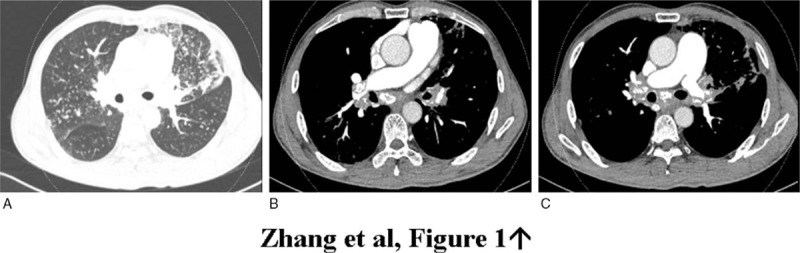
(A) Partially fibrotic lungs with a disseminated nodular pattern observed in the computer tomography pulmonary angiography. (B) Enlarged and calcified right hilar lymph nodes encompassed proximal upper right pulmonary artery, causing abrupt stenosis. (C) Enlarged and calcified left hilar lymph nodes encompassed proximal lower left pulmonary artery, causing abrupt stenosis.
Pulmonary angiograms were performed to observe the stenosis of pulmonary artery. A 6F guiding catheter and 5F pigtail catheter were advanced into the pulmonary artery through the right femoral vein. The angiograms showed severe stenosis of the proximal upper right and lower left pulmonary artery. Moderate stenosis occured in a branch of the lower right pulmonary artery and a branch of the upper left pulmonary artery. In the upper right pulmonary artery, predilation of the stenotic lesion with 4 × 20 mm balloon was performed, then a 5 × 15 mm stent was placed. In the lower left pulmonary artery, predilation of the stenotic lesion with 2 balloons (4 × 40 mm and 7 × 40 mm) was performed, whereafter, a 8 × 40 mm stent was deployed. The branch of the upper left pulmonary artery stenosis was expanded repeatedly by using 7 × 40 mm balloon. The final angiograms showed a significantly increased pulmonary artery caliber of the lesion and normal distal flow (Fig. 2).
Figure 2.
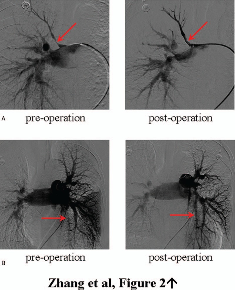
(A) The preoperation angiograms showed severe stenosis of the proximal upper right and lower left pulmonary artery. (B) The postoperation angiograms showed a significantly increased pulmonary artery caliber and normal distal flow.
Anticoagulation was administered after stent implantation. The clinical symptom of the patient was much improved in the fourth day and the 6-minute walk distance was 450 m 1 week later. The patient was discharged without adverse events.
3. Discussion
Exposure to silica promotes the development of several diseases such as silicosis, lung cancer, and chronic obstructive pulmonary diseases.[2,6–7] Inhaled silica particles are engulfed by macrophages and release intracellular silica. This macrophage activation produces cytokines such as IL-1β, IL-6, TNF-α, oxidants and nitrogen species, and the development of cellular events contributing to pulmonary fibrosis.[8] Meanwhile, these inflammatory mediators may produce vascular injury. Some research in silica-exposed mice had showed signs of vascular remodeling with pulmonary artery muscularization, vascular occlusion, and medial thickening. The upregulated expression of proinflammatory genes such as TNF-α and MCP-1 and attenuated expression of vasculature-specific genes involved in the regulation of endothelial function showed partial mechanistic link between silicosis and pulmonary hypertension.[9]
In a previous study, some investigations demonstrated that pulmonary hypertension in patients with silicosis was linked with poorer prognosis. They found some risk factors related to the death rate and length of survival for these patients, including the mean pulmonary artery pressure, pressure gradient across pulmonary vascular bed, pulmonary vascular resistance, arterial oxygen saturation, and forced expiratory volume. Moreover, 2 main risk factors determining the prognosis has been identified by the method of discriminating analysis: forced expiratory volume and pulmonary artery pressure.[10]
Usually, the fibrotic change in silicosis is associated with a high incidence of pulmonary hypertension and this kind of silicosis associated pulmonary hypertension could been categorized into the pulmonary hypertension because of lung diseases and/or hypoxia-related group in the current classification of pulmonary hypertension.[11] The severity of pulmonary hypertension is usually poorly associated with the severity of the underlying lung disease and the most common indicators in these patients are a disproportionally low DLCO and a low pCO2.[12] In this case, the right heart catheterization showed severe pulmonary arterial hypertension but the lung function test showed mild diffusing disfunction. Meanwhile, the clinical symptoms and 6-minute walking test improved significantly following stent implantation and balloon dilatation. We think it is because of multifactorial mechanisms, including pulmonary artery stenosis and hypoxia associated with fibrosis and vascular injury.
Only a few investigators have reported pulmonary hypertension stenosis because of enlarged hilar lymph nodes, in some patients with anthracosis, sarcoidosis, and fibrosing mediastinitis (FM).[13–15] Hilar lymphadenopathy as a single finding in silicosis has been described previously, but these changes had not caused pulmonary hypertension and clinical symptoms.[16] We suspect the pulmonary artery stenosis of this patient is because of the dense, often calcified hilar lymphnodes which cause severe distortion of normal tissue architecture.
Pulmonary artery stenting and/or balloon angioplasty are usually performed in cardiac catheterization laboratories treating children with congenital heart disease. In adults, literature focuses on percutaneous stenting of stenotic pulmonary artery as an effective treatment option for several other conditions, such as anthracosis and FM.[14,15] However, percutaneous treatment for silicosis-induced pulmonary artery stenosis has not previously been reported. Because of its rarity, there is no general recommendation for treatment of this condition.
Indeed, although percutaneous stenting and balloon angioplasty treatment are now preferred to relieve vascular obstruction in many diseases, complications must be aware. A multiinstitutional prospective registry research for congenital cardiac catheterization project which performed balloon angioplasty and stenting of branch pulmonary arteries showed the complications, including vascular/cardiac trauma, stent malposition/embolization, arrhythmias, hemodynamic, bleeding via endotracheal tube/reperfusion injury, and others.[17] A retrospective review of FM patients undergoing cardiac catheterization was conducted in 58 patients which been placed 24 stents in 17 right pulmonary arteries and 11 stents in 9 left pulmonary arteries. They found complications of intravascular stent placement including vessel injury and stent malposition may occur more frequently in patients with FM because of normal tissue architecture surrounding with dense, often calcified compressing mass can cause severe distortion.[15] We think it is similar to our patient.
The results of our patient encouraged us that percutaneous treatment can relief pulmonary artery caliber and clinical symptom. However, the patient was followed up for a short period and long-term outcomes have not yet been sufficiently evaluated. There is limited data on long-term outcome for stent implantation as treatment for large vessel obstruction. Some studies for FM and congenital heart disease patients which evaluated more than 5 years follow-up data showed these treatments can provide not only significant reduction in pressure gradients and effective improvement in vessel caliber at areas of stenosis but also sustained symptomatic relief in most patients. These reports also suggest low rates of symptomatic recurrent stenosis requiring further intervention. Recurrent obstruction in these cases was primarily related to neointimal proliferation, with or without progression of tissue growth distal to the stent.[15,18,19]
Surgical treatment may offer an alternative therapeutic option. In some limited experience, operative procedures were performed to bypass or reconstruct an obstructed pulmonary artery. Indeed, the dense, fibrosis, and calcification could cause the surgical morbidity and mortality, and limit the surgical options for direct repair. The surgeon recommended that for severely obstructive pulmonary artery, hybrid techniques of both concomitant or delayed conduit placement operation and stenting should be considered for these difficult patients.[20]
As far as we know, medical therapies have not yet been shown effective for these groups’ pulmonary hypertension.
4. Conclusion
To our knowledge, this is the first reported case of percutaneous treatment for silicosis-induced PA stenosis and pulmonary hypertension. The clinical symptoms, 6-minute walking test, and vessel caliber at areas of stenosis improved significantly following stent implantation and balloon dilatation. However, the patient was followed up for a short period and long-term outcomes have not yet been sufficiently evaluated.
Footnotes
Abbreviations: CT = computed tomography, CTPA = computer tomography pulmonary angiography, DLCO = carbon monoxide diffusing capacity, FM = fibrosing mediastinitis, HRCT = high resolution computed tomography.
Ethical approval was given by the ethics committee of Sir Run Run Shaw Hospital, medical school of Zhejiang University and the informed consent was given by the patient.
This study was supported by research grant 81570043 from the National Natural Science Foundation of China, the research grant 2016A67 from the Health Science and Technology Plan of Hangzhou City in China, the research grant LQ18H010001 from the Nature Science Foundation of Zhejiang Province in China; the research grant 2017KY415 from the Medical Science and Technology Foundation of Zhejiang Province in China; the research grant 2016YFC0901502 from the National Key Research and Development Program of China.
The authors report no conflicts of interest.
References
- [1].Hughes JM, Weill H, Checkoway H, et al. Radiographic evidence of silicosis risk in the diatomaceous earth industry. Am J Respir Crit Care Med 1998;158:807–14. [DOI] [PubMed] [Google Scholar]
- [2].Castranova V, Vallyathan V. Silicosis and coal workers’ pneumoconiosis. Environ Health Perspect 2000;108(Suppl 4):675–84. [DOI] [PMC free article] [PubMed] [Google Scholar]
- [3].Bessis L, Callard P, Gotheil C, et al. High-resolution CT of parenchymal lung disease: precise correlation with histologic findings. Radiographics 1992;12:45–58. [DOI] [PubMed] [Google Scholar]
- [4].Nemmar A, Vanbilloen H, Hoylaerts MF, et al. Passage of intratracheally instilled ultrafine particles from the lung into the systemic circulation in hamster. Am J Respir Crit Care Med 2001;164:1665–8. [DOI] [PubMed] [Google Scholar]
- [5].Seaton A, MacNee W, Donaldson K, et al. Particulate air pollution and acute health effects. Lancet 1995;345:176–8. [DOI] [PubMed] [Google Scholar]
- [6].Liu Y, Steenland K, Rong Y, et al. Exposure-response analysis and risk assessment for lung cancer in relationship to silica exposure: a 44-year cohort study of 34,018 workers. Am J Epidemiol 2013;178:1424–33. [DOI] [PMC free article] [PubMed] [Google Scholar]
- [7].Cullinan P. Occupation and chronic obstructive pulmonary disease (COPD). Br Med Bull 2012;104:143–61. [DOI] [PubMed] [Google Scholar]
- [8].Mossman BT, Churg A. Mechanisms in the pathogenesis of asbestosis and silicosis. Am J Respir Crit Care Med 1998;157:1666–80. [DOI] [PubMed] [Google Scholar]
- [9].Zelko IN, Zhu J, Ritzenthaler JD, et al. Pulmonary hypertension and vascular remodeling in mice exposed to crystalline silica. Respir Res 2016;17:160. [DOI] [PMC free article] [PubMed] [Google Scholar]
- [10].Jandova R, Widimsky J, Eisler L, et al. Long-term prognosis of pulmonary hypertension in silicosis. Cor Vasa 1980;22:221–37. [PubMed] [Google Scholar]
- [11].Galiè N, Humbert M, Vachiery JL, et al. 2015 ESC/ERS Guidelines for the diagnosis and treatment of pulmonary hypertension. Rev Esp Cardiol (Engl Ed) 2016;69:177. [DOI] [PubMed] [Google Scholar]
- [12].Chaouat A, Bugnet AS, Kadaoui N, et al. Severe pulmonary hypertension and chronic obstructive pulmonary disease. Am J Respir Crit Care Med 2005;172:189–94. [DOI] [PubMed] [Google Scholar]
- [13].Ahn JH, Ahn JM, Lee SW, et al. Percutaneous treatment for pulmonary hypertension caused by pulmonary artery stenosis due to anthracosis. Int J Tuberc Lung Dis 2015;19:747–8. [DOI] [PubMed] [Google Scholar]
- [14].Honda T, Hayasaka M, Hachiya T, et al. Unilateral hilar lymphadenopathy of sarcoidosis or sarcoid reaction compressing the trunk of the right pulmonary artery. Respiration 2000;67:330–2. [DOI] [PubMed] [Google Scholar]
- [15].Albers EL, Pugh ME, Hill KD, et al. Percutaneous vascular stent implantation as treatment for central vascular obstruction due to fibrosing mediastinitis. Circulation 2011;123:1391–9. [DOI] [PMC free article] [PubMed] [Google Scholar]
- [16].Baldwin DR, Lambert L, Pantin CF, et al. Silicosis presenting as bilateral hilar lymphadenopathy. Thorax 1996;51:1165–7. [DOI] [PMC free article] [PubMed] [Google Scholar]
- [17].Holzer RJ, Gauvreau K, Kreutzer J, et al. Balloon angioplasty and stenting of branch pulmonary arteries: adverse events and procedural characteristics: results of a multi-institutional registry. Circ Cardiovasc Interv 2011;4:287–96. [DOI] [PubMed] [Google Scholar]
- [18].Law MA, Shamszad P, Nugent AW, et al. Pulmonary artery stents: long-term follow-up. Catheter Cardiovasc Interv 2010;75:757–64. [DOI] [PubMed] [Google Scholar]
- [19].Stanfill R, Nykanen DG, Osorio S, et al. Stent implantation is effective treatment of vascular stenosis in young infants with congenital heart disease: acute implantation and long-term follow-up results. Catheter Cardiovasc Interv 2008;71:831–41. [DOI] [PubMed] [Google Scholar]
- [20].Brown ML, Cedeño AR, Edell ES, et al. Operative strategies for pulmonary artery occlusion secondary to mediastinal fibrosis. Ann Thorac Surg 2009;88:233–7. [DOI] [PubMed] [Google Scholar]