Traditional flexible endoscopes, which have been in use since the 1950s, rely on rear-push mechanical actuation to advance through the gastrointestinal tract. This necessitates a semi-rigid insertion tube to prevent buckling that may induce patient discomfort or trauma due to tissue stress [1] [2]. To overcome this limitation, magnetic fields have been utilized for endoscope actuation. Unfortunately, manual operation of magnetic actuation is not intuitive and therefore computer assistance has been shown to be beneficial [3]. The use of computers and robotics facilitates autonomy, which may be used to assist the operator during repetitive or complex maneuvers through relief of cognitive burden and potential learning curve reduction.
In our academic laboratory, we have developed a highly compliant magnetic flexible endoscope (MFE) platform (with diagnostic and therapeutic capability) that relies on actuation using an actuating permanent magnet (APM) manipulated by a robot that is external to the patient—thus it does not require push-actuation. Using proprioceptive sensing and software algorithms, we are able to control MFE motion and enact autonomous function.
Within endoscopy, colonoscopy is ripe for autonomous control due to the repetitive nature of some maneuvers and the skill/experience necessary to achieve excellent technique. We focus our demonstration of autonomy on retroflexion as it is a common endoscopic maneuver that is skill-intensive, repetitive, and technically challenging when using magnetic actuation. The ability to safely retroflex the MFE in any area of the colon may potentially increase polyp detection and reduce the incidence of colorectal cancer [4].
Our team has developed an autonomous control algorithm for MFE retroflexion. We conducted 30 autonomous retroflexions in-vivo in a 40 Kg female Yorkshire-Landrace cross swine (Supplemental Material: Retroflexion study design). All of the autonomous retroflexion maneuvers were successful (100%; n=30) with a mean maneuver time of 11.3±2.4 seconds. The visible difference in trajectories and difference in APM position respective to the starting point indicate that the APM did not follow a pre-computed trajectory—but was instead autonomously reacting to external input, in this case the MFE’s motion (Video). All of the trials in this study were completed without tissue perforation or trauma (gross or microscopic), or premature animal demise during the trial. The study was approved by the local Institutional Animal Care and Use Committee (IACUC).
In addition, feasibility of the diagnostic capabilities of the MFE was assessed through a series of preliminary experiments on lesion detection and lesion-targeting (Supplemental Material: Lesion detection and targeting study design). The mean lesion detection miss rate for the MFE was 21.7% (completion time 575 s) compared with a miss rate of 5% (completion time 257 s) for the traditional endoscope (p=0.17). For the lesion targeting experiments, all lesions were successfully “tagged” with biopsy forceps using the MFE and the traditional endoscope (time: 251 s v. 32 s; p<0.01). Despite the differences in time between the MFE and traditional endoscope, likely impacted by endoscopist familiarity with traditional endoscopy, the low polyp miss rate and ability to circumferentially examine the colon lumen suggest promise for continued development and refinement of the MFE.
Description of technology
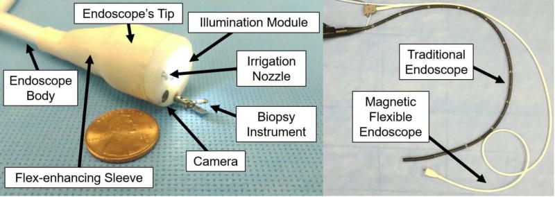
The MFE system, Figure 1, consists of a flexible endoscope with a magnet-embedded tip, an APM external to the patient that is manipulated by a robot, and a software control system that is described in detail in Ref. [5]. The MFE (Figure 2) maintains functionality of a traditional endoscope (i.e. therapeutic channel, illumination, viewing, irrigation, suction, lens cleansing, and insufflation) and contains proprioceptive sensors that facilitate magnetic interaction estimation harnessed for MFE retroflexion. Knowledge of magnetic field properties allows for precise device movement all while maintaining an applied tissue stress of no more than 0.25 bar, or 12 times less pressure than is necessary to induce tissue damage [5][6].
Figure 1.
The MFE platform consists of a magnet-embedded custom endoscope, a serial robot with an APM mounted at its end-effector, and control software. The system is shown during an in-vivo trial.
Figure 2.
The compliant MFE contains a camera, illumination module, therapeutic channel, an irrigation and insufflation channel, and a flexible sleeve joint that joins the endoscope’s tip to its body (left).
Video description
The video presents an overall description of the MFE platform and functions followed by demonstration of in-vivo autonomous retroflexion in real time. The robot arm with APM is shown in the upper right of the screen during the demonstration and the corresponding footage of the MFE is shown in the frame below where perspective footage is obtained via an auxiliary endoscope. Furthermore, we demonstrate unique MFE trajectories during retroflexion—a product of the use of autonomy, the use of biopsy forceps during MFE retroflexion, as well as in-vivo use of therapeutic tools as operated from the MFE.
Take home message
Our team has demonstrated the first use of in-vivo autonomous control for completion of an endoscopic maneuver in a reliable, efficient, and safe manner. This is also the first study to demonstrate closed-loop magnetic control of a device in-vivo and autonomous maneuvering of an endoscope that has the clinical capability of a traditional flexible endoscope.
We expect the cost of the MFE to be approximately $1000 USD with a one-time cost of $40,000 USD for the actuating robot. Although we have developed the technology for use in the colon, we anticipate that the platform, once further miniaturized, can be used in the upper GI tract and, in general, other areas of the body where there is physical space for maneuvering. In summary, our findings suggest promise in the use of autonomy to assist in endoscopic tasks and in magnetic actuation of endoscopic devices/instruments.
Supplementary Material
Acknowledgments
Grant Support: This research was supported by the National Institute of Biomedical Imaging and Bioengineering, USA of the National Institutes of Health under award no. R01EB018992, by the National Science Foundation, USA under grant no. CNS-1239355 and no. IIS-1453129, by the National Science Foundation Graduate Research Fellowship Program under grant no. 1445197, by the Royal Society, UK, and by the Engineering and Physical Sciences Research Council, UK. Any opinions, findings, conclusions, or recommendations expressed in this material are those of the authors and do not necessarily reflect the views of the National Institutes of Health, the National Science Foundation, the Royal Society, or the Engineering and Physical Sciences Research Council
Acronyms
- ASGE
American Society for Gastrointestinal Endoscopy
- MFE
Magnetic Flexible Endoscope
- EM
Embedded Magnet
- APM
Actuating Permanent Magnet
- IACUC
Institutional Animal Care and Use Committee
Footnotes
Publisher's Disclaimer: This is a PDF file of an unedited manuscript that has been accepted for publication. As a service to our customers we are providing this early version of the manuscript. The manuscript will undergo copyediting, typesetting, and review of the resulting proof before it is published in its final citable form. Please note that during the production process errors may be discovered which could affect the content, and all legal disclaimers that apply to the journal pertain.
Location where work was performed: Department of Mechanical Engineering, Vanderbilt University, Nashville, TN, USA
Disclosures: The authors (Piotr R. Slawinski, Addisu Z. Taddese, Pietro Valdastri, Keith L. Obstein) have submitted an invention disclosure to Vanderbilt University.
Kyle B. Musto has nothing to disclose.
Shabnam Sarker has nothing to disclose.
Conflicts of Interest:
Piotr R. Slawinski has no conflicts of interest.
Addisu Z. Taddese has no conflicts of interest.
Kyle B. Musto has no conflicts of interest.
Shabnam Sarker has no conflicts of interest.
Pietro Valdastri has no conflicts of interest.
Keith L. Obstein has no conflicts of interest.
Writing Assistance: None
Author Contributions:
Piotr Slawinski: Study concept and design, application and technology development, acquisition of data, analysis and interpretation of data, drafting the manuscript, critical revision of the manuscript for important intellectual content
Addisu Taddese: Study concept and design, application and technology development, acquisition of data, analysis and interpretation of data, critical revision of the manuscript for important intellectual content
Kyle Musto: Application and technology development, critical revision of the manuscript for important intellectual content
Shabnam Sarker: Study concept and design, acquisition of data, critical revision of the manuscript for important intellectual content
Pietro Valdastri: Study concept and design, application and technology development, analysis and interpretation of data, critical revision of the manuscript for important intellectual content
Keith Obstein: Study concept and design, application and technology development, analysis and interpretation of data, drafting of the manuscript, critical revision of the manuscript for important intellectual content, study supervision
References
- 1.Obstein KL, et al. World J Gastroenterol. 2013;19(4):431–439. doi: 10.3748/wjg.v19.i4.431. [DOI] [PMC free article] [PubMed] [Google Scholar]
- 2.Sliker LJ, et al. Expert Review of Medical Devices. 2014;11(6):649–666. doi: 10.1586/17434440.2014.941809. [DOI] [PubMed] [Google Scholar]
- 3.Ciuti G, et al. Endoscopy. 2010;42(2):148–152. doi: 10.1055/s-0029-1243808. [DOI] [PubMed] [Google Scholar]
- 4.Cohen J, et al. J Clin Gastroenterol. 2016;85(5):AB525. [Google Scholar]
- 5.Slawinski PR, et al. IEEE Robotics and Automation Letters. 2017;2(3):1352–1360. doi: 10.1109/LRA.2017.2668459. [DOI] [PMC free article] [PubMed] [Google Scholar]
- 6.Moshkowitz M, et al. Endoscopy. 2010;42(10):834–836. doi: 10.1055/s-0030-1255777. [DOI] [PubMed] [Google Scholar]
Associated Data
This section collects any data citations, data availability statements, or supplementary materials included in this article.


