Abstract
miR-9 and miR-9* (miR-9/9*) were first shown to be expressed in the nervous system and to function as versatile regulators of neurogenesis. The variable expression levels of miR-9/9* in human cancer prompted researchers to investigate whether these small RNAs may also have an important role in the deregulation of physiological and biochemical networks in human disease. In this review, we present a comprehensive overview of the involvement of miR-9/9* in various human malignancies focusing on their opposing roles in supporting or suppressing tumor development and metastasis. Importantly, it is shown that the capacity of miR-9/9* to impact tumor formation is independent from their influence on the metastatic potential of tumor cells. Moreover, data suggest that miR-9/9* may increase malignancy of one cancer cell population at the expense of another. The functional versatility of miR-9/9* emphasizes the complexity of studying miRNA function and the importance to perform functional studies of both miRNA strands in a relevant cellular context. The possible application of miR-9/9* as targets for miRNA-based therapies is discussed, emphasizing the need to obtain a better understanding of the functional properties of these miRNAs and to develop safe delivery methods to target specific cell populations.
Keywords: miRNA, miR-9, miR-9*, human cancer, miRNA-based therapies
INTRODUCTION
MiRNAs are short non-coding RNAs that by binding to target mRNAs decrease protein levels and in this way regulate crucial cellular processes. [1–3] miRNA transcripts are expressed as hairpin-like precursor structures that undergo stepwise maturation into double-stranded miRNA/miRNA* duplexes. In the past, it was proposed that one of the strands, called the mature miRNA, is stabilized and becomes functional, whereas another, referred to as the passenger strand or miRNA*, is degraded. Recently, it has been shown that miRNA*s can also display functionality and play complementary roles to their related miRNAs. [4–6]
miR-9 (miR-9-5p) and miR-9* (miR-9-3p) are two miRNAs that originate from the same precursor and are highly conserved during evolution from flies to humans. [7] All vertebrate miR-9/9* orthologs have an identical mature sequence. In mammals, miR-9/9* are encoded by three genes: MIR9-1, MIR9-2 and MIR9-3. In humans, these genes are located on the chromosomes 1 (1q22), 5 (5q14.3) and 15 (15q26.1), respectively. miR-9/9* are mainly expressed in the nervous system and were initially studied as regulators of neurogenesis. [8] Interestingly, aberrant expression of miR-9/9* has been found in various types of human cancer revealing an unanticipated functional versatility. [9–11] The high level of sequence conservation and the fact that miR-9/9* are encoded by three different genomic loci points to important functional roles of these miRNAs that may be exploited by cancer cells.
In the past years, several studies have reported on the relationship of miR-9/9* expression with different cellular processes, such as differentiation, proliferation, migration and metastasis. [11–14] Interestingly, miR-9 and miR-9*, although concomitantly expressed from one precursor miRNA, may be preferentially retained and can play synergistic or opposite roles within one malignancy. [15–17] Here, we summarize the diverse functions of miR-9/9* in the biology of human cancer. We outline the mechanisms through which miR-9/9* are involved in tumorigenesis and the cellular context in which these miRNAs operate. Although most of the reported findings still need validation under physiological (in vivo) conditions, they underscore the complexity of miRNA functionality within the heterogeneous population of cancer cells. This review may serve as the basis for a broader dispute about the often counteracting functions of a particular miRNA in the pathobiology of human cancer and their implications for future treatment opportunities.
GLIOBLASTOMA MULTIFORME
Glioblastoma multiforme (GBM; grade IV astrocytoma) is the most common and aggressive brain tumor. [18] It has been proposed that GBM originates from the cancer cell population with stem cell-like properties that is characterized by CD133 expression. [19] GBM can be divided into clinically and genetically distinct groups based on the similarity of miRNA and mRNA expression signatures to different neural precursor cell types: radial glia, oligoneuronal precursors, neuronal precursors, neuroepithelial/neural crest precursors or astrocyte precursors. [20]
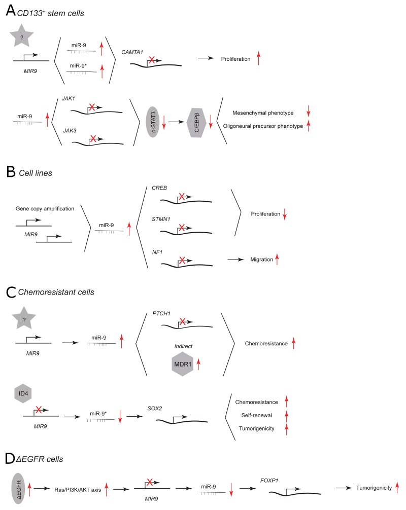
In CD133+ GBM stem cells, miR-9/9* are highly expressed and needed for stem cell renewal. [17] Inhibition of miR-9 as well as miR-9* using 2’-O-methylated antisense inhibitors results in reduced colony numbers (Figure 1A). Both miRNAs directly target a tumor suppressor calmodulin binding transcription activator 1 (CAMTA1), of which overexpression mimics the phenotype of miR-9/9* inhibition. Additionally, R28 GBM cells that overexpress CAMTA1 form smaller tumors in vivo than control cells.
Figure 1. miR-9 and miR-9* functions in human glioblastoma multiforme.
Each graph schematically depicts the reported levels of expression of miR-9/9* as well as their functional significance including relevant target genes and phenotypical effects in (A) CD133+ stem cells, (B) glioblastoma cell lines, (C) chemoresistant glioblastoma cells, (D) ΔEGFR cells.
The highest expression of miR-9 has been found in the oligoneural subclass of GBM. [20] miR-9 is considered a regulator of a subtype-specific gene expression network and drives subtype-specific cell decisions. [20] Overexpression of miR-9 using a mimic in CD133+ GBM stem cells promotes oligoneural and suppresses a more aggressive mesenchymal phenotype by downregulating expression of Janus kinases (JAK1 and JAK3), inhibiting activation of signal transducer and activator of transcription 3 (STAT3) and decreasing expression of the STAT3 transcriptional target CCAAT/enhancer-binding protein β (C/EBPβ) (Figure 1A). [20, 21]
In GBM cell lines, miR-9 has been reported to play a critical role in determination of the so-called “go or grow” phenotype. [13] miR-9 is part of a feedback minicircuitry that allows a tight control of the expression levels of target genes that coordinate the proliferation and migration of GBM cells (Figure 1B). In contrast to increasing colony numbers of CD133+ GBM stem cells via CAMTA1, miR-9 has been shown to inhibit proliferation of GBM cell lines by targeting the cyclic AMP response element-binding protein (CREB) but to promote migration by targeting neurofibromin 1 (NF1). Additionally, the transcription of both miR-9 and NF1 is under CREB’s control. Gene copy amplification of miR-9 hinders the balance of this regulatory minicircuitry and contributes to motility of GBM cells. Another miR-9 target that contributes to reduced proliferation and tumor growth is stathmin (STMN1), which regulates microtubule formation dynamics during cell-cycle progression. [22, 23] U87MG GBM cells transfected with miR-9 mimic are characterized by decreased expression of STMN1 and form smaller tumors than control cells.
In GBM cells that are resistant against alkylating agents, miR-9 is highly expressed and miR-9* is downregulated. [15, 16, 24, 25] miR-9 has been shown to contribute to the chemoresistance of GBM cells by direct targeting of patched homolog 1 protein (PTCH1) and subsequent activation of sonic hedgehog (SHH) signaling pathway (Figure 1C). [25] Additionally, the delivery of anti-miR-9 to the resistant GBM cells indirectly downregulates the expression of the multidrug transporter (MDR1) and sensitizes the GBM cells to chemotherapy. [15] miR-9* is part of an ID4-miR-9*-SOX2-ABCC3/ABCC6 regulatory pathway. [16] Inhibitor of differentiation 4 (ID4) suppresses miR-9* expression and upregulates the direct target of this miRNA SRY (sex determining region Y)-box 2 (SOX2). SOX2 is highly expressed in patients with GBM. [26] Its upregulation leads to increased chemoresistance, self-renewal and tumorigenicity of GBM cell lines and patient-derived CD133+ GBM stem cells. [16]
40% to 50% of primary GBM cases exhibit epidermal growth factor receptor (EGFR) amplification, overexpression, and/or mutations. [27] An EGFR mutant that lacks exons 2-7 (ΔEGFR) is constitutively active and present in a high proportion of GBM cases with EGFR amplification. This EGFR mutant confers a strong tumor-enhancing effect by promoting growth, cell invasion and chemoresistance. [28–30] In GBM cells that express ΔEGFR, miR-9 acts as a tumor suppressor that downregulates transcription factor forkhead box P1 (FOXP1) (Figure 1D). [31] Viral overexpression of miR-9 or silencing of FOXP1 antagonizes ΔEGFR-dependent tumor growth in vivo. ΔEGFR activates Ras/PI3K/AKT, which in turn suppresses miR-9. Of note, the viral transduction as used here likely results in overexpression of both miR-9 and miR-9* making it difficult to discern whether both or only a single miRNA display activity. However, as the presented outcome is in line with the previously mentioned reports concerning the function of miR-9* in chemoresistant GBM cells the expression of miR-9* and its influence on tumorigenicity of ΔEGFR GBM cells needs to be further investigated. [16]
BREAST CANCER
Breast cancer (BC) is a heterogeneous malignancy that can be classified by estrogen receptor (ESR1) expression (ER+), human epidermal growth factor receptor 2 (ERBB2) expression (HER2+), the absence of ESR1, ERBB2 and the progesterone receptor in triple-negative breast cancer (TNBC) or the expression of driver oncogenes (e.g. MYC). [32–35] A vast amount of data concerning the diverse roles of miR-9/9* have been obtained for breast cancer.
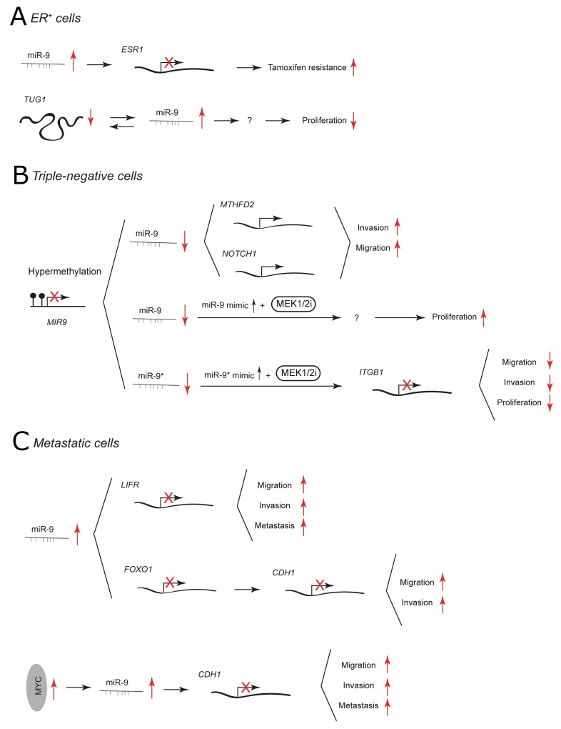
Because of the availability of endocrine-targeted therapy (e.g. tamoxifen treatment), patients with BC that express ER have better prognosis. [36] Nonetheless, therapeutic resistance eventually occurs in a large number of cases. In the ER+ MCF-7 cell line, miR-9 has been shown to directly target ER and to influence, not only ER signaling but also other steroid receptor pathways (Figure 2A). [37] miR-9 levels are reduced in most of ER+ BC cases compared to ER-. However, when upregulated it is associated with worse patient outcome and its viral overexpression in MCF-7 cells contributes to tamoxifen resistance. [37, 38] The expression of miR-9 in ER+ BC has recently been linked to the level of lncRNA taurine-upregulated gene 1 (TUG1). It has been proposed that TUG1 and miR-9 may co-regulate each other to impact cell proliferation [39].
Figure 2. miR-9 and miR-9* functions in human breast cancer.
Each graph schematically depicts the reported levels of expression of miR-9/9* as well as their functional significance including relevant target genes and phenotypical effects in (A) ER+ cells, (B) triple-negative cells, (C) metastatic cells.
In TNBC cells, miR-9/9* are expressed at low levels due to promoter hypermethylation of the MIR-9 loci. [40] miR-9 has been suggested to play a tumor suppressive role by targeting mitochondrial bifunctional enzyme MTHFD2 and NOTCH1 receptor (Figure 2B). [14, 41] Overexpression using pre-miR-9 or lentiviral constructs decreases the invasiveness and migration of TNBC MDA-MB-231 cells. [14, 41] In line with this, knockdown of MTHFD2 recapitulates the anti-invasive effect of miR-9. NOTCH1 is known to be involved in the pathogenesis of TNBC and its inhibition reduces the migratory potential of MDA-MB-231 cells. [42, 43] Interestingly, the downregulation of NOTCH1 with γ-secretase inhibitors in ER+ MCF-7 cell line stimulates migration in vitro and promotes tumor growth in vivo. [43] Recently, it has been reported that miR-9 may influence TNBC aggressiveness by taking part in cross-talk between cancer cells and cancer-associated fibroblasts [44].
Mitogen-activated protein kinase enzymes 1 and 2 (MEK1/2) inhibitors have been used in cancer therapy but can become ineffective due to acquired drug resistance. [45] In TNBC cells, treatment with a MEK1/2 inhibitor together with a miR-9 mimic increases cell proliferation, whereas treatment together with a miR-9* mimic suppresses growth, migration and invasion of tumor cells (Figure 2B). [40] miR-9* activity is mediated through downregulation of β1 integrin(ITGB1), which is important for growth factor receptor and extracellular matrix-related signaling.
The expression of miR-9 has been widely related to BC metastasis. In non-metastatic SUM159 cells, miR-9-mediated downregulation of leukemia inhibitory factor receptor (LIFR) induces migration, invasion and metastatic colonization through deregulation of the Hippo-YAP pathway. [46] Additionally, miR-9 has been reported to be higher expressed in metastatic than in non-metastatic primary human breast cancer. In MCF-7 and MDA-MB-231 cells, miR-9 has been shown to downregulate the expression of another tumor suppressor gene FOXO1 that belongs to the FOXO family of Forkhead transcription factors. [47] FOXO1 3’ UTR may sequester miR-9 from E-cadherin 3’ UTR. Overexpression of FOXO1 leads to upregulation of E-cadherin and decreases the migration and invasiveness of BC cell lines. In 2010, Ma et al. reported that miR-9 plays an important role in metastasis of MYC-driven breast tumors. [11] MYC oncoprotein activates miR-9 expression, which consequently causes downregulation of miR-9 direct target E-cadherin (Figure 2C). This leads to increased cell motility and invasiveness of BC cells in vitro. E-cadherin is an epithelial cell adhesion molecule that forms the core of adherens junctions between adjacent epithelial cells and its inactivation enables dissociation of carcinoma cells. [48] By targeting E-cadherin in breast tumor cells, miR-9 enables non-metastatic cells to form pulmonary micrometastasis. [11] In summary, the data show that in BC miR-9 can target two alternative metastatic suppressors: LIFR (which activates Hippo signaling, leading to inactivation of the transcriptional co-activator YAP) and E-cadherin (that maintains adherens junctions) [11, 46].
CERVICAL CANCER
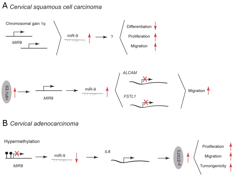
Cervical cancer can be classified into two prevailing subtypes: cervical squamous cell carcinoma (CSCC; about 80% of cases) and cervical adenocarcinoma (CA; about 5-20% of cases). [49] In CSCC, a chromosomal gain of 1q results in upregulation of miR-9 (1q23.3) and is linked with malignant progression (Figure 3A). [50] Overexpression of miR-9 in normal keratinocytes blocks epithelial differentiation, and induces proliferation and migration. Beside chromosomal gain, an elevated expression of miR-9 in CSCC is caused by human papillomavirus (HPV) infection (Figure 3A). [51] miR-9 expression is activated by HPV E6 – an essential oncogene in cervical cancer development. In normal keratinocytes, overexpression of HPV E6 and miR-9 leads to downregulation of miR-9 target genes involved in cell migration, such as activated leukocyte cell adhesion molecule (ALCAM) and follistatin-related protein 1 (FSTL1). [51–53] This leads to increase in cell motility [51].
Figure 3. miR-9 and miR-9* functions in human cervical cancer.
Each graph schematically depicts the reported levels of expression of miR-9/9* as well as their functional significance including relevant target genes and phenotypical effects in (A) cervical squamous cell carcinoma cells, (B) cervical adenocarcinoma cells.
In CA, miR-9 is downregulated due to frequent promoter-hypermethylation and has been shown to act as a tumor suppressor (Figure 3B). [54] Ectopic expression of miR-9 inhibits the JAK/STAT3 pathway by targeting interleukin 6 (IL-6). This results in decreased proliferation and migration of HeLa cells in vitro and reduced tumor growth in vivo. IL-6 is highly expressed in human cervical cancer promoting tumorigenesis by activation of the JAK/STAT3 pathway, subsequent upregulation of vascular endothelial growth factor (VEGF) and increased angiogenesis [55].
SQUAMOUS CELL CARCINOMA OF SKIN AND ORAL CAVITY
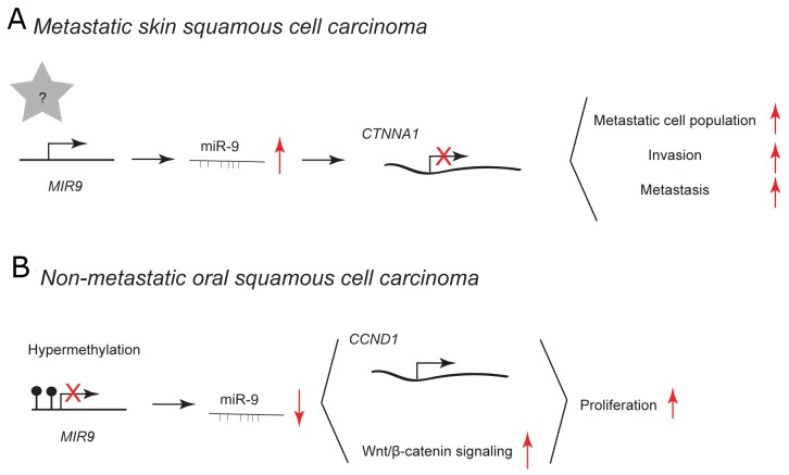
Squamous cell carcinoma (SCC) is a type of cancer that develops from squamous epithelial cells in diverse tissues, e.g. within skin and oral cavity. Cells of skin epithelium undergo constant self-renewal throughout life, therefore it is believed that SCC originates from keratin 15-expressing stem cells (K15+) that harbor pro-proliferative mutations in KrasG12D. [56] Additional deletion of Smad4 in these cells leads to the spontaneous development of multi-lineage tumors, including metastatic squamous cell carcinoma. [57, 58] In murine K15.KrasG12D.Smad4–/– cancer stem cell-enriched population, viral overexpression of miR-9 leads to the expansion of metastatic cell population resulting in increased invasion and metastasis (Figure 4A). [58] In primary human SCC cells, high expression of miR-9 correlates with metastasis and the loss of a predicted direct target α-catenin. However, α-catenin depletion alone does not cause SCC metastasis suggesting that additional targets are required for miR-9-mediated effect. [59] miR-9 has been reported to be expressed at high levels in patients with recurrent head and neck SCC [60].
Figure 4. miR-9 and miR-9* functions in human skin and oral cavity squamous cell carcinoma.
Each graph schematically depicts the reported levels of expression of miR-9/9* as well as their functional significance including relevant target genes and phenotypical effects in (A) metastatic skin squamous cell carcinoma cells, (B) non-metastatic oral squamous cell carcinoma cells.
In non-metastatic human oral SCC specimens, miR-9 is downregulated probably due to frequent promoter hypermethylation. [61, 62] Overexpression using miR-9 mimic in human the UM-SCC22A cell line inhibits cell proliferation (Figure 4B). [61] Curcumin has been reported to have growth-suppressive potential in different types of cancer, as well as in oral SCC. [62, 63] In the human SCC-9 cell line, curcumin treatment leads to upregulation of miR-9, which in turn inhibits cell proliferation via downregulation of cyclin D1 and suppression of Wnt/β-catenin signaling (Figure 4B). [62] Cyclin D1 and the Wnt/β-catenin signaling pathway are frequently deregulated in human cancer and may play essential roles in the process of tumorigenesis [64, 65].
HEMATOLOGICAL MALIGNANCIES
Hematopoiesis is a hierarchical differentiation process in which hematopoietic stem cells (HSCs) undergo step-wise maturation into various types of blood cells. [66, 67] During this process, HSCs lose their self-renewal and multi-lineage differentiation capability to give rise to lymphoid and myeloid progeny. Deregulation of normal hematopoiesis may result in development of hematological tumors. [68, 69] Acute and chronic myelogenous leukemia, myelodysplastic syndromes, and myeloproliferative disorders are tumors derived from the myeloid line, whereas lymphomas, lymphocytic leukemias, and myeloma have a lymphoid origin. Hematological malignancies are heterogeneous disorders that are characterized by frequent chromosomal abnormalities, genetic mutations and aberrations in epigenetic regulation. [68, 69]
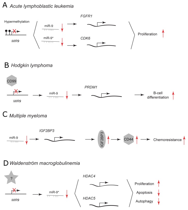
In acute lymphoblastic leukemia (ALL), low miR-9 expression is associated with hypermethylation of MIR9 gene family (Figure 5A). [70] This epigenetic downregulation leads to upregulation of predicted miR-9 and miR-9* targets, fibroblast growth factor receptor 1 (FGFR1) and cyclin-dependent kinase 6 (CDK6). FGFR1 and CDK6 are involved in cell proliferation and survival. [71, 72] Treatment with FGFR1 and CDK6 inhibitors suppresses the proliferation of ALL cells. [70] MIR9 genes have been reported to be also frequently methylated in chronic lymphocytic leukemia (CLL) and overexpression of miR-9 using a mimic decreases CLL cell proliferation. [73]
Figure 5. miR-9 and miR-9* functions in human lymphoid malignancies.
Each graph schematically depicts the reported levels of expression of miR-9/9* as well as their functional significance including relevant target genes and phenotypical effects in (A) acute lymphoblastic leukemia cells, (B) Hodgkin lymphoma cells, (C) multiple myeloma cells, (D) Waldenström macroglobulinemia cells.
CD99 is a transmembrane glycoprotein that is implicated in cell migration, adhesion and differentiation. [74–76] It is expressed at low levels in Hodgkin/Reed-Sternberg (HRS) cells of Hodgkin lymphoma (HL). [77] CD99 downregulates the expression of miR-9 and upregulates a direct miR-9 target: positive regulatory domain 1 (PRDM1/BLIMP-1) (Figure 5B). [10, 77] PRDM1 is the master regulator of terminal B-cell differentiation. miR-9 is highly expressed in HL cells and its downregulation by CD99 overexpression or a direct knockdown using miR-9 inhibitor augments PRDM1 levels that trigger B-cell differentiation into plasma cells. [77] During normal B-cell development within the germinal centers, B cells closely interact with follicular dendritic cells (FDC). [78] Only B cells that bind to these cells survive in the germinal centers and differentiate. It has been shown that direct cell-cell contact between follicular dendritic cells and B cells leads to downregulation of miR-9 and upregulation of PRDM1. This subsequently may promote B-cell differentiation.
In multiple myeloma (MM), insulin-like growth factor 2 mRNA binding protein 3 (IGF2BP3) stabilizes the expression of a cell surface glycoprotein CD44 that is involved in drug resistance of MM cells. [79] Histone deacetylase (HDAC) inhibitors are promising novel chemotherapeutics in MM since they downregulate CD44 expression. HDAC inhibitors treatment leads to upregulation of miR-9 and downregulation of its direct target IGF2BP3 (Figure 5C). Subsequent downregulation of CD44 sensitizes the resistant MM cell to lenalidomide treatment.
miR-9*, has been reported to have a tumor suppressive role in Waldenström macroglobulinemia (WM) (Figure 5D). [80] WM is a B-cell low-grade lymphoma characterized by the accumulation of B cells in the bone marrow. miR-9* is expressed at reduced levels in WM CD19+ cells compared to normal CD19+ counterparts. Its overexpression using pre-miR-9* in WM cells inhibits the unbalanced HDAC activity by downregulation of HDAC4 and 5. This results in decreased proliferation, increased apoptosis and autophagy. Neither adherence to primary BM stromal cells nor growth factors protected against the miR-9*-dependent growth inhibition. Aberrant HDAC activity has been reported to have a tumorigenic effect in many malignancies by influencing the expression of genes controlling cellular proliferation, differentiation and apoptosis [81].
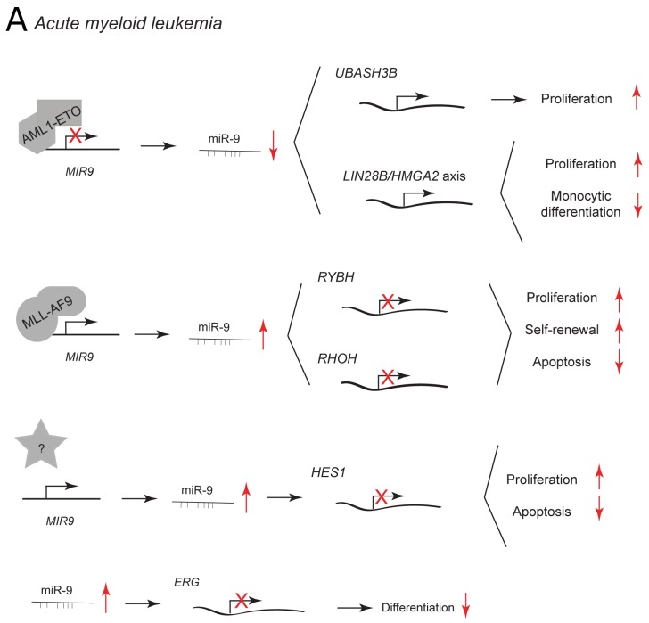
In acute myeloid leukemia (AML), miR-9 has been reported to be differentially expressed between AML subtypes. [12, 82, 83] Dependent on the type of leukemic cell, it may suppress or promote leukemic development. The t(8;21) rearrangement is the most common chromosomal translocation in AML resulting in the formation of AML1-ETO fusion protein. [84] AML1-ETO downregulates miR-9 and in this way promotes the expression of UBASH3B/Sts-1, a tyrosine phosphatase that inhibits CBL and enhances STAT5/AKT/ERK/Src signaling to promote myeloid proliferation (Figure 6A). Ectopic expression of miR-9 in t(8;21) AML cells reduces leukemic growth and enhances monocytic differentiation induced by calcitrol by direct repression of the oncogenic LIN28B/HMGA2 axis. [82] LIN28 and HMGA2 are expressed in undifferentiated proliferating cells during embryogenesis and their upregulation in adult cells leads to oncogenic transformation [85, 86].
Figure 6. miR-9 and miR-9* functions in human myeloid malignancies.
Each graph schematically depicts the reported levels of expression of miR-9/9* as well as their functional significance including relevant target genes and phenotypical effects in (A) acute myeloid leukemia cells.
miR-9 is highly upregulated in MLL-rearranged leukemic cells as compared to non-MLL-rearranged cells and normal controls (Figure 6A). [12, 83] MLL fusion proteins may promote miR-9 expression by direct binding to the promoter regions of MIR9 genes. Knockdown of endogenous miR-9 expression with a miR-9 sponge inhibits MLL fusion–induced immortalization/transformation of normal hematopoietic progenitor cell, whereas its viral overexpression has the opposite effect. miR-9 function may be mediated by the two predicted targets: RING1 and YY1-binding protein (RYBH) and Ras homolog family member H (RHOH). RYBP is a polycomb complex-associated protein that can stabilize p53 and has tumor suppressor activity. [87] RHOH is a member of the Rho GTPase protein family and it can function as an oncogene or tumor suppressor depending on the context [88].
In AML patients with a normal karyotype, miR-9 is expressed at higher levels in leukemic stem/progenitor cells (LSPCs) than in normal hematopoietic stem cells derived from the same patient. [89] Additionally, miR-9 expression is inversely correlated to the levels of hairy and enhancer of split-1 (HES1), a known tumor-suppressor (Figure 6A). [90, 91] Knockdown of miR-9 by lentiviral infection decreases leukemic cell proliferation and survival by increasing HES1 expression in vitro and in vivo [89].
miR-9/9* are both aberrantly upregulated in most of human AML cases. [12] In normal hematopoietic stem and progenitor cells, ectopic expression of miR-9/9* inhibits myeloid differentiation by post-transcriptional regulation of ETS-related gene (ERG) (Figure 6A). ERG is a transcription factor that is essential for definitive hematopoiesis and its functional activity depends on its expression level. [12, 92, 93] In patients with AML, expression of miR-9 has no prognostic significance, whereas miR-9* predicts favorable outcome. [94] Recently, it has been proposed that miR-9* may sensitize tumor cells to chemotherapy in chronic myelogenous leukemia [95].
CONCLUSIONS AND OUTLOOK
Initially discovered as versatile regulators of neurogenesis, miR-9/9* quickly became a focus of attention in cancer research. In the past years, multiple studies have reported on the deregulated expression of miR-9/9* in various types of human cancer and the relation of their aberrant expression levels with different processes, e.g. self-renewal, proliferation and differentiation. Furthermore, these miRNAs have been shown to have important regulatory roles in cancer biology regulating processes such as tumor initiation, tumor progression and chemosensitivity. Table 1 summarizes the different reported functions of miR-9/9* in various cell and tumor types. It also provides information on the up- or downregulation of miR-9/9* and lists putative mRNA targets and target-related pathways according to www.genecards.org. It is evident that miR-9/9* expression affects many biochemical pathways commonly deregulated in human cancer such as the PI3K/AKT, JAK/STAT, NOTCH1, Wnt/β-catenin, Ras and ERK signaling pathways. This underscores the relevance and intricate involvement of miR-9/9* in human cancer biology. The picture that emerges from the current literature is still fragmentary impeding firm conclusions about the role(s) of miR-9/9* in cancer. More research is needed that incorporates: 1) systems biology to delineate and integrate the miR-9/9* regulatory networks; 2) in vivo experiments performed under physiological conditions and 3) the need to address miR-9 and miR-9* functions separately. Interestingly, miR-9 and miR-9* serve as an example of miRNAs that, although co-transcribed and derived from the same precursor, may fulfill different and sometimes opposing functions. As of yet, not much is known about the functional relationship between miR-9 and miR-9* and which factors determine their individual stability and functionality. These insights are critical to improve our understanding of the functional significance of miR-9/9* in the context of cancer.
Table 1. Summary of the reported oncogenic or tumor suppressor functions of miR-9 and 9* in human cancer.
Tumor types and functions affected are listed in alphabetical order. It is indicated whether miR-9 levels are increased (↑) or decreased (↓) together with a list of direct targets when miR-9 or 9* is expressed or re-introduced in the given cell type. The information about the possible pathways involved has been added according to the literature based on the reported targets.
| Function | Apoptosis | Autophagy | Cell frequency | Chemo/drug resistance | Differentiation | Invasion | Metastasis | Migration | Proliferation | Self-renewal | Tumori-genicity | ||
|---|---|---|---|---|---|---|---|---|---|---|---|---|---|
| Tumor | Cell type | Feature | |||||||||||
| BC | ER+ | Direction | ↑ | ↓ | |||||||||
| Target | ESR1 | TUG1 | |||||||||||
| Pathway* | ER signaling ERK |
||||||||||||
| Metastatic | Direction | ↑ | ↑ | ↑ | |||||||||
| Target |
LIFR CDH1 |
LIFR CDH1 FOXO1 |
LIFR CDH1 FOXO1 |
||||||||||
| Pathway | Ras ERK E-cadherin |
Ras ERK E-cadherin PI3K/AKT |
Ras ERK E-cadherin PI3K/AKT |
||||||||||
| TNBC | Direction | ↓ | ↓ | ||||||||||
| Target |
MTHFD2 NOTCH1 |
MTHFD2 NOTCH1 |
|||||||||||
| Pathway | ERK NOTCH1 |
ERK NOTCH1 |
|||||||||||
| GBM | CD133+ | Direction | # | ↑↑ | |||||||||
| Target |
JAK1 JAK3 |
CAMTA1 CAMTA1 |
|||||||||||
| Pathway | ERK JAK/STAT EGFR PI3K/AKT |
||||||||||||
| Cell lines | Direction | ↑ | ↓ | ||||||||||
| Target | NF1 |
CREB STMN1 |
|||||||||||
| Pathway | EGFR ERK Ras |
NOTCH1 JAK/STAT EGFR ERK |
|||||||||||
| Chemo-resistant | Direction | ↑↓ | ↓ | ↓ | |||||||||
| Target |
PTCH1 SOX2 |
SOX2 | SOX2 | ||||||||||
| Pathway | ERK Wnt |
ERK Wnt |
ERK Wnt |
||||||||||
| ΔEGFR | Direction | ↓ | |||||||||||
| Target | FOXP1 | ||||||||||||
| Pathway | Wnt | ||||||||||||
| CC | CA | Direction | ↓ | ↓ | ↓ | ||||||||
| Target | IL6 | IL6 | IL6 | ||||||||||
| Pathway | JAK/STAT ERK |
JAK/STAT ERK |
JAK/STAT ERK |
||||||||||
| CSCC | Direction | ↓ | ↑ | ↑ | |||||||||
| Target |
ALCAM FSTL1 |
||||||||||||
| Pathway | CAM | ||||||||||||
| HM | ALL | Direction | ↓↓ | ||||||||||
| Target |
FGFR1 CDK6 |
||||||||||||
| Pathway | ERK Ras PI3K/AKT |
||||||||||||
| AML | Direction | ↓ | ↑↓ | ↑↓ | ↑ | ||||||||
| Target |
RYBH RHOH HES1 |
LIN28B/HMGA2 ERG |
UBASH3B LIN28B/HMGA2 RYBH RHOH HES1 |
RYBH RHOH |
|||||||||
| Pathway | ERK AKT NOTCH1 |
Wnt | ERK AKT NOTCH1 Wnt |
ERK | |||||||||
| HL | Direction | ↓ | |||||||||||
| Target | PRDM1 | ||||||||||||
| Pathway | TP53 NF-kappaB |
||||||||||||
| MM | Direction | ↓ | |||||||||||
| Target | IGF2BP3 | ||||||||||||
| Pathway | IGF2BP | ||||||||||||
| WM | Direction | ↑ | ↑ | ↓ | |||||||||
| Target |
HDAC4 HDAC5 |
HDAC4 HDAC5 |
HDAC4 HDAC5 |
||||||||||
| Pathway | JAK/STAT NOTCH1 HDAC |
JAK/STAT NOTCH1 HDAC |
JAK/STAT NOTCH1 HDAC |
||||||||||
| SCC | Oral | Direction | ↓ | ||||||||||
| Target | CCND1 | ||||||||||||
| Pathway | ERK JAK/STAT AKT Wnt |
||||||||||||
| Skin | Direction | ↑ | ↑ | ↑ | |||||||||
| Target | CTNNA1 | CTNNA1 | CTNNA1 | ||||||||||
| Pathway | Wnt ERK E-cadherin |
Wnt ERK E-cadherin |
Wnt ERK E-cadherin |
||||||||||
*: Possible affected target-related pathways according to www.genecards.org.
#: miR-9 has been reported to influence the direction of differentiation – it promotes oligoneural and suppresses more aggressive mesenchymal phenotype.
Functions attributed to miR-9* are marked in red.
Abbreviations: ALL, acute lymphoblastic leukemia; AML, acute myeloid leukemia; BC, breast cancer; CA, cervical adenocarcinoma; CC, cervical cancer; CSCC, cervical squamous cell carcinoma; ΔEGFR, mutant epidermal growth factor receptor; ER, estrogen receptor; GBM, glioblastoma multiforme; HL, Hodgkin lymphoma; HM, hematological malignancies; MM, multiple myeloma; SCC, squamous cell carcinoma; TNBC, triple-negative breast cancer; WM, Waldenström macroglobulinemia.
Recently, several miRNA-based therapeutics have entered clinical trials in humans, e.g. miR-122 and miR-155. [96–100] As demonstrated in this review, miR-9/9* may exert gross functional effects and change cellular phenotypes. The use of such miRNAs in human-cancer therapy might theoretically attenuate oncogenic effects and offer potential novel therapeutic avenues for treatment of human cancer. The precise functional role of miR-9/9*, however, depends on a specific cellular context and may consequently vary in different cell populations within one malignancy. Moreover, the capacity of miR-9/9* to impact tumor formation does not necessarily predict their influence on the metastatic potential of tumor cells. These facts make future miR-9/9*-based anticancer therapies challenging. Furthermore, the potency of miR-9/9* requires careful toxicity studies complemented with development of reliable and safe delivery methods to specifically target distinct cancer cell populations with miRNA mimics or antimiRs. Only when these technical issues are adequately addressed and we have a better understanding of miR-9/9* biology both in health and disease, we can consider the full therapeutic potential of these miRNAs.
Footnotes
CONFLICTS OF INTEREST
The Authors declare no conflicts of interest.
FUNDING
This study was supported in part by an Erasmus MC grant (to M.J.L.) and Dutch Cancer Society grant (EMCR2009-4472 to M.J.L.).
REFERENCES
- 1.Bartel DP. MicroRNAs: genomics, biogenesis, mechanism, and function. Cell. 2004;116:281–97. doi: 10.1016/s0092-8674(04)00045-5. [DOI] [PubMed] [Google Scholar]
- 2.Pulikkan JA, Dengler V, Peramangalam PS, Peer Zada AA, Muller-Tidow C, Bohlander SK, Tenen DG, Behre G. Cell-cycle regulator E2F1 and microRNA-223 comprise an autoregulatory negative feedback loop in acute myeloid leukemia. Blood. 2010;115:1768–78. doi: 10.1182/blood-2009-08-240101. https://doi.org/10.1182/blood-2009-08-240101. [DOI] [PMC free article] [PubMed] [Google Scholar]
- 3.Bousquet M, Quelen C, Rosati R, Mansat-De Mas V, La Starza R, Bastard C, Lippert E, Talmant P, Lafage-Pochitaloff M, Leroux D, Gervais C, Viguie F, Lai JL, et al. Myeloid cell differentiation arrest by miR-125b-1 in myelodysplastic syndrome and acute myeloid leukemia with the t(2;11)(p21;q23) translocation. J Exp Med. 2008;205:2499–506. doi: 10.1084/jem.20080285. https://doi.org/10.1084/jem.20080285. [DOI] [PMC free article] [PubMed] [Google Scholar]
- 4.Kuchenbauer F, Mah SM, Heuser M, McPherson A, Ruschmann J, Rouhi A, Berg T, Bullinger L, Argiropoulos B, Morin RD, Lai D, Starczynowski DT, Karsan A, et al. Comprehensive analysis of mammalian miRNA* species and their role in myeloid cells. Blood. 2011;118:3350–8. doi: 10.1182/blood-2010-10-312454. https://doi.org/10.1182/blood-2010-10-312454. [DOI] [PubMed] [Google Scholar]
- 5.Zhou H, Huang X, Cui H, Luo X, Tang Y, Chen S, Wu L, Shen N. miR-155 and its star-form partner miR-155* cooperatively regulate type I interferon production by human plasmacytoid dendritic cells. Blood. 2010;116:5885–94. doi: 10.1182/blood-2010-04-280156. https://doi.org/10.1182/blood-2010-04-280156. [DOI] [PubMed] [Google Scholar]
- 6.Bhayani MK, Calin GA, Lai SY. Functional relevance of miRNA sequences in human disease. Mutat Res. 2012;731:14–9. doi: 10.1016/j.mrfmmm.2011.10.014. https://doi.org/10.1016/j.mrfmmm.2011.10.014. [DOI] [PMC free article] [PubMed] [Google Scholar]
- 7.Yuva-Aydemir Y, Simkin A, Gascon E, Gao FB. MicroRNA-9: functional evolution of a conserved small regulatory RNA. RNA Biol. 2011;8:557–64. doi: 10.4161/rna.8.4.16019. https://doi.org/10.4161/rna.8.4.16019. [DOI] [PMC free article] [PubMed] [Google Scholar]
- 8.Coolen M, Katz S, Bally-Cuif L. miR-9: a versatile regulator of neurogenesis. Front Cell Neurosci. 2013;7:220. doi: 10.3389/fncel.2013.00220. https://doi.org/10.3389/fncel.2013.00220. [DOI] [PMC free article] [PubMed] [Google Scholar]
- 9.Jongen-Lavrencic M, Sun SM, Dijkstra MK, Valk PJ, Lowenberg B. MicroRNA expression profiling in relation to the genetic heterogeneity of acute myeloid leukemia. Blood. 2008;111:5078–85. doi: 10.1182/blood-2008-01-133355. https://doi.org/10.1182/blood-2008-01-133355. [DOI] [PubMed] [Google Scholar]
- 10.Nie K, Gomez M, Landgraf P, Garcia JF, Liu Y, Tan LH, Chadburn A, Tuschl T, Knowles DM, Tam W. MicroRNA-mediated down-regulation of PRDM1/Blimp-1 in Hodgkin/Reed-sternberg cells: a potential pathogenetic lesion in hodgkin lymphomas. Am J Pathol. 2008;173:242–52. doi: 10.2353/ajpath.2008.080009. https://doi.org/10.2353/ajpath.2008.080009. [DOI] [PMC free article] [PubMed] [Google Scholar]
- 11.Ma L, Young J, Prabhala H, Pan E, Mestdagh P, Muth D, Teruya-Feldstein J, Reinhardt F, Onder TT, Valastyan S, Westermann F, Speleman F, Vandesompele J, et al. miR-9, a MYC/MYCN-activated microRNA, regulates E-cadherin and cancer metastasis. Nat Cell Biol. 2010;12:247–56. doi: 10.1038/ncb2024. https://doi.org/10.1038/ncb2024. [DOI] [PMC free article] [PubMed] [Google Scholar]
- 12.Nowek K, Sun SM, Bullinger L, Bindels EM, Exalto C, Dijkstra MK, van Lom K, Döhner H, Erkeland SJ, Lowenberg B, Jongen-Lavrencic M. Aberrant expression of miR-9/9* in myeloid progenitors inhibits neutrophil differentiation by post-transcriptional regulation of ERG. Leukemia. 2016;30:229–37. doi: 10.1038/leu.2015.183. https://doi.org/10.1038/leu.2015.183. [DOI] [PubMed] [Google Scholar]
- 13.Tan X, Wang S, Yang B, Zhu L, Yin B, Chao T, Zhao J, Yuan J, Qiang B, Peng X. The CREB-miR-9 negative feedback minicircuitry coordinates the migration and proliferation of glioma cells. PLoS One. 2012;7:e49570. doi: 10.1371/journal.pone.0049570. https://doi.org/10.1371/journal.pone.0049570. [DOI] [PMC free article] [PubMed] [Google Scholar]
- 14.Selcuklu SD, Donoghue MT, Rehmet K, de Souza Gomes M, Fort A, Kovvuru P, Muniyappa MK, Kerin MJ, Enright AJ, Spillane C. MicroRNA-9 inhibition of cell proliferation and identification of novel miR-9 targets by transcriptome profiling in breast cancer cells. J Biol Chem. 2012;287:29516–28. doi: 10.1074/jbc.M111.335943. https://doi.org/10.1074/jbc.M111.335943. [DOI] [PMC free article] [PubMed] [Google Scholar]
- 15.Munoz JL, Bliss SA, Greco SJ, Ramkissoon SH, Ligon KL, Rameshwar P. Delivery of functional anti-miR-9 by mesenchymal stem cell-derived exosomes to glioblastoma multiforme cells conferred chemosensitivity. Mol Ther Nucleic Acids. 2013;2:e126. doi: 10.1038/mtna.2013.60. https://doi.org/10.1038/mtna.2013.60. [DOI] [PMC free article] [PubMed] [Google Scholar]
- 16.Jeon HM, Sohn YW, Oh SY, Kim SH, Beck S, Kim S, Kim H. ID4 imparts chemoresistance and cancer stemness to glioma cells by derepressing miR-9*-mediated suppression of SOX2. Cancer Res. 2011;71:3410–21. doi: 10.1158/0008-5472.CAN-10-3340. https://doi.org/10.1158/0008-5472.CAN-10-3340. [DOI] [PubMed] [Google Scholar]
- 17.Schraivogel D, Weinmann L, Beier D, Tabatabai G, Eichner A, Zhu JY, Anton M, Sixt M, Weller M, Beier CP, Meister G. CAMTA1 is a novel tumour suppressor regulated by miR-9/9* in Glioblastoma stem cells. EMBO J. 2011;30:4309–22. doi: 10.1038/emboj.2011.301. https://doi.org/10.1038/emboj.2011.301. [DOI] [PMC free article] [PubMed] [Google Scholar]
- 18.DeAngelis LM. Brain tumors. N Engl J Med. 2001;344:114–23. doi: 10.1056/NEJM200101113440207. https://doi.org/10.1056/NEJM200101113440207. [DOI] [PubMed] [Google Scholar]
- 19.Tabatabai G, Weller M. Glioblastoma stem cells. Cell Tissue Res. 2011;343:459–65. doi: 10.1007/s00441-010-1123-0. https://doi.org/10.1007/s00441-010-1123-0. [DOI] [PubMed] [Google Scholar]
- 20.Kim TM, Huang W, Park R, Park PJ, Johnson MD. A developmental taxonomy of glioblastoma defined and maintained by microRNAs. Cancer Res. 2011;71:3387–99. doi: 10.1158/0008-5472.CAN-10-4117. https://doi.org/10.1158/0008-5472.CAN-10-4117. [DOI] [PMC free article] [PubMed] [Google Scholar]
- 21.Carro MS, Lim WK, Alvarez MJ, Bollo RJ, Zhao X, Snyder EY, Sulman EP, Anne SL, Doetsch F, Colman H, Lasorella A, Aldape K, Califano A, et al. The transcriptional network for mesenchymal transformation of brain tumours. Nature. 2010;463:318–25. doi: 10.1038/nature08712. https://doi.org/10.1038/nature08712. [DOI] [PMC free article] [PubMed] [Google Scholar]
- 22.Song Y, Mu L, Han X, Li Q, Dong B, Li H, Liu X. MicroRNA-9 inhibits vasculogenic mimicry of glioma cell lines by suppressing stathmin expression. J Neurooncol. 2013;115:381–90. doi: 10.1007/s11060-013-1245-9. https://doi.org/10.1007/s11060-013-1245-9. [DOI] [PubMed] [Google Scholar]
- 23.Rubin CI, Atweh GF. The role of stathmin in the regulation of the cell cycle. J Cell Biochem. 2004;93:242–50. doi: 10.1002/jcb.20187. https://doi.org/10.1002/jcb.20187. [DOI] [PubMed] [Google Scholar]
- 24.Munoz JL, Rodriguez-Cruz V, Rameshwar P. High expression of miR-9 in CD133+ glioblastoma cells in chemoresistance to temozolomide. J Cancer Stem Cell Res. 2015:3. doi: 10.14343/JCSCR.2015.3e1003. https://doi.org/10.14343/JCSCR.2015.3e1003. [DOI] [PMC free article] [PubMed] [Google Scholar]
- 25.Munoz JL, Rodriguez-Cruz V, Ramkissoon SH, Ligon KL, Greco SJ, Rameshwar P. Temozolomide resistance in glioblastoma occurs by miRNA-9-targeted PTCH1, independent of sonic hedgehog level. Oncotarget. 2015;6:1190–201. doi: 10.18632/oncotarget.2778. https://doi.org/10.18632/oncotarget.2778. [DOI] [PMC free article] [PubMed] [Google Scholar]
- 26.Gangemi RM, Griffero F, Marubbi D, Perera M, Capra MC, Malatesta P, Ravetti GL, Zona GL, Daga A, Corte G. SOX2 silencing in glioblastoma tumor-initiating cells causes stop of proliferation and loss of tumorigenicity. Stem Cells. 2009;27:40–8. doi: 10.1634/stemcells.2008-0493. https://doi.org/10.1634/stemcells.2008-0493. [DOI] [PubMed] [Google Scholar]
- 27.Nishikawa R, Sugiyama T, Narita Y, Furnari F, Cavenee WK, Matsutani M. Immunohistochemical analysis of the mutant epidermal growth factor, deltaEGFR, in glioblastoma. Brain Tumor Pathol. 2004;21:53–6. doi: 10.1007/BF02484510. [DOI] [PubMed] [Google Scholar]
- 28.Mukasa A, Wykosky J, Ligon KL, Chin L, Cavenee WK, Furnari F. Mutant EGFR is required for maintenance of glioma growth in vivo, and its ablation leads to escape from receptor dependence. Proc Natl Acad Sci U S A. 2010;107:2616–21. doi: 10.1073/pnas.0914356107. https://doi.org/10.1073/pnas.0914356107. [DOI] [PMC free article] [PubMed] [Google Scholar]
- 29.Li M, Mukasa A, Inda MM, Zhang J, Chin L, Cavenee W, Furnari F. Guanylate binding protein 1 is a novel effector of EGFR-driven invasion in glioblastoma. J Exp Med. 2011;208:2657–73. doi: 10.1084/jem.20111102. https://doi.org/10.1084/jem.20111102. [DOI] [PMC free article] [PubMed] [Google Scholar]
- 30.Munoz JL, Rodriguez-Cruz V, Greco SJ, Ramkissoon SH, Ligon KL, Rameshwar P. Temozolomide resistance in glioblastoma cells occurs partly through epidermal growth factor receptor-mediated induction of connexin 43. Cell Death Dis. 2014;5:e1145. doi: 10.1038/cddis.2014.111. https://doi.org/10.1038/cddis.2014.111. [DOI] [PMC free article] [PubMed] [Google Scholar]
- 31.Gomez GG, Volinia S, Croce CM, Zanca C, Li M, Emnett R, Gutmann DH, Brennan CW, Furnari FB, Cavenee WK. Suppression of microRNA-9 by mutant EGFR signaling upregulates FOXP1 to enhance glioblastoma tumorigenicity. Cancer Res. 2014;74:1429–39. doi: 10.1158/0008-5472.CAN-13-2117. https://doi.org/10.1158/0008-5472.CAN-13-2117. [DOI] [PMC free article] [PubMed] [Google Scholar]
- 32.Hynes NE, Stoelzle T. Key signalling nodes in mammary gland development and cancer: Myc. Breast Cancer Res. 2009;11:210. doi: 10.1186/bcr2406. https://doi.org/10.1186/bcr2406. [DOI] [PMC free article] [PubMed] [Google Scholar]
- 33.Dai X, Li T, Bai Z, Yang Y, Liu X, Zhan J, Shi B. Breast cancer intrinsic subtype classification, clinical use and future trends. Am J Cancer Res. 2015;5:2929–43. [PMC free article] [PubMed] [Google Scholar]
- 34.Prat A, Parker JS, Karginova O, Fan C, Livasy C, Herschkowitz JI, He X, Perou CM. Phenotypic and molecular characterization of the claudin-low intrinsic subtype of breast cancer. Breast Cancer Res. 2010;12:R68. doi: 10.1186/bcr2635. https://doi.org/10.1186/bcr2635. [DOI] [PMC free article] [PubMed] [Google Scholar]
- 35.Perou CM, Sorlie T, Eisen MB, van de Rijn M, Jeffrey SS, Rees CA, Pollack JR, Ross DT, Johnsen H, Akslen LA, Fluge O, Pergamenschikov A, Williams C, et al. Molecular portraits of human breast tumours. Nature. 2000;406:747–52. doi: 10.1038/35021093. https://doi.org/10.1038/35021093. [DOI] [PubMed] [Google Scholar]
- 36.Musgrove EA, Sutherland RL. Biological determinants of endocrine resistance in breast cancer. Nat Rev Cancer. 2009;9:631–43. doi: 10.1038/nrc2713. https://doi.org/10.1038/nrc2713. [DOI] [PubMed] [Google Scholar]
- 37.Pillai MM, Gillen AE, Yamamoto TM, Kline E, Brown J, Flory K, Hesselberth JR, Kabos P. HITS-CLIP reveals key regulators of nuclear receptor signaling in breast cancer. Breast Cancer Res Treat. 2014;146:85–97. doi: 10.1007/s10549-014-3004-9. https://doi.org/10.1007/s10549-014-3004-9. [DOI] [PMC free article] [PubMed] [Google Scholar]
- 38.Bastos EP, Brentani H, Pereira CA, Polpo A, Lima L, Puga RD, Pasini FS, Osorio CA, Roela RA, Achatz MI, Trape AP, Gonzalez-Angulo AM, Brentani MM. A set of miRNAs, their gene and protein targets and stromal genes distinguish early from late onset ER positive breast cancer. PLoS One. 2016;11:e0154325. doi: 10.1371/journal.pone.0154325. https://doi.org/10.1371/journal.pone.0154325. [DOI] [PMC free article] [PubMed] [Google Scholar]
- 39.Zhao XB, Ren GS. LncRNA taurine-upregulated gene 1 promotes cell proliferation by inhibiting microRNA-9 in MCF-7 cells. J Breast Cancer. 2016;19:349–57. doi: 10.4048/jbc.2016.19.4.349. https://doi.org/10.4048/jbc.2016.19.4.349. [DOI] [PMC free article] [PubMed] [Google Scholar]
- 40.Zawistowski JS, Nakamura K, Parker JS, Granger DA, Golitz BT, Johnson GL. MicroRNA 9-3p targets beta1 integrin to sensitize claudin-low breast cancer cells to MEK inhibition. Mol Cell Biol. 2013;33:2260–74. doi: 10.1128/MCB.00269-13. https://doi.org/10.1128/MCB.00269-13. [DOI] [PMC free article] [PubMed] [Google Scholar]
- 41.Mohammadi-Yeganeh S, Mansouri A, Paryan M. Targeting of miR9/NOTCH1 interaction reduces metastatic behavior in triple-negative breast cancer. Chem Biol Drug Des. 2015;86:1185–91. doi: 10.1111/cbdd.12584. https://doi.org/10.1111/cbdd.12584. [DOI] [PubMed] [Google Scholar]
- 42.Zhu H, Bhaijee F, Ishaq N, Pepper DJ, Backus K, Brown AS, Zhou X, Miele L. Correlation of notch1, pAKT and nuclear Nf-kappaB expression in triple negative breast cancer. Am J Cancer Res. 2013;3:230–9. [PMC free article] [PubMed] [Google Scholar]
- 43.Bolos V, Mira E, Martinez-Poveda B, Luxan G, Canamero M, Martinez AC, Manes S, de la Pompa JL. Notch activation stimulates migration of breast cancer cells and promotes tumor growth. Breast Cancer Res. 2013;15:R54. doi: 10.1186/bcr3447. https://doi.org/10.1186/bcr3447. [DOI] [PMC free article] [PubMed] [Google Scholar]
- 44.Baroni S, Romero-Cordoba S, Plantamura I, Dugo M, D’Ippolito E, Cataldo A, Cosentino G, Angeloni V, Rossini A, Daidone MG, Iorio MV. Exosome-mediated delivery of miR-9 induces cancer-associated fibroblast-like properties in human breast fibroblasts. Cell Death Dis. 2016;7:e2312. doi: 10.1038/cddis.2016.224. https://doi.org/10.1038/cddis.2016.224. [DOI] [PMC free article] [PubMed] [Google Scholar]
- 45.Duncan JS, Whittle MC, Nakamura K, Abell AN, Midland AA, Zawistowski JS, Johnson NL, Granger DA, Jordan NV, Darr DB, Usary J, Kuan PF, Smalley DM, et al. Dynamic reprogramming of the kinome in response to targeted MEK inhibition in triple-negative breast cancer. Cell. 2012;149:307–21. doi: 10.1016/j.cell.2012.02.053. https://doi.org/10.1016/j.cell.2012.02.053. [DOI] [PMC free article] [PubMed] [Google Scholar]
- 46.Chen D, Sun Y, Wei Y, Zhang P, Rezaeian AH, Teruya-Feldstein J, Gupta S, Liang H, Lin HK, Hung MC, Ma L. LIFR is a breast cancer metastasis suppressor upstream of the hippo-YAP pathway and a prognostic marker. Nat Med. 2012;18:1511–7. doi: 10.1038/nm.2940. https://doi.org/10.1038/nm.2940. [DOI] [PMC free article] [PubMed] [Google Scholar]
- 47.Yang J, Li T, Gao C, Lv X, Liu K, Song H, Xing Y, Xi T. FOXO1 3’UTR functions as a ceRNA in repressing the metastases of breast cancer cells via regulating mirna activity. FEBS Lett. 2014;588:3218–24. doi: 10.1016/j.febslet.2014.07.003. https://doi.org/10.1016/j.febslet.2014.07.003. [DOI] [PubMed] [Google Scholar]
- 48.Onder TT, Gupta PB, Mani SA, Yang J, Lander ES, Weinberg RA. Loss of E-cadherin promotes metastasis via multiple downstream transcriptional pathways. Cancer Res. 2008;68:3645–54. doi: 10.1158/0008-5472.CAN-07-2938. https://doi.org/10.1158/0008-5472.CAN-07-2938. [DOI] [PubMed] [Google Scholar]
- 49.Wang SS, Sherman ME, Hildesheim A, Lacey Jr JV, Devesa S. Cervical adenocarcinoma and squamous cell carcinoma incidence trends among white women and black women in the united states for 1976-2000. Cancer. 2004;100:1035–44. doi: 10.1002/cncr.20064. https://doi.org/10.1002/cncr.20064. [DOI] [PubMed] [Google Scholar]
- 50.Wilting SM, Snijders PJ, Verlaat W, Jaspers A, van de Wiel MA, van Wieringen WN, Meijer GA, Kenter GG, Yi Y, le Sage C, Agami R, Meijer CJ, Steenbergen RD. Altered microRNA expression associated with chromosomal changes contributes to cervical carcinogenesis. Oncogene. 2013;32:106–16. doi: 10.1038/onc.2012.20. https://doi.org/10.1038/onc.2012.20. [DOI] [PubMed] [Google Scholar]
- 51.Liu W, Gao G, Hu X, Wang Y, Schwarz JK, Chen JJ, Grigsby PW, Wang X. Activation of miR-9 by human papillomavirus in cervical cancer. Oncotarget. 2014;5:11620–30. doi: 10.18632/oncotarget.2599. https://doi.org/10.18632/oncotarget.2599. [DOI] [PMC free article] [PubMed] [Google Scholar]
- 52.Wang J, Gu Z, Ni P, Qiao Y, Chen C, Liu X, Lin J, Chen N, Fan Q. NF-kappaB p50/p65 hetero-dimer mediates differential regulation of CD166/ALCAM expression via interaction with micoRNA-9 after serum deprivation, providing evidence for a novel negative auto-regulatory loop. Nucleic Acids Res. 2011;39:6440–55. doi: 10.1093/nar/gkr302. https://doi.org/10.1093/nar/gkr302. [DOI] [PMC free article] [PubMed] [Google Scholar]
- 53.Chan QK, Ngan HY, Ip PP, Liu VW, Xue WC, Cheung AN. Tumor suppressor effect of follistatin-like 1 in ovarian and endometrial carcinogenesis: a differential expression and functional analysis. Carcinogenesis. 2009;30:114–21. doi: 10.1093/carcin/bgn215. https://doi.org/10.1093/carcin/bgn215. [DOI] [PubMed] [Google Scholar]
- 54.Zhang J, Jia J, Zhao L, Li X, Xie Q, Chen X, Wang J, Lu F. Down-regulation of microRNA-9 leads to activation of IL-6/Jak/STAT3 pathway through directly targeting IL-6 in HeLa cell. Mol Carcinog. 2015 doi: 10.1002/mc.22317. https://doi.org/10.1002/mc.22317. [DOI] [PMC free article] [PubMed] [Google Scholar]
- 55.Wei LH, Kuo ML, Chen CA, Chou CH, Lai KB, Lee CN, Hsieh CY. Interleukin-6 promotes cervical tumor growth by VEGF-dependent angiogenesis via a STAT3 pathway. Oncogene. 2003;22:1517–27. doi: 10.1038/sj.onc.1206226. https://doi.org/10.1038/sj.onc.1206226. [DOI] [PubMed] [Google Scholar]
- 56.Lapouge G, Youssef KK, Vokaer B, Achouri Y, Michaux C, Sotiropoulou PA, Blanpain C. Identifying the cellular origin of squamous skin tumors. Proc Natl Acad Sci U S A. 2011;108:7431–6. doi: 10.1073/pnas.1012720108. https://doi.org/10.1073/pnas.1012720108. [DOI] [PMC free article] [PubMed] [Google Scholar]
- 57.Bornstein S, White R, Malkoski S, Oka M, Han G, Cleaver T, Reh D, Andersen P, Gross N, Olson S, Deng C, Lu SL, Wang XJ. Smad4 loss in mice causes spontaneous head and neck cancer with increased genomic instability and inflammation. J Clin Invest. 2009;119:3408–19. doi: 10.1172/JCI38854. https://doi.org/10.1172/JCI38854. [DOI] [PMC free article] [PubMed] [Google Scholar]
- 58.White RA, Neiman JM, Reddi A, Han G, Birlea S, Mitra D, Dionne L, Fernandez P, Murao K, Bian L, Keysar SB, Goldstein NB, Song N, et al. Epithelial stem cell mutations that promote squamous cell carcinoma metastasis. J Clin Invest. 2013;123:4390–404. doi: 10.1172/JCI65856. https://doi.org/10.1172/JCI65856. [DOI] [PMC free article] [PubMed] [Google Scholar]
- 59.Kobielak A, Fuchs E. Links between alpha-catenin, NF-kappaB, and squamous cell carcinoma in skin. Proc Natl Acad Sci U S A. 2006;103:2322–7. doi: 10.1073/pnas.0510422103. https://doi.org/10.1073/pnas.0510422103. [DOI] [PMC free article] [PubMed] [Google Scholar]
- 60.Citron F, Armenia J, Franchin G, Polesel J, Talamini R, D’Andrea S, Sulfaro S, Croce CM, Klement W, Otasek D, Pastrello C, Tokar T, Jurisica I, et al. An integrated approach identifies mediators of local recurrence in Head & Neck Squamous Carcinoma. Clin Cancer Res. 2017 doi: 10.1158/1078-0432.CCR-16-2814. https://doi.org/10.1158/1078-0432.CCR-16-2814. [DOI] [PMC free article] [PubMed] [Google Scholar]
- 61.Minor J, Wang X, Zhang F, Song J, Jimeno A, Wang XJ, Lu X, Gross N, Kulesz-Martin M, Wang D, Lu SL. Methylation of microRNA-9 is a specific and sensitive biomarker for oral and oropharyngeal squamous cell carcinomas. Oral Oncol. 2012;48:73–8. doi: 10.1016/j.oraloncology.2011.11.006. https://doi.org/10.1016/j.oraloncology.2011.11.006. [DOI] [PMC free article] [PubMed] [Google Scholar]
- 62.Xiao C, Wang L, Zhu L, Zhang C, Zhou J. Curcumin inhibits oral squamous cell carcinoma SCC-9 cells proliferation by regulating miR-9 expression. Biochem Biophys Res Commun. 2014;454:576–80. doi: 10.1016/j.bbrc.2014.10.122. https://doi.org/10.1016/j.bbrc.2014.10.122. [DOI] [PubMed] [Google Scholar]
- 63.Li Y, Zhang T. Targeting cancer stem cells by curcumin and clinical applications. Cancer Lett. 2014;346:197–205. doi: 10.1016/j.canlet.2014.01.012. https://doi.org/10.1016/j.canlet.2014.01.012. [DOI] [PubMed] [Google Scholar]
- 64.Zheng L, Qi T, Yang D, Qi M, Li D, Xiang X, Huang K, Tong Q. microRNA-9 suppresses the proliferation, invasion and metastasis of gastric cancer cells through targeting cyclin D1 and Ets1. PLoS One. 2013;8:e55719. doi: 10.1371/journal.pone.0055719. https://doi.org/10.1371/journal.pone.0055719. [DOI] [PMC free article] [PubMed] [Google Scholar]
- 65.Duchartre Y, Kim YM, Kahn M. The Wnt signaling pathway in cancer. Crit Rev Oncol Hematol. 2016;99:141–9. doi: 10.1016/j.critrevonc.2015.12.005. https://doi.org/10.1016/j.critrevonc.2015.12.005. [DOI] [PMC free article] [PubMed] [Google Scholar]
- 66.Eaves CJ. Hematopoietic stem cells: concepts, definitions, and the new reality. Blood. 2015;125:2605–13. doi: 10.1182/blood-2014-12-570200. https://doi.org/10.1182/blood-2014-12-570200. [DOI] [PMC free article] [PubMed] [Google Scholar]
- 67.Reya T, Morrison SJ, Clarke MF, Weissman IL. Stem cells, cancer, and cancer stem cells. Nature. 2001;414:105–11. doi: 10.1038/35102167. https://doi.org/10.1038/35102167. [DOI] [PubMed] [Google Scholar]
- 68.Vardiman JW, Thiele J, Arber DA, Brunning RD, Borowitz MJ, Porwit A, Harris NL, Le Beau MM, Hellstrom-Lindberg E, Tefferi A, Bloomfield CD. The 2008 revision of the world health organization (WHO) classification of myeloid neoplasms and acute leukemia: rationale and important changes. Blood. 2009;114:937–51. doi: 10.1182/blood-2009-03-209262. https://doi.org/10.1182/blood-2009-03-209262. [DOI] [PubMed] [Google Scholar]
- 69.Jaffe ES, Harris NL, Stein H, Isaacson PG. Classification of lymphoid neoplasms: the microscope as a tool for disease discovery. Blood. 2008;112:4384–99. doi: 10.1182/blood-2008-07-077982. https://doi.org/10.1182/blood-2008-07-077982. [DOI] [PMC free article] [PubMed] [Google Scholar]
- 70.Rodriguez-Otero P, Roman-Gomez J, Vilas-Zornoza A, Jose-Eneriz ES, Martin-Palanco V, Rifon J, Torres A, Calasanz MJ, Agirre X, Prosper F. Deregulation of FGFR1 and CDK6 oncogenic pathways in acute lymphoblastic leukaemia harbouring epigenetic modifications of the MIR9 family. Br J Haematol. 2011;155:73–83. doi: 10.1111/j.1365-2141.2011.08812.x. https://doi.org/10.1111/j.1365-2141.2011.08812.x. [DOI] [PubMed] [Google Scholar]
- 71.Dienstmann R, Rodon J, Prat A, Perez-Garcia J, Adamo B, Felip E, Cortes J, Iafrate AJ, Nuciforo P, Tabernero J. Genomic aberrations in the FGFR pathway: opportunities for targeted therapies in solid tumors. Ann Oncol. 2014;25:552–63. doi: 10.1093/annonc/mdt419. https://doi.org/10.1093/annonc/mdt419. [DOI] [PMC free article] [PubMed] [Google Scholar]
- 72.Choi YJ, Anders L. Signaling through cyclin D-dependent kinases. Oncogene. 2014;33:1890–903. doi: 10.1038/onc.2013.137. https://doi.org/10.1038/onc.2013.137. [DOI] [PubMed] [Google Scholar]
- 73.Wang LQ, Kwong YL, Kho CS, Wong KF, Wong KY, Ferracin M, Calin GA, Chim CS. Epigenetic inactivation of miR-9 family microRNAs in chronic lymphocytic leukemia--implications on constitutive activation of NFkappaB pathway. Mol Cancer. 2013;12:173. doi: 10.1186/1476-4598-12-173. https://doi.org/10.1186/1476-4598-12-173. [DOI] [PMC free article] [PubMed] [Google Scholar]
- 74.Schenkel AR, Mamdouh Z, Chen X, Liebman RM, Muller WA. CD99 plays a major role in the migration of monocytes through endothelial junctions. Nat Immunol. 2002;3:143–50. doi: 10.1038/ni749. https://doi.org/10.1038/ni749. [DOI] [PubMed] [Google Scholar]
- 75.Cerisano V, Aalto Y, Perdichizzi S, Bernard G, Manara MC, Benini S, Cenacchi G, Preda P, Lattanzi G, Nagy B, Knuutila S, Colombo MP, Bernard A, et al. Molecular mechanisms of CD99-induced caspase-independent cell death and cell-cell adhesion in ewing’s sarcoma cells: actin and zyxin as key intracellular mediators. Oncogene. 2004;23:5664–74. doi: 10.1038/sj.onc.1207741. https://doi.org/10.1038/sj.onc.1207741. [DOI] [PubMed] [Google Scholar]
- 76.Rocchi A, Manara MC, Sciandra M, Zambelli D, Nardi F, Nicoletti G, Garofalo C, Meschini S, Astolfi A, Colombo MP, Lessnick SL, Picci P, Scotlandi K. CD99 inhibits neural differentiation of human ewing sarcoma cells and thereby contributes to oncogenesis. J Clin Invest. 2010;120:668–80. doi: 10.1172/JCI36667. https://doi.org/10.1172/JCI36667. [DOI] [PMC free article] [PubMed] [Google Scholar]
- 77.Huang X, Zhou X, Wang Z, Li F, Liu F, Zhong L, Li X, Han X, Wu Z, Chen S, Zhao T. CD99 triggers upregulation of mir-9-modulated PRDM1/BLIMP1 in hodgkin/reed-sternberg cells and induces redifferentiation. Int J Cancer. 2012;131:E382–94. doi: 10.1002/ijc.26503. https://doi.org/10.1002/ijc.26503. [DOI] [PubMed] [Google Scholar]
- 78.Lin J, Lwin T, Zhao JJ, Tam W, Choi YS, Moscinski LC, Dalton WS, Sotomayor EM, Wright KL, Tao J. Follicular dendritic cell-induced microRNA-mediated upregulation of PRDM1 and downregulation of BCL-6 in non-hodgkin’s B-cell lymphomas. Leukemia. 2011;25:145–52. doi: 10.1038/leu.2010.230. https://doi.org/10.1038/leu.2010.230. [DOI] [PMC free article] [PubMed] [Google Scholar]
- 79.Canella A, Cordero Nieves H, Sborov DW, Cascione L, Radomska HS, Smith E, Stiff A, Consiglio J, Caserta E, Rizzotto L, Zanesi N, Stefano V, Kaur B, et al. HDAC inhibitor AR-42 decreases CD44 expression and sensitizes myeloma cells to lenalidomide. Oncotarget. 2015;6:31134–50. doi: 10.18632/oncotarget.5290. https://doi.org/10.18632/oncotarget.5290. [DOI] [PMC free article] [PubMed] [Google Scholar]
- 80.Roccaro AM, Sacco A, Jia X, Azab AK, Maiso P, Ngo HT, Azab F, Runnels J, Quang P, Ghobrial IM. microRNA-dependent modulation of histone acetylation in waldenstrom macroglobulinemia. Blood. 2010;116:1506–14. doi: 10.1182/blood-2010-01-265686. https://doi.org/10.1182/blood-2010-01-265686. [DOI] [PMC free article] [PubMed] [Google Scholar]
- 81.Esteller M. Epigenetics provides a new generation of oncogenes and tumour-suppressor genes. Br J Cancer. 2006;94:179–83. doi: 10.1038/sj.bjc.6602918. https://doi.org/10.1038/sj.bjc.6602918. [DOI] [PMC free article] [PubMed] [Google Scholar]
- 82.Emmrich S, Katsman-Kuipers JE, Henke K, Khatib ME, Jammal R, Engeland F, Dasci F, Zwaan CM, den Boer ML, Verboon L, Stary J, Baruchel A, de Haas V, et al. miR-9 is a tumor suppressor in pediatric AML with t(8;21) Leukemia. 2014;28:1022–32. doi: 10.1038/leu.2013.357. https://doi.org/10.1038/leu.2013.357. [DOI] [PubMed] [Google Scholar]
- 83.Chen P, Price C, Li Z, Li Y, Cao D, Wiley A, He C, Gurbuxani S, Kunjamma RB, Huang H, Jiang X, Arnovitz S, Xu M, et al. miR-9 is an essential oncogenic microRNA specifically overexpressed in mixed lineage leukemia-rearranged leukemia. Proc Natl Acad Sci U S A. 2013;110:11511–6. doi: 10.1073/pnas.1310144110. https://doi.org/10.1073/pnas.1310144110. [DOI] [PMC free article] [PubMed] [Google Scholar]
- 84.Goyama S, Schibler J, Gasilina A, Shrestha M, Lin S, Link KA, Chen J, Whitman SP, Bloomfield CD, Nicolet D, Assi SA, Ptasinska A, Heidenreich O, et al. UBASH3B/Sts-1-CBL axis regulates myeloid proliferation in human preleukemia induced by AML1-ETO. Leukemia. 2015 doi: 10.1038/leu.2015.275. https://doi.org/10.1038/leu.2015.275. [DOI] [PMC free article] [PubMed] [Google Scholar]
- 85.Yu J, Vodyanik MA, Smuga-Otto K, Antosiewicz-Bourget J, Frane JL, Tian S, Nie J, Jonsdottir GA, Ruotti V, Stewart R, Slukvin II, Thomson JA. Induced pluripotent stem cell lines derived from human somatic cells. Science. 2007;318:1917–20. doi: 10.1126/science.1151526. https://doi.org/10.1126/science.1151526. [DOI] [PubMed] [Google Scholar]
- 86.Schoenmakers EF, Wanschura S, Mols R, Bullerdiek J, Van den Berghe H, Van de Ven WJ. Recurrent rearrangements in the high mobility group protein gene, HMGI-C, in benign mesenchymal tumours. Nat Genet. 1995;10:436–44. doi: 10.1038/ng0895-436. https://doi.org/10.1038/ng0895-436. [DOI] [PubMed] [Google Scholar]
- 87.Chen D, Zhang J, Li M, Rayburn ER, Wang H, Zhang R. RYBP stabilizes p53 by modulating MDM2. EMBO Rep. 2009;10:166–72. doi: 10.1038/embor.2008.231. https://doi.org/10.1038/embor.2008.231. [DOI] [PMC free article] [PubMed] [Google Scholar]
- 88.Iwasaki T, Katsumi A, Kiyoi H, Tanizaki R, Ishikawa Y, Ozeki K, Kobayashi M, Abe A, Matsushita T, Watanabe T, Amano M, Kojima T, Kaibuchi K, et al. Prognostic implication and biological roles of rhoH in acute myeloid leukaemia. Eur J Haematol. 2008;81:454–60. doi: 10.1111/j.1600-0609.2008.01132.x. https://doi.org/10.1111/j.1600-0609.2008.01132.x. [DOI] [PubMed] [Google Scholar]
- 89.Tian C, You MJ, Yu Y, Zhu L, Zheng G, Zhang Y. MicroRNA-9 promotes proliferation of leukemia cells in adult CD34-positive acute myeloid leukemia with normal karyotype by downregulation of Hes1. Tumour Biol. 2015 doi: 10.1007/s13277-015-4581-x. https://doi.org/10.1007/s13277-015-4581-x. [DOI] [PubMed] [Google Scholar]
- 90.Tian C, Zheng G, Cao Z, Li Q, Ju Z, Wang J, Yuan W, Cheng T. Hes1 mediates the different responses of hematopoietic stem and progenitor cells to T cell leukemic environment. Cell Cycle. 2013;12:322–31. doi: 10.4161/cc.23160. https://doi.org/10.4161/cc.23160. [DOI] [PMC free article] [PubMed] [Google Scholar]
- 91.Kato T, Sakata-Yanagimoto M, Nishikii H, Ueno M, Miyake Y, Yokoyama Y, Asabe Y, Kamada Y, Muto H, Obara N, Suzukawa K, Hasegawa Y, Kitabayashi I, et al. Hes1 suppresses acute myeloid leukemia development through FLT3 repression. Leukemia. 2015;29:576–85. doi: 10.1038/leu.2014.281. https://doi.org/10.1038/leu.2014.281. [DOI] [PubMed] [Google Scholar]
- 92.Loughran SJ, Kruse EA, Hacking DF, de Graaf CA, Hyland CD, Willson TA, Henley KJ, Ellis S, Voss AK, Metcalf D, Hilton DJ, Alexander WS, Kile BT. The transcription factor Erg is essential for definitive hematopoiesis and the function of adult hematopoietic stem cells. Nat Immunol. 2008;9:810–9. doi: 10.1038/ni.1617. https://doi.org/10.1038/ni.1617. [DOI] [PubMed] [Google Scholar]
- 93.Thoms JA, Birger Y, Foster S, Knezevic K, Kirschenbaum Y, Chandrakanthan V, Jonquieres G, Spensberger D, Wong JW, Oram SH, Kinston SJ, Groner Y, Lock R, et al. ERG promotes T-acute lymphoblastic leukemia and is transcriptionally regulated in leukemic cells by a stem cell enhancer. Blood. 2011;117:7079–89. doi: 10.1182/blood-2010-12-317990. https://doi.org/10.1182/blood-2010-12-317990. [DOI] [PubMed] [Google Scholar]
- 94.Nowek K, Sun SM, Dijkstra MK, Bullinger L, Dohner H, Erkeland SJ, Löwenberg B, Jongen-Lavrencic M. Expression of a passenger miR-9* predicts favorable outcome in adults with acute myeloid leukemia less than 60 years of age. Leukemia. 2016;30:303–9. doi: 10.1038/leu.2015.282. https://doi.org/10.1038/leu.2015.282. [DOI] [PubMed] [Google Scholar]
- 95.Li Y, Zhao L, Li N, Miao Y, Zhou H, Jia L. miR-9 regulates the multidrug resistance of chronic myelogenous leukemia by targeting ABCB1. Oncol Rep. 2017 doi: 10.3892/or.2017.5464. https://doi.org/10.3892/or.2017.5464. [DOI] [PubMed] [Google Scholar]
- 96.Haussecker D, Kay MA. miR-122 continues to blaze the trail for microrna therapeutics. Mol Ther. 2010;18:240–2. doi: 10.1038/mt.2009.313. https://doi.org/10.1038/mt.2009.313. [DOI] [PMC free article] [PubMed] [Google Scholar]
- 97.Bandiera S, Pfeffer S, Baumert TF, Zeisel MB. miR-122--a key factor and therapeutic target in liver disease. J Hepatol. 2015;62:448–57. doi: 10.1016/j.jhep.2014.10.004. https://doi.org/10.1016/j.jhep.2014.10.004. [DOI] [PubMed] [Google Scholar]
- 98.Elton TS, Selemon H, Elton SM, Parinandi NL. Regulation of the miR155 host gene in physiological and pathological processes. Gene. 2013;532:1–12. doi: 10.1016/j.gene.2012.12.009. https://doi.org/10.1016/j.gene.2012.12.009. [DOI] [PubMed] [Google Scholar]
- 99.Rupaimoole R, Slack FJ. MicroRNA therapeutics: towards a new era for the management of cancer and other diseases. Nat Rev Drug Discov. 2017;16:203–22. doi: 10.1038/nrd.2016.246. https://doi.org/10.1038/nrd.2016.246. [DOI] [PubMed] [Google Scholar]
- 100.Wallace JA, O’Connell RM. MicroRNAs and acute myeloid leukemia: therapeutic implications and emerging concepts. Blood. 2017;130:1290–301. doi: 10.1182/blood-2016-10-697698. https://doi.org/10.1182/blood-2016-10-697698. [DOI] [PMC free article] [PubMed] [Google Scholar]


