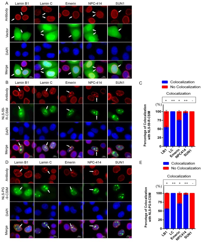
Figure 3.
Disorganized distributions of NE proteins. (A) Immunocytochemistry was performed on EGFP vector-, (B) EGFP–NLS-59-R-CSIM- and (D) EGFP–NLS-PG-9-CSIM-transfected HeLa cells. In panels A, B, and D, cells were stained with anti-lamin B1, lamin C, emerin, NPC-414, and SUN1 antibodies. Chromatin was stained with DAPI. Representative images of lamin B1, lamin C, emerin, NPC-414, and SUN1 staining in the indicated cells. Colocalization of lamin B1, lamin C, emerin, and NPC-414 with EGFP signals at the NE and NE invaginations are as indicated with arrowheads. Scale bar, 10 µm. (C,E) Percentages of cells displaying colocalization of the NE protein and EGFP signal in the images shown. More than 300 EGFP-positive cells for each construct were counted, n = 3.

