Abstract
Strain and physical trauma to mechanically active cells, such as skeletal muscle myofibers, injures their plasma membranes, and mitochondrial function is required for their repair. Here we found that mitochondrial function was also needed for plasma membrane repair in myoblasts as well as nonmuscle cells, which depended on mitochondrial uptake of calcium through the mitochondrial calcium uniporter (MCU). Calcium uptake transiently increased the production of mitochondrial reactive oxygen species (ROS), which locally activated the GTPase RhoA, triggering F-actin accumulation at the site of injury and facilitating membrane repair. Blocking mitochondrial calcium uptake or ROS production prevented injury-triggered RhoA activation, actin polymerization, and plasma membrane repair. This repair mechanism was shared between myoblasts, nonmuscle cells, and mature skeletal myofibers. Quenching mitochondrial ROS in myofibers during eccentric exercise ex vivo caused increased damage to myofibers, resulting in a greater loss of muscle force. These results suggest a physiological role for mitochondria in plasma membrane repair in injured cells, a role that highlights a beneficial effect of ROS.
Introduction
Skeletal muscles produce the force for all our voluntary movements. The muscle cell (myofiber) produces this force using energy generated, in part, by mitochondria. This energy allows the actin cytoskeleton to contract and force to be transmitted across the plasma membrane to the basement membrane and adjacent myofibers. Stress due to this force transmission routinely injures myofibers (1). If a myofiber is damaged beyond repair, it is regenerated in the following weeks through a series of steps involving muscle stem cells (2). However, the mechanism by which injured myofibers repair damage to the plasma membrane in the minutes following injury is poorly studied. Plasma membrane repair is a ubiquitous and vital cellular process that has been studied mostly using invertebrate eggs and mono-nucleated mammalian cells. These studies have identified that plasma membrane repair depends on entry of extracellular calcium into the injured cell (3, 4). Calcium entry facilitates plasma membrane repair by triggering vesicle exocytosis, shedding of the damaged portion of the plasma membrane, and polymerization of cortical F-actin, all of which contribute to repair (4–9). Vesicle fusion provides endomembrane and lipid modifying enzymes, which together with plasma membrane scission machinery remodels the injured membrane (7, 8, 10–12). Injury-triggered F-actin polymerization, which is mediated by the GTPases Cdc42 and Rho, provides structural support to the newly remodeled plasma membrane, allowing successful wound closure (5).
A growing recognition of the relevance of plasma membrane repair deficit in muscle and other diseases raises the need to better understand this process (13–15). We have previously performed a proteomic screen and identified that functional mitochondria are required for skeletal myofiber plasma membrane repair (16). We have demonstrated that a defect in this mechanism contributes to initiation and progression of disease in the mouse model for Duchenne muscular dystrophy (17). However, the mechanism for mitochondria-mediated plasma membrane repair and its potential relevance for non-muscle cells have not been resolved. Here we investigated both of these aspects of mitochondria-mediated plasma membrane repair. We found that mitochondria facilitated plasma membrane repair in both muscle and non-muscle cells. Because mitochondria have roles in cellular metabolism, signaling, reactive oxygen species (ROS) production, and calcium homeostasis, we investigated which of these functions of mitochondria facilitated plasma membrane repair. We found that mitochondria facilitate plasma membrane repair by providing the link between injury-triggered cellular calcium increase and F-actin formation. This role of mitochondria required calcium uptake through the mitochondrial calcium uniporter (MCU) and activation of the GTPase RhoA by mitochondrial ROS signaling. Inhibiting this pathway not only compromised plasma membrane repair, but also resulted in greater eccentric exercise-induced damage and force loss for skeletal myofibers.
Results
De novo ATP production by mitochondria is not required for plasma membrane repair
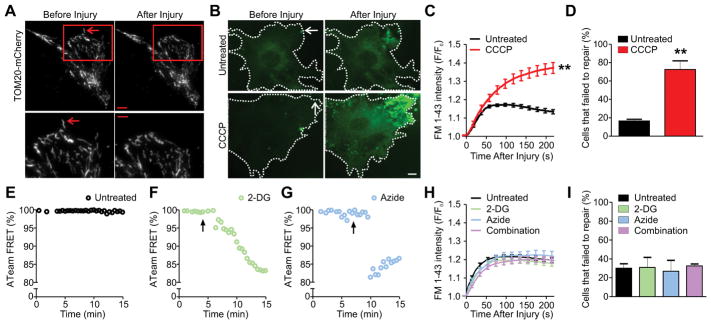
Active mitochondria accumulate at the site of plasma membrane repair in myofibers (16). To examine if mitochondria accumulation was also required for plasma membrane repair in myoblasts we monitored the localization of mitochondria after plasma membrane injury. Similar to cultured myotubes (16), mitochondria did not accumulate at the site of repair in cultured myoblasts and non-muscle cells (Fig. S1A–D). Using total internal reflection fluorescence microscopy (TIRFM) we found that mitochondria were localized within 150 nm of the plasma membrane in cultured cells and that they maintained this close proximity even during plasma membrane repair (Fig. 1A, Fig. S1E). Such a close association of mitochondria with the plasma membrane suggested that a further increase in mitochondria at the site of injury might not be required for mitochondria-mediated plasma membrane repair in these cells. To examine if mitochondria facilitated plasma membrane repair in cultured cells we used a laser injury assay that we have previously established for monitoring plasma membrane repair (18). In this assay, laser-induced focal plasma membrane injury in the presence of membrane impermeant (FM 1-43) dye allows the dye to enter the cell and bind to endomembranes, leading to increase in dye fluorescence until the plasma membrane is repaired (18). As we have demonstrated previously (7, 8), injured myoblast plasma membrane was repaired within a minute of injury (Fig. 1B, C). To test whether mitochondrial activity contributed to plasma membrane repair we used carbonyl cyanide m-chlorophenyl hydrazone (CCCP), a protonophore that blocks oxidative phosphorylation and other mitochondrial activity by uncoupling the mitochondrial transmembrane potential (19). We have previously used it to determine the role of mitochondria in myofiber plasma membrane repair (16).
Figure 1. Mitochondria facilitate plasma membrane repair independently of their role in ATP synthesis.
(A) Image of mitochondria localizing near the site of membrane injury by Total Internal Reflection Fluorescence (TIRF) microscopy. Mitochondria are marked by Translocase of the Outer Membrane 20 (TOM20), a structural protein of the outer mitochondrial membrane. Red box (top) indicates magnified areas shown below. Arrows mark the injury site (3 independent experiments). (B and C) Images (B) and quantification (C) of FM 1-43 dye entry in untreated and 5 μM CCCP treated myoblasts (n≥15 myoblasts for each group from 3 independent experiments). Dotted lines mark the cell boundary and arrows mark the injury site. (D) Quantification of myoblasts that failed to repair after laser injury (n≥25 myoblasts for each group from 3 independent experiments). (E–G) Change in YFP/CFP FRET for the cytosolic ATP sensor ATeam monitored in myoblasts that were either untreated (E), treated with 10 mM 2-Deoxyglucose (2-DG) (F), or 10 mM Sodium Azide (Azide) (G). Arrow indicates the time at which drug was added (n≥5 myoblasts for each group from 2 independent experiments). (H) Plot showing kinetics of FM 1-43 dye entry after laser injury of myoblasts pre-treated for 30 minutes (n≥15 myoblasts for each group from 3 independent experiments) with both 10 mM 2-DG and Azide 1 hour before injury. (I) Quantification of myoblasts that failed to repair after laser injury (n≥19 myoblasts for each group from 3 independent experiments). Scale bar=10 μm or 5 μm for magnified image. ** P-value <0.01 by unpaired T-test compared to untreated sample.
Upon treatment with CCCP, FM-dye entry continued unabated (Fig. 1B, C), which resulted in a nearly five-fold increase in the number of cells that failed to repair (Fig. 1D). CCCP treatment also resulted in a three-fold increase in the number of cells that failed to repair from mechanical injury by glass beads (Fig. S2A, B). To examine if mitochondria also facilitated plasma membrane repair in non-muscle cells, we used CCCP to treat HeLa cells. CCCP-treatment in HeLa cells resulted in unabated FM-dye entry following laser injury and caused a four-fold increase in the number of cells that failed to repair from laser injury (Fig. S2C–E). Thus, mitochondria are involved in plasma membrane repair independent of cell-type and of mitochondrial accumulation at the site of repair.
Mitochondria help meet the energy demand associated with repair and regeneration of axons following axonal injury (20). Thus, we examined if de novo production of ATP in response to plasma membrane damage aids in muscle cell plasma membrane repair. Using the FRET-based ATP biosensor ATeam (21), we confirmed that ATP synthesis was rapidly inhibited by acutely blocking either oxidative phosphorylation using Sodium Azide (Azide), which is a mitochondrial respiratory chain Complex IV inhibitor, or glycolysis using 2-Deoxyglucose (2-DG) (Fig. 1E–G). Inhibiting de novo ATP synthesis by oxidative phosphorylation using Azide did not affect the kinetics or the success of myoblast plasma membrane repair (Fig. 1H, I). Similarly, inhibition of glycolytic ATP production by 2-DG, either alone or in combination with Azide, also did not affect plasma membrane repair (Fig. 1H, I). Together these observations rule out a bioenergetic role of mitochondria in mediating muscle cell plasma membrane repair.
MCU-dependent calcium uptake is required for mitochondria-mediated plasma membrane repair
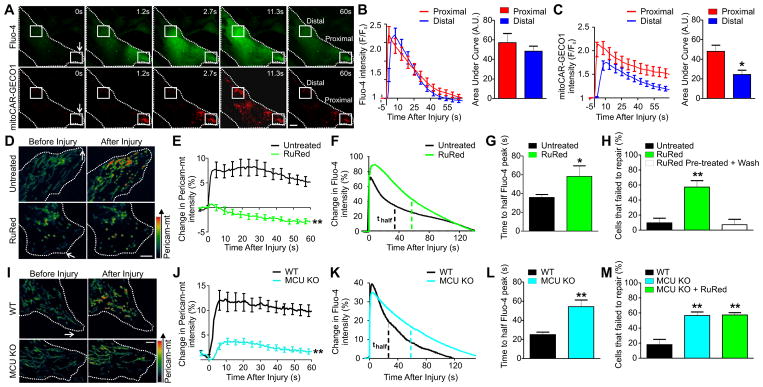
A non-bioenergetic role of mitochondria is regulation of cellular calcium homeostasis and signaling (22, 23). Entry of extracellular calcium following plasma membrane injury is required for plasma membrane repair, but chronically high cytosolic calcium is lethal (24, 25). To examine if mitochondria aid in the clearance of injury-triggered cytosolic calcium ([Ca2+]C) increase, we directly monitored mitochondrial matrix calcium ([Ca2+]M) in response to plasma membrane injury. To simultaneously visualize [Ca2+]M and [Ca2+]C changes during plasma membrane repair we used a red [Ca2+]M sensor mito-CAR-GECO1 (26) and the green [Ca2+]C sensor Fluo-4 (Fig. 2A). These sensors showed that the increase in cytosolic and mitochondrial calcium following injury occurred in a wave like manner, and that calcium remained increased in mitochondria proximal to the injury site even 60s post-injury. To capture the profile of both calcium uptake and retention in the cytosol and mitochondria, we calculated the area under the curve in regions both proximal and distal to the site of plasma membrane injury. While the kinetics of [Ca2+]C increase did not differ (Fig. 2B), mitochondria proximal to the injury site took up more calcium and retained calcium longer than those distal to the injury site (Fig. 2C, left panel). This resulted in a nearly two-fold increase in calcium load in these mitochondria, as indicated by two-fold greater area under the curve for the kinetics of [Ca2+]M change in proximal compared to distal mitochondria (Fig. 2C, right panel).
Figure 2. MCU-mediated calcium uptake is required for plasma membrane repair.
(A) Images showing change in Fluo-4 ([Ca2+]C) and mito-CAR-GECO1 ([Ca2+]M) intensity during myoblast plasma membrane repair. Boxes mark regions that are proximal or distal to the injury site. (B and C) Kinetics of Fluo-4 (B) or mito-CAR-GECO1 (C) intensity after laser injury and the area under each curve (n=17 myoblasts for each group from 2 independent experiments). (D and E) Pseudocolor images (D) and the kinetics of change in the [Ca2+]M sensor Pericam-mt intensity (E) in untreated or 500 μM RuRed treated myoblasts (n≥11 myoblasts for each group from 3 independent experiments). (F and G) Kinetics of change in Fluo-4 intensity (F) and averaged T1/2max (G) in untreated or 500 μM RuRed treated myoblasts (n≥10 myoblasts for each group from 2 independent experiments). Dotted lines mark the time for Fluo-4 intensity to reach half maximal value (T1/2max) post-injury. (H) Quantification of myoblasts that failed to repair laser injury after being pre-treated with 500 μM RuRed and injured in either presence or absence of RuRed (n≥26 myoblasts for each group from 3 independent experiments). (I and J) Pseudocolor images (I) and the kinetics of change in the [Ca2+]M sensor Pericam-mt intensity (J) in WT or MCU KO fibroblasts (n≥17 fibroblasts for each group from 3 independent experiments). (K and L) Kinetics of change in Fluo-4 intensity (K) and averaged T1/2max (L) in WT or MCU KO fibroblasts (n≥11 fibroblasts for each group from 3 independent experiments). Dotted lines mark the time for Fluo-4 intensity to reach half maximal value (T1/2max) post-injury. (M) Quantification of WT and MCU KO fibroblasts (injured either with or without 500 μM RuRed) that failed to repair laser injury (n≥26 fibroblasts for each group from 3 independent experiments). Dotted lines mark the cell boundary and arrows mark the injury site. Scale bar=10 μm (A), or 5 μm (D, I). *P-value<0.05 and **P-value <0.01 by unpaired T-test or ANOVA followed by Dunnett’s post-test for multiple comparisons
To assess the role of mitochondrial activity in clearance of Ca2+ from the cytosol we treated myoblasts with CCCP, which prevented mitochondrial uptake of Ca2+ (Fig. S2F, G). Immediately following injury in untreated cells, [Ca2+]C peaked and then decreased to its half maximal value by 35.8±3.1 seconds, which was delayed by two-fold in CCCP-treated cells (Fig. S2H, I). Thus, mitochondrial uptake of Ca2+ is required for the rapid clearance of the injury-triggered increase in [Ca2+]C (Fig. S2H, I). Such spatial and temporal regulation of [Ca2+]M suggested a potential involvement of the mitochondrial calcium uniporter (MCU) in this process. To assess the role of MCU in this process we acutely inhibited MCU activity in myoblasts by using Ruthenium Red (RuRed), an inhibitor of MCU that is impermeant to intact plasma membrane (27). Thus, RuRed can only enter the cell and inhibit MCU after plasma membrane integrity has been compromised due to injury. Injury in the presence of RuRed blocked [Ca2+]M increase and delayed the return of the injury-triggered increase in [Ca2+]C to pre-injury values (Fig. 2D–G). Consequently, when cells pre-treated with RuRed were washed free of the drug prior to injury, they repaired normally (Fig. 2H, white bar). However, when cells were injured in the continued presence of RuRed the number of myoblasts that failed to repair increased significantly (Fig. 2H, green bar). To establish that MCU-mediated plasma membrane repair occurred independent of the cell type, we injured primary fibroblasts prepared from MCU knockout mice (MCU KO). Similar to RuRed treated myoblasts, the injury-triggered increase in [Ca2+]M was reduced in the MCU KO fibroblasts and this led to a delayed return of [Ca2+]C to baseline (Fig. 2I–L). MCU deficiency also significantly compromised plasma membrane repair and resulted in a 4-fold increase in the number of fibroblasts that failed to repair as compared to wild-type fibroblasts (Fig. 2M). Treating the MCU KO fibroblasts with RuRed did not change the plasma membrane repair capacity of these cells, establishing that reduced myoblast plasma membrane repair caused by RuRed treatment (Fig. 2H) was mediated by inhibition of the MCU activity (Fig. 2M).
Acute mitochondrial ROS signaling facilitates plasma membrane repair
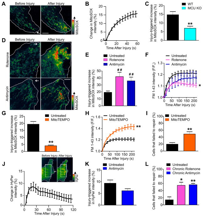
An increase in [Ca2+]M stimulates oxidative phosphorylation, causing increased ATP and reactive oxygen species (ROS) production (28–31). Because we found de novo ATP synthesis to be dispensable for plasma membrane repair, we next examined the role of cellular redox status, which is also implicated in plasma membrane repair (32, 33). Using the mitochondrial matrix superoxide indicator (mitoSOX) we observed a rapid increase in mitochondrial matrix ROS in mitochondria near the injury site (Fig. 3A, B). MCU KO fibroblasts produced significantly less mitochondrial ROS in response to injury, suggesting that an injury-triggered increase in [Ca2+]M resulted in mitochondrial ROS production (Fig. 3C). To assess if mitochondrial ROS facilitated plasma membrane repair, we pharmacologically increased its production with the mitochondrial respiratory chain Complex I inhibitor Rotenone and the Complex III inhibitor Antimycin A (Fig. S3A). These inhibitors, which block mitochondrial ATP production, also increased injury-triggered mitochondrial ROS production as shown by mitoSOX staining (Fig. 3D, E). Concomitant with the greater mitochondrial ROS increase caused by Rotenone as compared to Antimycin, Rotenone significantly reduced FM dye entry (indicating faster plasma membrane repair) while Antimycin treatment resulted in a similar trend, although the decrease was not statistically significant (Fig. 3F). Together these results showed a dose-dependent beneficial role of mROS production on plasma membrane repair. 2-DG and Azide, which did not affect plasma membrane repair- (Fig. 1H, I), also did not affect basal or injury-triggered mitochondrial ROS production (Fig. S3B, C) confirming that the increase in mitochondrial ROS production was not due to inhibition of de novo ATP synthesis. These findings further suggested that it was the injury-triggered mitochondrial ROS production and not ATP synthesis that was required for plasma membrane repair. To directly assess the requirement of mitochondrial ROS for plasma membrane repair, we used the mitochondria matrix targeted antioxidant mitoTEMPO to quench mitochondrial ROS. Treatment with mitoTEMPO significantly reduced injury-triggered mitochondrial ROS production (Fig. 3G), increased injury-triggered FM dye entry into the cell (Fig. 3H), and caused an over two-fold increase in the number of cells that failed to repair (Fig. 3I) further confirming the requirement of mitochondrial ROS for plasma membrane repair. To examine if the injury-triggered mitochondrial ROS increase was caused by an increase in [Ca2+]M, we assessed the effect of mitochondrial ROS agonists Rotenone and Antimycin on plasma membrane repair in MCU KO fibroblasts. Neither of these mitochondrial ROS agonists improved plasma membrane repair in MCU KO cells (Fig. S3D, S3E). These findings demonstrated that the beneficial effect of Rotenone and Antimycin on plasma membrane repair is due to their ability to increase injury-triggered mitochondrial ROS production in a manner that requires an injury-triggered increase in [Ca2+]M.
Figure 3. Mitochondrial ROS production is required for plasma membrane repair.
(A and B) Pseudocolored images (A) and kinetics of change in mitoSOX intensity (B) in untreated myoblasts (n=33 cells from 5 independent experiments). (C) Maximal injury-triggered increase in mitoSOX intensity at the site of plasma membrane repair in WT or MCU KO fibroblasts (n≥10 fibroblasts for each group from 3 independent experiments). (D and E) Pseudocolored images showing injury-triggered change in mitoSOX intensity (D) and maximal injury-triggered increase in mitoSOX intensity at the site of plasma membrane repair (E) in myoblasts treated with 5 μM Rotenone (n=16) or Antimycin (n=12) (3 independent experiments). (F) Quantification of FM 1-43 dye entry following laser injury in myoblasts either untreated or treated as in (E) (n≥15 myoblasts for each group from 3 independent experiments). (G) Maximal injury-triggered increase in mitoSOX intensity at the site of plasma membrane repair in myoblasts treated with 25 μM mitoTEMPO (n≥11 myoblasts for each group from 3 independent experiments). (H) Quantification of FM 1-43 dye entry following laser injury in myoblasts treated with 25 μM mitoTEMPO (n≥16 myoblasts for each group from 4 independent experiments). (I) Quantification of myoblasts that failed to repair after laser injury (n≥21 myoblasts for each group from 4 independent experiments). (J) Pseudocolor image of a portion of a myoblast near the site of injury and quantification showing change in HyPer-3 intensity after injury in myoblasts (n=11 myoblasts from 3 independent experiments). White box indicates region near site of injury used for quantification. (K) Maximal injury-triggered increase in HyPer-3 intensity at the site of plasma membrane repair in untreated and Antimycin treated myoblasts (n≥11 myoblasts for each group from 3 independent experiments). (L) Quantification of myoblasts that failed to repair after laser injury after chronic (8 hour) treatment with 5 μM Rotenone or Antimycin (n≥17 myoblasts for each group from 3 independent experiments). Dotted lines mark the cell boundary and arrows indicate site of injury. Scale bar=5 μm. *P-value<0.05 and **P-value <0.01 by unpaired T-test compared to untreated sample or ANOVA followed by Dunnett’s post-test for multiple comparisons. ##P-value<0.01 for Kruskal-Wallice followed by Dunn’s post-test for multiple comparisons.
Because cytosolic ROS is involved in plasma membrane repair (32, 33), we next examined the effect of injury-triggered mitochondrial ROS increase on cytosolic ROS. Using the genetically encoded ROS (H2O2) sensor HyPer-3, we monitored the effect of plasma membrane injury on cytosolic ROS. Plasma membrane injury caused rapid, transient, and local increase in cytosolic ROS at the site of injury (Fig. 3J). This transient increase in cytosolic ROS was not enhanced by Antimycin treatment (Fig. 3K), suggesting that it was the increase in mitochondrial ROS and not a global increase in cytosolic ROS that facilitated plasma membrane repair. These data agree with the detrimental effect of chronic cytosolic ROS increase on plasma membrane repair, which can be reversed by the use of antioxidants (33, 34). To test this we treated cells with Antimycin and Rotenone for 8 hours to cause a chronic increase in cellular ROS concentrations. In contrast to the improved plasma membrane repair observed following acute mitochondrial ROS increase by Antimycin and Rotenone treatment (Fig. 3F), a chronic increase in cellular ROS led to a three-fold increase in the number of cells that failed to repair (Fig. 3L). Together, these results establish that an acute, localized increase in mitochondrial ROS at the site of injury did not significantly increase global cytosolic ROS and facilitated plasma membrane repair, while chronic and global increase in cellular ROS inhibited plasma membrane repair.
Mitochondria enable plasma membrane repair by activating F-actin dependent wound closure
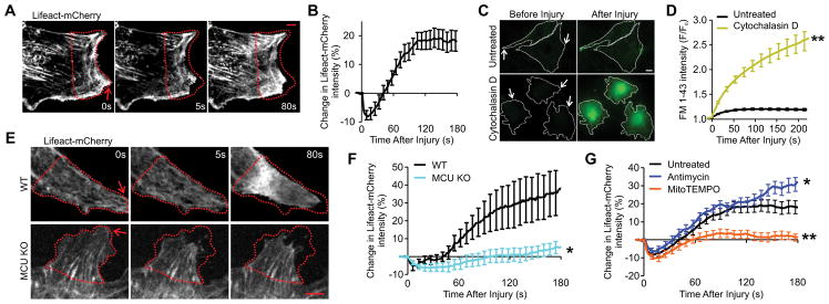
Formation of F-actin at the wound site is required for plasma membrane repair and is also sensitive to cellular redox changes (5, 6, 9, 35–37). Our previous proteomic analyses of myofibers and myoblasts has shown that the abundance of actin and actin-associated proteins increases at the injured plasma membrane (8, 16). We directly monitored actin responses during plasma membrane repair in myoblasts using β-actin-GFP and found that plasma membrane injury triggered a transient and robust actin accumulation at the injury site (Fig. S4A, B). To examine if this accumulation was due to a local change in F-actin we used the reporter Lifeact, an F-actin binding peptide that can be used in live cells (38). We found that plasma membrane repair in myoblasts expressing Lifeact-mCherry (Fig. 4A, B) was associated with robust and rapid F-actin buildup at the site of injury, that preceded wound closure. Inhibition of F-actin polymerization using Cytochalasin D resulted in increased FM dye uptake and compromised the ability of cells to repair (Fig. 4C, D; Fig. S4C). These observations, together with previous studies on myofiber repair (35, 36), confirmed the requirement of F-actin for muscle cell plasma membrane repair.
Figure 4. Mitochondria mediate F-actin buildup at the site of plasma membrane repair.
(A and B) Time-lapse images (A) and quantification of change in Lifeact-mCherry intensity at the site of plasma membrane repair after laser injury in myoblasts (B) (n=26 myoblasts from 4 independent experiments). (C and D) Images (C) and quantification of FM 1-43 dye entry (D) in untreated and 2 μM Cytochalasin D treated myoblasts (n≥12 myoblasts for each group from 2 independent experiments). (E and F) Time-lapse images (E) and quantification of change in Lifeact-mCherry intensity at the site of plasma membrane repair after laser injury (F) in WT or MCU KO fibroblasts (n≥9 fibroblasts for each group from 3 independent experiments). (G) Quantification of change in Lifeact-mCherry intensity at the site of plasma membrane repair in myoblasts either untreated or treated with 5 μM Antimycin or 25 μM mitoTEMPO (n≥17 myoblasts for each group from 3 independent experiments). Arrows indicate site of injury. Dotted red line marks the site of F-actin accumulation. Dotted white line indicates cell boundary. Scale bar=10 μM (C) or 5 μM (A, E). *P-value<0.05 and **P-value<0.01 by unpaired T-test compared to untreated sample or ANOVA followed by Dunnett’s post-test for multiple comparisons.
To examine if injury-triggered changes in mitochondrial activity regulated F-actin accumulation at the site of plasma membrane repair we treated myoblasts with CCCP, which prevented injury-triggered F-actin buildup (Fig. S4D, E). To establish if this effect was due to the injury-triggered increase in [Ca2+]M we monitored F-actin buildup in MCU KO fibroblasts. In contrast to wild-type fibroblasts, MCU KO fibroblasts did not accumulate F-actin after plasma membrane injury (Fig. 4E, F). Similarly, quenching mitochondrial ROS in the healthy myoblasts with mitoTEMPO prevented F-actin accumulation (Fig. 4G). In a complimentary approach, treating healthy myoblasts with the mitochondrial ROS agonist Antimycin enhanced the injury-triggered increase in F-actin at the site of plasma membrane repair (Fig. 4G). We assessed if plasma membrane repair required an accumulation of microtubules by using the GFP-tagged microtubule end-capping protein EB3 (39). Unlike F-actin, microtubules did not show polarized growth towards the site of injury and preventing microtubule polymerization with Nocodazole had no effect on plasma membrane repair in myoblasts (Fig. S4F–H). Together, these results showed that mitochondria ROS-mediated plasma membrane repair required selective activation of F-actin buildup at the site of repair.
Mitochondrial ROS-dependent RhoA activation mediates plasma membrane repair by F-actin accumulation
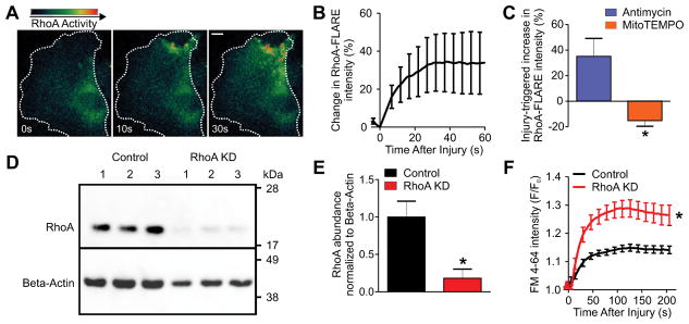
Although oxidation inhibits F-actin polymerization, oxidation of cysteine residues activates the GTPase RhoA (37, 40, 41). Further, RhoA activation upon plasma membrane injury is required for F-actin buildup (42). To test if injury-triggered mitochondrial ROS production facilitated F-actin buildup by activating RhoA, we used a FRET-based RhoA sensor, RhoA-FLARE (43). Plasma membrane injury caused immediate and local increase in RhoA activity at the site of injury, which peaked within 30 seconds, closely mirroring the spatial and temporal increase in mitochondrial ROS and preceding the peak in F-actin buildup (Fig. 5A, B). Treating cells with the mitochondrial ROS agonist Antimycin further enhanced injury-triggered increase in RhoA activity, while use of the mitochondrial ROS antagonist mitoTEMPO inhibited the injury-triggered activation of RhoA (Fig. 5C), which was similar to their respective effects on F-actin accumulation observed previously. To establish the relevance of RhoA in plasma membrane repair, we used an adenovirally delivered micro-RNA construct targeted against RhoA (40), which reduced the amount of RhoA protein five-fold in myoblasts (RhoA-KD) (Fig. 5D, E). RhoA-KD myoblasts showed increased FM dye entry as compared to control cells (Fig. 5F). This reduction in the ability of the RhoA-KD cells to efficiently repair plasma membrane injury suggested that RhoA was a mediator of mitochondrial ROS-triggered F-actin accumulation during plasma membrane repair.
Figure 5. Mitochondrial ROS-dependent RhoA activity is required for repair.
(A and B) Time-lapse images (A) and quantification of change in RhoA-FLARE intensity after laser injury in myoblasts (B) (n=12 myoblasts from 4 independent experiments). (C) Injury-triggered change in the RhoA-FLARE FRET value (RhoA activity) following treatment with 5 μM Antimycin, or 25 μM mitoTEMPO (n=5 or 7 myoblasts respectively from 3 independent experiments). (D) Western blot analysis of RhoA in control or RhoA-targeted miRNA (RhoA KD) treated myoblasts (n=3 independent replicates). (E) RhoA protein abundance in control and RhoA KD myoblasts normalized to loading control (β-actin). (F) Plot showing kinetics of FM 4-64 dye entry after laser injury in control and RhoA KD myoblasts (n≥21 myoblasts for each group from 2 independent experiments). Dotted white line indicates cell boundary. Scale bar=10 μM. *P-value<0.05 by unpaired T-test compared to untreated/control sample.
Mitochondrial ROS signaling repairs myofibers from exercise-induced injury
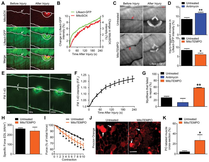
The biochemical functions of mitochondria are conserved across various cell types, but because of the differences in mitochondrial organization and injury-triggered mobilization between cultured cells and skeletal myofibers, we next examined if the link between mitochondrial ROS and plasma membrane repair was conserved between cultured cells and skeletal myofibers. Simultaneous imaging of mitoSOX and F-actin in myofibers from Lifeact-GFP transgenic mice revealed an association between myofiber plasma membrane repair and a rapid and local increase in mitochondrial ROS (Fig. 6A, B), which overlapped with the accumulation of F-actin at the site of repair. To examine the effect of injury-triggered mitochondrial ROS increase on F-actin accumulation in myofibers we pharmacologically altered mitochondrial ROS in isolated intact muscles from transgenic Lifeact-GFP mice. Similar to myoblasts, Antimycin treatment increased F-actin accumulation at the site of injury, while mitoTEMPO treatment significantly decreased F-actin accumulation at the site of injury (Fig. 6C, D). This reduction in injury-triggered F-actin accumulation was sufficient to prevent myofiber plasma membrane repair as assessed by the FM dye entry assay, which showed that quenching mitochondrial ROS by using mitoTEMPO caused over two-fold increase in the number of myofibers that failed to undergo plasma membrane repair while Antimycin treated myofibers showed a trend toward improved plasma membrane repair (Fig. 6E–G).
Figure 6. Mitochondrial ROS enables repair of focal and exercise-induced myofiber injury.
(A and B) Image (A) and plot (B) showing mitoSOX and Lifeact-GFP intensity at the injury site for a skeletal myofiber in an intact mouse bicep (2 independent experiments). (C and D) Images (C) and quantification of change in Lifeact-GFP intensity at the injury site in myofibers either untreated or treated with 30 μM Antimycin or 250 μM mitoTEMPO (D) (n≥22 myofibers for each group from 3 mice and 2 independent experiments). (E and F) Images (E) and kinetics of FM 1-43 dye uptake by myofibers after focal laser injury (F) (n=20 myofibers from 3 mice and 2 independent experiments). (G) Plot showing myofibers that failed to repair from focal laser injury. Myofibers were either untreated or treated with 30 μM Antimycin or 250 μM mitoTEMPO (n≥20 myofibers from 2 mice and 2 independent experiments). (H) Specific force of EDL muscles untreated or treated for 20 minutes with 250 μM mitoTEMPO (n=5 mice each from 5 independent experiments). (I) Reduction in EDL muscle contractile force following each round of 20% eccentric exercise (n=5 mice each from 5 independent experiments). (J and K) Images (J) and quantification of Procion Orange (PO) labeling of myofibers in EDL muscle cross section following 10 rounds of eccentric exercise of muscle treated as indicated (K) (n=5 mice each from 5 independent experiments). Arrows mark some of the PO positive fibers. Arrows in laser injury images mark the site of injury and the solid lines mark the myofiber boundary. Scale bar=10 μm (A, C, E) or 100 μm (J). *P-value<0.05 and **P-value<0.01 by unpaired T-test compared to untreated sample or ANOVA followed by Dunnett’s post-test for multiple comparisons.
Given the above requirement of mitochondrial ROS in facilitating myofiber plasma membrane repair, we hypothesized that mitochondrial ROS production may facilitate repair of myofibers during routine eccentric exercise-induced injury. To test this hypothesis we performed acute ex vivo exercise of the mouse Extensor Digitorum Longus (EDL) muscle. Since mitochondrial ATP synthesis is required for force production during exercise, this approach only allowed testing the role of mitochondrial ROS through the use of the mitochondrial ROS quencher mitoTEMPO and not the inhibitors of Complex I and III. Acute mitoTEMPO treatment did not alter the muscle contractile strength (Fig. 6H). However, upon eccentric exercise (by 20% stretching) the mitoTEMPO treated muscle suffered a greater decline in contractile force, such that after the 2nd eccentric contraction, mitoTEMPO treated muscles lost significantly more force than similarly exercised untreated muscles, a decline that continued with each additional eccentric contraction (Fig. 6I). To assess if the greater decline in contractile force of mitoTEMPO treated muscle was due to poor plasma membrane repair, these muscles were incubated with the membrane-impermeant dye Procion Orange (PO) following exercise. Indeed, significantly more myofibers in mitoTEMPO treated muscles were labeled with PO (Fig. 6J, K), indicating that repair of plasma membrane injury due to eccentric exercise required mitochondrial ROS.
Discussion
Our work here reveals a previously unknown cellular pathway that repairs injury to the plasma membrane of both mature skeletal myofibers and non-muscle cells, and describes a role of mitochondria during injury. In contrast to the mitochondrial response during ischemia/reperfusion injury, which is detrimental to cells (44), we found that mitochondrial function was required for the survival of cells following plasma membrane injury. Mitochondria also facilitate the repair of neuronal injury by helping to meet the bioenergetic demands of axonal regeneration (20). Healthy mitochondria are recruited to the injury site in axons (20), and similarly, we have previously shown that mitochondria accumulate at the site of plasma membrane injury in mature skeletal muscle fibers (16). Here we found that mitochondria were required for plasma membrane repair even in myoblasts and non-muscle cells where mitochondria are present adjacent to the plasma membrane and thus do not need to accumulate at the repair site (Fig. 1A, S1). Acute inhibition of de novo ATP production did not affect the success of plasma membrane repair, indicating that mitochondria did not facilitate plasma membrane repair through ATP production. Thus plasma membrane repair can proceed in a quasi-energy-deprived state without new ATP synthesis, provided the existing cellular pool of ATP is sufficient to supply the required energy-dependent processes. This ability to function in a reduced energy state would be a beneficial, if not an essential adaptation, for plasma membrane repair machinery since efflux of cytosol due to plasma membrane injury would inevitably cause loss of ATP and thus acute energy deprivation.
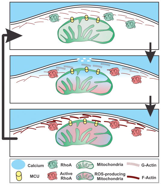
We have previously proposed that mitochondria could facilitate plasma membrane repair through uptake of [Ca2+]C to reduce the increase in [Ca2+]C due to the influx of extracellular calcium (16). We found that mitochondria near the site of plasma membrane injury retain calcium at higher amounts for a greater duration during the repair period (Fig. 2A, C). Calcium retention at the site of plasma membrane repair also occurs in Xenopus oocytes (12). Here, we demonstrated how this locally high [Ca2+]M did not induce calcium-induced death in mammalian cells. MCU-mediated uptake of the increase in [Ca2+]C was part of the role of mitochondria in plasma membrane repair (Fig. 2, 7), enabling mitochondria to work as a biochemical capacitor that continues to signal well past the rapid burst of calcium following plasma membrane injury. The output of this biochemical capacitor was the mitochondrial ROS signal, which locally activated RhoA and initiated F-actin remodeling at the site of plasma membrane injury. Thus, mitochondria serve dual roles by aiding in removal of cytosolic calcium and simultaneously using calcium to initiate local and acute mitochondrial ROS signaling to facilitate repair (Fig. 7).
Figure 7. Model for mitochondrially-mediated plasma membrane repair.
Before membrane disruption (top panel), mitochondria are found in close proximity to the plasma membrane. In response to plasma membrane injury (middle panel), mitochondria located near the injury take up injury-triggered cytosolic calcium increase (blue) in an MCU-dependent manner. Calcium uptake stimulates production of reactive oxygen species (ROS) within mitochondria, which activates RhoA. RhoA initiates F-actin polymerization resulting in buildup of F-actin at the site of plasma membrane injury (bottom panel), facilitating membrane repair.
Chronic mitochondrial oxidation is broadly implicated in tissue degeneration during ischemic injury, ageing, and metabolic and genetic diseases (44, 45). Antioxidants are used to address ageing, disease, and muscle exercise-induced damage (45, 46). Our findings refine this broadly detrimental role of ROS by identifying that unlike a chronic ROS increase, which caused poor plasma membrane repair (Fig. 3L), acute and localized increases in ROS were required for signaling that facilitated plasma membrane repair in muscle and other cells. This finding is critical for a basic function of the muscle cell, which is surviving contraction-induced injury. While further studies are needed to fully realize the implication of our findings for diseases associated with mitochondrial dysfunction, we have found that mitochondrial dysfunction caused by the lack of the muscle-specific protein dystrophin impairs the ability of myofibers to undergo repair, contributing to the initiation of muscle pathology in the mouse model of Duchenne muscular dystrophy (17). A beneficial role of mitochondrial ROS in repair has also been identified in repair of epidermal injury in C. elegans (47). However, unlike the epidermal injury in C. elegans where injury causes a rapid initial decline followed by increase in mitochondrial ROS production, mammalian cells showed increased mitochondrial ROS within seconds of injury. Further, unlike C. elegans where F-actin accumulates an hour post-wounding, mammalian cells and myofibers showed wound closure and F-actin accumulation within minutes of being injured (Figs. 4, 6). These differences highlight that despite using similar regulators, contrasting mechanisms can be employed by these regulators to facilitate repair of injured mammalian and C. elegans cells.
Mitochondria promote inflammation and the repair of injured tissue (48), and our findings show that the signaling role of mitochondria in repair extends even to the level of single cells. We have previously proposed that signaling may be among the earliest functions of mitochondria (49). The conserved role of mitochondria in mediating repair of different types of cells and tissues across species supports such an ancient role of mitochondrial signaling in organismal survival. In summary, our findings suggest that in conditions of muscle and other tissue injury there is a need to take a nuanced view of mitochondrial ROS production, distinguishing between the potential benefits of acute and localized changes in ROS and the detriment of chronic and global increase in cellular ROS.
Materials and Methods
Cell Culture, Reagents, and Plasmids
Mouse C2C12 myoblasts (ATCC-CRL1772), WT and MCU knockout mouse embryonic fibroblasts, and HeLa cells were routinely cultured in Dulbecco’s Modified Eagle Medium (DMEM) with Glutamax, high glucose, 110 mg/L sodium pyruvate (Invitrogen, USA) supplemented with 10% fetal bovine serum (Hyclone, Thermo Fisher Scientific, USA) and with 100 mg/mL Penicillin and 100 mg/mL Streptomycin (Invitrogen, USA) at 37°C and 5% CO2. Cells were transfected with the indicated plasmid using Lipofectamine LTX reagent (Life Technologies) as per manufacturer’s instructions. Expression was allowed for 24 hours prior to imaging. All pharmacologic inhibitors including CCCP, Cytochalasin D and Nocodazole were obtained from Sigma-Aldrich with the exception of Ruthenium Red (Santa-Cruz Biotechnology). All pharmacological treatments, unless stated otherwise, were carried out in Cellular Imaging Medium (CIM; HBSS with 10 mM HEPES, 2 mM Ca2+, pH 7.4) at 37°C. All the cultured cells were injured in the presence of the indicated drug.
The plasmids used in this study were obtained from Addgene or were gifts from the investigator: Dr. Vsevolod V. Belousov (Hyper-3 H2O2 Biosensor) (50), Gyorgy Hajnoczky (Pericam-mt) (51), Robert Campbell (CMV-mito-CAR-GECO1; Addgene plasmid # 46022) (26), Michael Davidson (mCherry-Lifeact; Addgene Plasmid #54491), Klaus Hahn (pTriEx-RhoA FLARE.sc Biosensor; Addgene plasmid # 12150) (43), and Hiromi Imamura (ATeam, ATP Biosensor) (21).
Plasma Membrane Repair Assays
Cells cultured on coverslips were transferred to CIM and placed in a Tokai Hit microscopy stage-top ZILCS incubator (Tokai Hit Co., Fujinomiya-shi, Japan) maintained at 37°C. For laser injury, a 1–2 mm2 area was irradiated for 10 ms with a pulsed laser (Ablate!, 3i Intelligent Imaging Innovations, Inc. Denver, CO, USA). Cells were imaged using an inverted IX81 Olympus microscope (Olympus America, Center Valley, PA, USA) custom equipped with a CSUX1 spinning disc confocal unit (Yokogawa Electric Corp., Tokyo, Japan). Images were acquired using Evolve 512 EMCCD (Photometrics, Tucson, AZ) at 1 Hz. Image acquisition and laser injury was controlled using Slidebook 6.0 (Intelligent Imaging Innovations, Inc., Denver, CO). For quantification of plasma membrane repair cells were pre-incubated with the indicated treatment before 1 mg/ml FM1-43 dye or FM4-64 dye (Life Technologies) was added to CIM prior to injury. FM dye intensity (F/F0 - where F0 represents baseline fluorescence) was used to calculate the kinetics of repair. Successful repair was determined by the entry of FM dye into the cell interior, where a plateau in FM dye increase indicated successful repair and failure to repair was indicated by unabated FM dye increase.
Cells cultured on coverslips were transferred to CIM or PBS (Sigma-Aldrich) containing 2 mg/ml of lysine-fixable FITC-dextran (Life Technologies). Cells were injured by rolling glass beads (Sigma-Aldrich) over them. Cells were then allowed to heal at 37°C for 5 min, followed by incubation at 37°C for 5 min in CIM or PBS buffer containing 2 mg/ml of lysine-fixable TRITC dextran (Life Technologies). Cells were fixed in 4% PFA, nuclei counterstained with Hoechst 33342, mounted in fluorescence mounting medium (Dako), and imaged and quantified as previously described (18). The number of FITC-positive cells (injured and repaired) and TRITC-positive cells (injured and not repaired) were counted. The details of these two assays are as previously described (18).
TIRF Imaging
Cells were subjected to laser injury and live cell TIRF imaging as described (52, 53). Cells we transfected with Tom20-mCherry and 24 hours later the cells were imaged in CIM. During imaging cells were maintained at 37°C in microscope stage top ZILCS incubator (Tokai Hit Co., Japan). For TIRF imaging the penetration depth was set at 150nm. Images were acquired using IX81 Olympus microscope (Olympus America, PA) equipped with, 60X/1.45NA oil objective and 561 nm laser (Cobolt, Sweden) and Evolve 512 EMCCD (Photometrics, Tucson, AZ) camera. Acquisition and analysis was carried out using Slidebook 6.0 (Intelligent Imaging Innovations, Inc. Denver, CO).
Measurement of Calcium Increase Following Injury
For measurement of cytosolic calcium, cells were incubated in DMEM with 10 μM Fluo-4 AM (Life Technologies) for 20 min at 37°C. After washing with pre-warmed CIM, cells were laser-injured as described above. Cytosolic Ca2+ was measured as the change in fluorescence over the initial Fluo-4 fluorescence (F/F0). Mitochondrial calcium was measured using the ratiometric calcium sensor Pericam-mt (54). Calcium increase was quantified by taking the ratio F488/F405 where 488 emission indicates bound calcium. Within each cell, calcium increase was averaged between three regions containing one or few mitochondria to determine a single value per cell, which was then averaged across multiple cells. For simultaneous acquisition of cytosolic and mitochondrial calcium, Fluo-4 was paired with the mitochondrial localized CAR-GECO1 calcium sensor (26).
ROS Detection and Quantification
To detect mitochondrial ROS, cells were incubated in 2.5 μM mitoSOX (Molecular Probes) for 10 min at 37°C. After washing with pre-warmed CIM, cells were laser-injured as described above. Mitochondrial ROS was quantified as the change in fluorescence over the initial mitoSOX fluorescence (F/F0). Cytosolic ROS was measured using the ratiometric hydrogen peroxide-specific sensor HyPer 3. ROS increase was quantified by taking the ratio F488/F405.
RhoA and ATP FRET Imaging and Analysis
Cells were transfected with the FRET based RhoA-FLARE biosensor sensitive to active RhoA (43) or with ATP sensor ATeam (21). Three channel FRET imaging was performed by monitoring the donor (445nm laser excitation), acceptor (515nm laser excitation) and transfer (FRET) channels being imaged. Using Slidebook 6.0 the emission from these 3 channels were corrected for bleed through and used to measure corrected FRET: Fc=Transfer-Acceptor-Donor, and generate a corrected FRET image.
RhoA Knockdown and Western Blot
The adenovirally-packaged miRNA target to RhoA was a gift of Dr. Keith Burridge (40). Virus was produced using a HEK293 cell line according to the manufacturer’s recommended protocol (ViraPower Adenoviral Expression System, Invitrogen). To infect myoblasts for RhoA knockdown, crude virus-containing supernatant harvested from HEK293 cells was added to C2C12 myoblasts growing in their normal growth medium. Cells were incubated for 3 days prior to use for experiments or RhoA protein quantification by Western Blot. For control cells, crude supernatant from HEK293 cells lacking adenovirus was added to cells as above. For quantification of RhoA protein knockdown, protein lysates (30 μg) harvested from C2C12 myoblasts were resolved by electrophoresis, transferred to nitrocellulose membrane and incubated overnight at 4°C with primary antibody against RhoA (1:200, RhoA monoclonal antibody 26C4, Santa Cruz Biotechnology). Anti-mouse secondary antibody conjugated to horseradish peroxidase (1:1000, Sigma-Aldrich) was incubated for 1 hour at room temperature. Protein bands were revealed with ECL chemi-luminescence substrate (Thermo Fisher Scientific, USA). Blots were scanned digitally with a ChemiDoc Touch Imager and densitometric analysis was carried out using Image Lab software (Bio-Rad, Hercules, CA, USA). Data are presented as ratio of RhoA protein normalized to loading control (β-actin).
Quantification of Injury-Triggered Actin Dynamics
For measurement of actin dynamics in mononucleate cells, β-actin-GFP and Lifeact-mCherry (F-actin binding peptide) were used. In each case, transfection was carried out as described above and fluorescence intensity is presented as F/F0. For measurement of F-actin dynamics in myofibers, we used Lifeact-GFP transgenic mice previously described (55). Myofibers were prepared for imaging as described below.
Isolation and Treatment of Muscle Tissue for ex vivo Injury
For live myofiber imaging, mice were euthanized by CO2 asphyxiation and muscles were immediately dissected. For myofiber laser injury, whole biceps muscles were mounted in pre-warmed Tyrode’s buffer (119 mM NaCl, 5 mM KCl, 25 mM HEPES buffer, 2 mM CaCl2, 2 mM MgCl2, 6 g/liter glucose, pH - 7.4) and imaged using the IX81 Olympus microscope as described above. To assess myofiber repair, cells were pre-incubated in the indicated treatment before being injured in the presence of 1–2 mg/mL of FM1-43 dye. Repair kinetics and successful repair were determined as described above.
Eccentric Exercise by Lengthening Contractions
After anesthetizing C57Bl/6 mice with a mixture of ketamine and xylazine, EDL muscles were carefully exposed and 6 silk sutures were firmly attached to proximal and distal tendons. The EDL was placed in a bath containing buffered mammalian Ringer’s solution (137mM NaCl, 24mM NaHCO3, 11mM glucose, 5mM KCl, 2mM CaCl2, 1mM MgSO4, 1mM NaH2PO4 and 0.025mM tubocurarine chloride) at 25°C and bubbled with 95% O2-5% CO2 to stabilize the pH at 7.4. Prior to lengthening-contractions, one EDL per mouse was allowed to incubate for 20 min in 250 μM MitoTEMPO. In the bath the distal tendon was securely connected to a fixed bottom plate and the proximal tendon was attached to the arm of a servomotor (800A in vitro muscle apparatus, Aurora Scientific). The vertically aligned EDL muscle was flanked by two stainless steel plate electrodes. Using single 0.2mm square simulation pulses, the muscle was adjusted to the optimal muscle length for force generation. At optimal length, with isometric tetanic contractions 300ms in duration at frequencies up to 250Hz separated by 2 min of rest intervals, the maximal force was determined. Specific force, defined as maximal force normalized for the muscle cross sectional area, was calculated as the maximal force/(muscle mass * (density of muscle tissue * fiber length)-1). The fiber length was based on the fiber to muscle length ratio of 0.45. The muscle tissue density is 1.056 kg/L. To study contraction-induced plasma membrane injury, EDL muscles were subjected to 9 lengthening contractions with 20% strain at a velocity of 2 fiber lengths per second. Each contraction was separated by a 1 min rest interval. Lengthening contraction induced force deficits were expressed as percentage of first contraction. The values were represented as mean ± SEM. After the protocol of lengthening contractions muscles were pinned on cork at optimal length and placed in 0.2% procion orange (PO) dye Ringer-solution for 1 hour at room temperature. After removing the excess dye by washing with Ringer’s solution, the muscle was frozen using isopentane pre-chilled in liquid nitrogen. Frozen tissues were sectioned (10 μm) and imaged using a Nikon Eclipse E800 (Nikon, Japan) microscope that was fitted with a SPOT digital color camera with SPOT advanced software (Diagnostic Instruments, Sterling Heights, MI). Representative images (20X) were taken and procion orange (PO) positive fibers/image was quantified for each section. Data are represented as the averaged area of PO positive fibers per image.
Statistical Analysis
Image acquisition and analysis was performed using Slidebook 6.0 (Intelligent Imaging Innovations Inc.). The statistical analysis was carried out using the GraphPad Prism Software (GraphPad, LaJolla, CA, USA). For all kinetic trace data, individual time points were compared across all cells in a given treatment to determine significance by unpaired t-test. For all data, the D’Agostino and Pearson omnibus normality test was performed prior to determining the appropriate statistical test. For pairwise comparisons, unpaired t-tests were used for all normally distributed data (indicated by *) while Mann-Whitney tests were used for nonparametric data (indicated by #). For multiple comparisons, ANOVA followed by Dunnett’s post-hoc test was used to determine significance of normally distributed data (*). For nonparametric multiple comparisons, a Kruskal-Wallis test followed by Dunn’s Multiple Comparison Test was used to determine significance (indicated by #). In all cases, data not indicated as significant should be considered not statistically different unless otherwise stated. All data are presented as mean ± SEM with p< 0.05 considered statistically significant.
Supplementary Material
Fig. S1: Cultured cells have abundant mitochondria near the plasma membrane that do not move substantially during plasma membrane repair
Fig. S2: CCCP treatment causes poor plasma membrane repair and aberrant [Ca2+]M handling following injury
Fig. S3: Acute, Ca2+-dependent mitochondrial ROS production is required for plasma membrane repair
Fig. S4: Injury-triggered change in F-actin, but not microtubules, facilitates plasma membrane repair
Editor’s summary: Mitochondria for plasma membrane repair.
Mechanical strain on cells can cause plasma membrane damage that must be repaired. Horn et al. elucidated that mitochondria mediate a repair response in muscle cells (which experience mechanical strain during exercise) and nonmuscle cells. The influx of extracellular Ca2+ caused by plasma membrane injury triggered an increase in mitochondrial Ca2+ that initiated the generation of reactive oxygen species (ROS). Mitochondrially produced ROS activated actin polymerization and wound closure at the sites of plasma membrane injury. Quenching this source of ROS in mouse muscle exercised ex vivo resulted in greater damage to injured myofibers and reduced muscle force. These findings demonstrate that rampant quenching of ROS, such as with antioxidants (which are a popular nutritional supplement), may have detrimental effects that must be balanced with their potential benefits.
Acknowledgments
AH performed these studies as part of his doctoral studies at the Institute for Biomedical Sciences at the George Washington University, and the work here represents part of his PhD dissertation. We thank Dr. Toren Finkel (NIH NHLBI) for MCU knockout mouse embryonic fibroblasts, Dr. Judy Liu (Children’s National Health System) for help with the use of Lifeact-GFP mouse and EB3-GFP plasmid, Dr. Anamaris Colberg-Poley (Children’s National Health System) for suggestions and providing Tom20-mCherry plasmid, Dr. Vsevolod Belousov (Nizhny Novgorod State Medical Academy) for Hyper-3 sensor, Dr. Jesper Nylandsted (Danish Cancer Society) for discussions and for providing β-actin-GFP plasmid, Dr. Keith Burridge (University of North Carolina – Chapel Hill) for adenovirally-packaged RhoA-targeted and control miRNA, and Dr. Heather Gordish-Dressman (Children’s National Health System) for help and suggestions in statistical analysis.
Funding: This study was supported by grants from NIAMS (R01AR055686) and MDA (MDA277389) to JKJ, NHLBI (5P01HL071643) to NSC, and CRI microscopy core grant U54HD090257 by NICHD.
Footnotes
Author Contributions: JKJ conceived the study. AH, JKJ, NSC designed the study and AH performed all experiments, with help from JKJ, AR, and LS for cellular studies, and JVM, AD, MH, SS, and JKJ for mouse studies.
Competing Interests: The authors declare that they have no competing interests.
References and Notes
- 1.McNeil PL, Khakee R. Am J Pathol. 1992;140:1097. [PMC free article] [PubMed] [Google Scholar]
- 2.Charge SB, Rudnicki MA. Physiol Rev. 2004;84:209. doi: 10.1152/physrev.00019.2003. [DOI] [PubMed] [Google Scholar]
- 3.Bi GQ, Alderton JM, Steinhardt RA. J Cell Biol. 1995;131:1747. doi: 10.1083/jcb.131.6.1747. [DOI] [PMC free article] [PubMed] [Google Scholar]
- 4.Steinhardt RA, Bi G, Alderton JM. Science. 1994;263:390. doi: 10.1126/science.7904084. [DOI] [PubMed] [Google Scholar]
- 5.Abreu-Blanco MT, Verboon JM, Parkhurst SM. Curr Biol. 2014;24:144. doi: 10.1016/j.cub.2013.11.048. [DOI] [PMC free article] [PubMed] [Google Scholar]
- 6.Mandato CA, Bement WM. J Cell Biol. 2001;154:785. doi: 10.1083/jcb.200103105. [DOI] [PMC free article] [PubMed] [Google Scholar]
- 7.Defour A, Van der Meulen JH, Bhat R, Bigot A, Bashir R, Nagaraju K, Jaiswal JK. Cell Death Dis. 2014;5:e1306. doi: 10.1038/cddis.2014.272. [DOI] [PMC free article] [PubMed] [Google Scholar]
- 8.Scheffer LL, Sreetama SC, Sharma N, Medikayala S, Brown KJ, Defour A, Jaiswal JK. Nat Commun. 2014;5:5646. doi: 10.1038/ncomms6646. [DOI] [PMC free article] [PubMed] [Google Scholar]
- 9.Jaiswal JK, Lauritzen SP, Scheffer L, Sakaguchi M, Bunkenborg J, Simon SM, Kallunki T, Jaattela M, Nylandsted J. Nat Commun. 2014;5:3795. doi: 10.1038/ncomms4795. [DOI] [PMC free article] [PubMed] [Google Scholar]
- 10.Tam C, Idone V, Devlin C, Fernandes MC, Flannery A, He X, Schuchman E, Tabas I, Andrews NW. J Cell Biol. 2010;189:1027. doi: 10.1083/jcb.201003053. [DOI] [PMC free article] [PubMed] [Google Scholar]
- 11.Reddy A, Caler EV, Andrews NW. Cell. 2001;106:157. doi: 10.1016/s0092-8674(01)00421-4. [DOI] [PubMed] [Google Scholar]
- 12.Davenport NR, Sonnemann KJ, Eliceiri KW, Bement WM. Mol Biol Cell. 2016;27:2272. doi: 10.1091/mbc.E16-04-0223. [DOI] [PMC free article] [PubMed] [Google Scholar]
- 13.Demonbreun AR, McNally EM. Curr Top Membr. 2016;77:67. doi: 10.1016/bs.ctm.2015.10.006. [DOI] [PMC free article] [PubMed] [Google Scholar]
- 14.Duann P, Li H, Lin P, Tan T, Wang Z, Chen K, Zhou X, Gumpper K, Zhu H, Ludwig T, Mohler PJ, Rovin B, Abraham WT, Zeng C, Ma J. Sci Transl Med. 2015;7:279ra36. doi: 10.1126/scitranslmed.3010755. [DOI] [PMC free article] [PubMed] [Google Scholar]
- 15.Jaiswal JK, Nylandsted J. Cell Cycle. 2015;14:502. doi: 10.1080/15384101.2014.995495. [DOI] [PMC free article] [PubMed] [Google Scholar]
- 16.Sharma N, Medikayala S, Defour A, Rayavarapu S, Brown KJ, Hathout Y, Jaiswal JK. J Biol Chem. 2012;287:30455. doi: 10.1074/jbc.M112.354415. [DOI] [PMC free article] [PubMed] [Google Scholar]
- 17.Vila MC, Rayavarapu S, Hogarth MW, Van der Meulen JH, Horn A, Defour A, Takeda S, Brown KJ, Hathout Y, Nagaraju K, Jaiswal JK. Cell Death Differ. 2017;24:330. doi: 10.1038/cdd.2016.127. [DOI] [PMC free article] [PubMed] [Google Scholar]
- 18.Defour A, Sreetama SC, Jaiswal JK. J Vis Exp. 2014;85:e51106. doi: 10.3791/51106. [DOI] [PMC free article] [PubMed] [Google Scholar]
- 19.HEYTLER PG. Biochemistry. 1963;2:357. doi: 10.1021/bi00902a031. [DOI] [PubMed] [Google Scholar]
- 20.Zhou B, Yu P, Lin MY, Sun T, Chen Y, Sheng ZH. J Cell Biol. 2016 doi: 10.1083/jcb.201605101. [DOI] [PMC free article] [PubMed] [Google Scholar]
- 21.Imamura H, Nhat KP, Togawa H, Saito K, Iino R, Kato-Yamada Y, Nagai T, Noji H. Proc Natl Acad Sci U S A. 2009;106:15651. doi: 10.1073/pnas.0904764106. [DOI] [PMC free article] [PubMed] [Google Scholar]
- 22.Finkel T, Menazza S, Holmstrom KM, Parks RJ, Liu J, Sun J, Liu J, Pan X, Murphy E. Circ Res. 2015;116:1810. doi: 10.1161/CIRCRESAHA.116.305484. [DOI] [PMC free article] [PubMed] [Google Scholar]
- 23.Pan X, Liu J, Nguyen T, Liu C, Sun J, Teng Y, Fergusson MM, Rovira II, Allen M, Springer DA, Aponte AM, Gucek M, Balaban RS, Murphy E, Finkel T. Nat Cell Biol. 2013;15:1464. doi: 10.1038/ncb2868. [DOI] [PMC free article] [PubMed] [Google Scholar]
- 24.McNeil PL, Steinhardt RA. Annu Rev Cell Dev Biol. 2003;19:697. doi: 10.1146/annurev.cellbio.19.111301.140101. [DOI] [PubMed] [Google Scholar]
- 25.Jaiswal JK. J Biosci. 2001;26:357. doi: 10.1007/BF02703745. [DOI] [PubMed] [Google Scholar]
- 26.Wu J, Liu L, Matsuda T, Zhao Y, Rebane A, Drobizhev M, Chang YF, Araki S, Arai Y, March K, Hughes TE, Sagou K, Miyata T, Nagai T, Li WH, Campbell RE. ACS Chem Neurosci. 2013;4:963. doi: 10.1021/cn400012b. [DOI] [PMC free article] [PubMed] [Google Scholar]
- 27.Hajnoczky G, Csordas G, Das S, Garcia-Perez C, Saotome M, Sinha RS, Yi M. Cell Calcium. 2006;40:553. doi: 10.1016/j.ceca.2006.08.016. [DOI] [PMC free article] [PubMed] [Google Scholar]
- 28.Brookes PS, Yoon Y, Robotham JL, Anders MW, Sheu SS. Am J Physiol Cell Physiol. 2004;287:C817. doi: 10.1152/ajpcell.00139.2004. [DOI] [PubMed] [Google Scholar]
- 29.De SD, Rizzuto R, Pozzan T. Annu Rev Biochem. 2016;85:161. doi: 10.1146/annurev-biochem-060614-034216. [DOI] [PubMed] [Google Scholar]
- 30.Glancy B, Balaban RS. Biochemistry. 2012;51:2959. doi: 10.1021/bi2018909. [DOI] [PMC free article] [PubMed] [Google Scholar]
- 31.Peng TI, Jou MJ. Ann N Y Acad Sci. 2010;1201:183. doi: 10.1111/j.1749-6632.2010.05634.x. [DOI] [PubMed] [Google Scholar]
- 32.Cai C, Masumiya H, Weisleder N, Matsuda N, Nishi M, Hwang M, Ko JK, Lin P, Thornton A, Zhao X, Pan Z, Komazaki S, Brotto M, Takeshima H, Ma J. Nat Cell Biol. 2009;11:56. doi: 10.1038/ncb1812. [DOI] [PMC free article] [PubMed] [Google Scholar]
- 33.Howard AC, McNeil AK, McNeil PL. Nat Commun. 2011;2:597. doi: 10.1038/ncomms1594. [DOI] [PMC free article] [PubMed] [Google Scholar]
- 34.Howard AC, McNeil AK, Xiong F, Xiong WC, McNeil PL. Diabetes. 2011;60:3034. doi: 10.2337/db11-0851. [DOI] [PMC free article] [PubMed] [Google Scholar]
- 35.McDade JR, Archambeau A, Michele DE. FASEB J. 2014;28:3660. doi: 10.1096/fj.14-250191. [DOI] [PMC free article] [PubMed] [Google Scholar]
- 36.Demonbreun AR, Quattrocelli M, Barefield DY, Allen MV, Swanson KE, McNally EM. J Cell Biol. 2016;213:705. doi: 10.1083/jcb.201512022. [DOI] [PMC free article] [PubMed] [Google Scholar]
- 37.Gellert M, Hanschmann EM, Lepka K, Berndt C, Lillig CH. Biochim Biophys Acta. 2015;1850:1575. doi: 10.1016/j.bbagen.2014.10.030. [DOI] [PubMed] [Google Scholar]
- 38.Riedl J, Crevenna AH, Kessenbrock K, Yu JH, Neukirchen D, Bista M, Bradke F, Jenne D, Holak TA, Werb Z, Sixt M, Wedlich-Soldner R. Nat Methods. 2008;5:605. doi: 10.1038/nmeth.1220. [DOI] [PMC free article] [PubMed] [Google Scholar]
- 39.Stepanova T, Slemmer J, Hoogenraad CC, Lansbergen G, Dortland B, De Zeeuw CI, Grosveld F, van CG, Akhmanova A, Galjart N. J Neurosci. 2003;23:2655. doi: 10.1523/JNEUROSCI.23-07-02655.2003. [DOI] [PMC free article] [PubMed] [Google Scholar]
- 40.Aghajanian A, Wittchen ES, Campbell SL, Burridge K. PLoS ONE. 2009;4:e8045. doi: 10.1371/journal.pone.0008045. [DOI] [PMC free article] [PubMed] [Google Scholar]
- 41.Lassing I, Schmitzberger F, Bjornstedt M, Holmgren A, Nordlund P, Schutt CE, Lindberg U. J Mol Biol. 2007;370:331. doi: 10.1016/j.jmb.2007.04.056. [DOI] [PubMed] [Google Scholar]
- 42.Burkel BM, Benink HA, Vaughan EM, von DG, Bement WM. Dev Cell. 2012;23:384. doi: 10.1016/j.devcel.2012.05.025. [DOI] [PMC free article] [PubMed] [Google Scholar]
- 43.Pertz O, Hodgson L, Klemke RL, Hahn KM. Nature. 2006;440:1069. doi: 10.1038/nature04665. [DOI] [PubMed] [Google Scholar]
- 44.Lesnefsky EJ, Moghaddas S, Tandler B, Kerner J, Hoppel CL. J Mol Cell Cardiol. 2001;33:1065. doi: 10.1006/jmcc.2001.1378. [DOI] [PubMed] [Google Scholar]
- 45.Ames BN, Shigenaga MK, Hagen TM. Proc Natl Acad Sci U S A. 1993;90:7915. doi: 10.1073/pnas.90.17.7915. [DOI] [PMC free article] [PubMed] [Google Scholar]
- 46.Dekkers JC, van Doornen LJ, Kemper HC. Sports Med. 1996;21:213. doi: 10.2165/00007256-199621030-00005. [DOI] [PubMed] [Google Scholar]
- 47.Xu S, Chisholm AD. Dev Cell. 2014;31:48. doi: 10.1016/j.devcel.2014.08.002. [DOI] [PMC free article] [PubMed] [Google Scholar]
- 48.Krysko DV, Agostinis P, Krysko O, Garg AD, Bachert C, Lambrecht BN, Vandenabeele P. Trends Immunol. 2011;32:157. doi: 10.1016/j.it.2011.01.005. [DOI] [PubMed] [Google Scholar]
- 49.Chandel NS. Cell Metab. 2015;22:204. doi: 10.1016/j.cmet.2015.05.013. [DOI] [PubMed] [Google Scholar]
- 50.Bilan DS, Pase L, Joosen L, Gorokhovatsky AY, Ermakova YG, Gadella TW, Grabher C, Schultz C, Lukyanov S, Belousov VV. ACS Chem Biol. 2013;8:535. doi: 10.1021/cb300625g. [DOI] [PubMed] [Google Scholar]
- 51.Csordas G, Varnai P, Golenar T, Roy S, Purkins G, Schneider TG, Balla T, Hajnoczky G. Mol Cell. 2010;39:121. doi: 10.1016/j.molcel.2010.06.029. [DOI] [PMC free article] [PubMed] [Google Scholar]
- 52.Defour A, Sreetama SC, Jaiswal JK. J Vis Exp. 2014;85:e51106. doi: 10.3791/51106. [DOI] [PMC free article] [PubMed] [Google Scholar]
- 53.Jaiswal JK, Simon SM. Current Protocols in Cell Biology. 2003 doi: 10.1002/0471143030.cb0412s20. [DOI] [PubMed] [Google Scholar]
- 54.Nagai T, Sawano A, Park ES, Miyawaki A. Proc Natl Acad Sci U S A. 2001;98:3197. doi: 10.1073/pnas.051636098. [DOI] [PMC free article] [PubMed] [Google Scholar]
- 55.Riedl J, Flynn KC, Raducanu A, Gartner F, Beck G, Bosl M, Bradke F, Massberg S, Aszodi A, Sixt M, Wedlich-Soldner R. Nat Methods. 2010;7:168. doi: 10.1038/nmeth0310-168. [DOI] [PubMed] [Google Scholar]
Associated Data
This section collects any data citations, data availability statements, or supplementary materials included in this article.
Supplementary Materials
Fig. S1: Cultured cells have abundant mitochondria near the plasma membrane that do not move substantially during plasma membrane repair
Fig. S2: CCCP treatment causes poor plasma membrane repair and aberrant [Ca2+]M handling following injury
Fig. S3: Acute, Ca2+-dependent mitochondrial ROS production is required for plasma membrane repair
Fig. S4: Injury-triggered change in F-actin, but not microtubules, facilitates plasma membrane repair


