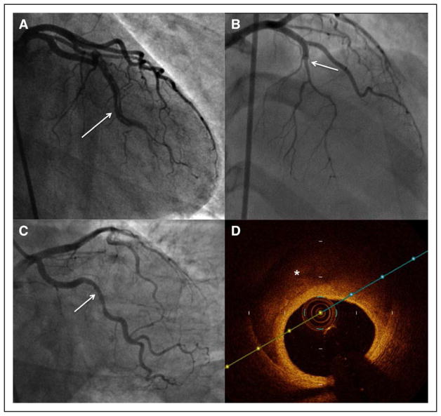
Figure 5. Angiographic features of spontaneous coronary artery dissection.
A, Type 1, multiple radiolucent lumens (arrow) or arterial wall contrast staining. B, Type 2, diffuse stenosis that can be of varying severity and length (dissection starting from arrow). C, Type 3: focal or tubular stenosis (arrow), usually <20 mm in length, that mimics atherosclerosis. Intracoronary imaging should be performed to confirm the presence of intramural hematoma or multiple lumens. D, Optical coherence tomography in type 3 (C) shows intramural hematoma (asterisk).

