Abstract
Introduction
The aim of this study was to compare kinetical data from gait analysis of patients who have undergone total and uni-condylar knee replacement.
Materials and methods
Thirteen patients with unilateral total knee arthroplasty (TKA) and 13 unicondylar knee arthroplasty (UKA), were included, all performed by the same surgeon more than one year prior. The Vicon gait analysis system was used. Statistical power was calculated using SPSS.
Results
No significant difference was found in the spatiotemporal parameters of gait and survival years of the knee prosthesis between the two groups. The UKA group was found to have significantly larger moments than the TKA group in knee adduction on the operated side and knee flexion moment on the unoperated side during the loading phase. The maximum and minimum sagittal plane moments of the operated sides in the TKA group were significantly lower than the unoperated side. The difference was most significant at pre-swing. The maximum and minimum moments on the operated sides in the UKA group were significantly lower for the knee flexion and adduction moments when compared with the unoperated side and were most prevalent during the loading phase.
Conclusions
These results are relevant in terms of prosthesis wear. The TKA knees had smaller magnitude moments than the UKA knees in the sagittal and coronal planes. This could explain the higher revision rates for UKA. In both groups, the non-operated knees had significantly larger moments than the operated knees, which implies that after unilateral knee replacement of either type, the non-operated knee is being put under greater stress.
Keywords: Oxford knee, LCS mobile bearing, Gait analysis, Unilateral knee arthroplasty
Introduction
Knee arthroplasty is a routine orthopaedic procedure. The most common types of knee arthroplasty in the UK are total knee arthroplasty (TKA) followed by unicondylar knee arthroplasty (UKA).1 The main indication is osteoarthritis. In the knee, this is due to the biomechanical stresses affecting the articular cartilage and the subchondral bone of the knee.2
Gait analysis is a technique which has progressed over the years and has become more readily available for research and clinical application due to the computer technology3,4 and new gait analysis equipment. A moment (or torque) is the rotatory effect of a force. The knee is subjected to many forces and moments during gait and it is this area which was explored during this study. The moments around the knee in the sagittal (X) and coronal (Y) planes were the focus of the study because there is so far no clear evidence of kinematic and kinetic advantages of UKA over TKA.5 In this study it is aimed to analyse patients’ gait who have undergone unilateral TKA and UKA and to discover the effects of two different types of knee replacement surgery on patient gait. In addition, the effect of unilateral knee replacement on the opposite clinically asymptomatic knee.
Patients and Methods
Participants
Unicondylar knee replacement is offered to those patients who have intact anterior and posterior cruciate ligaments, a well-preserved lateral compartment, varus and flexion deformity of less than 15 degrees each and at least 110 degrees of knee flexion.6 There were 32 patients eligible for this study, of whom 26 volunteered and were available for testing; these patients participated in the pilot study. Thirteen of the available patients had received unilateral TKA with a LCS® mobile-bearing prosthesis,7 and thirteen had received unilateral medial UKA with Oxford® partial knee prosthesis.6 To be included in this study, the patients were required to be at least one-year postoperative, have an asymptomatic and clinically normal contralateral knee and be able to walk independently. Patients were excluded if their contralateral knee had been replaced or if there was any impairment of their hip or ankle on either leg. These criteria were selected to ensure kinetic gait analysis could be carried out on patients with well-functioning knee replacements and no external factors affecting their gait.
Equipment
Vicon Nexus Software version 2.2.3 was used and the kinetic gait analysis data were collected using a combination of Vicon infra-red cameras and AMTI force platforms. Thirteen cameras were positioned around the gait laboratory to ensure that all markers could be seen throughout testing and therefore the patients’ gait could be captured entirely. Twenty retroreflective markers were placed on the patient’s pelvis and lower limbs. The three-dimensional coordinates of these markers allowed the joints to be orientated in space and the moments around them calculated. Two force platforms were situated in the centre of the gait laboratory so the patient could walk normally from one end of the laboratory to the other, making contact with the force platforms as they walked. Three successful steps in the centre of the force plate were required for each foot. Other equipment used were: scales to measure body mass, measuring tape to record height, distance between anterior superior iliac spines and leg length, calipers to measure knee width and ankle width, and a goniometer to assess the range of motion of the operated knee and as part of the knee score assessment.
Data analysis
SPSS version 22 was used for statistical analysis. The general linear model for repeated measurements was used to compare the variables between operated side and non-operated side, where the main factor was side and the between-subject factor was surgery (TKA and UKA). Considering that some of parameters were not normal distribution, non-parameter tests (Mann–Whitney or Kruskal–Wallis) were used to compare the two surgical groups. The significant level was set at P < 0.05. These groups were compared for two parameters: knee moments in the X (sagittal) and Y (coronal) planes. Maximum and minimum moments were compared for each parameter, together with the percentage of the stance phase at which they occurred. The parameters were also compared over seven events across the gait cycle: foot (heel) strike, initial contact, loading response, midstance, terminal stance, pre-swing and foot off. These parameters are listed chronologically and occur at 0, 2, 11, 35, 65, 90 and 100 percent of the stance phase respectively.
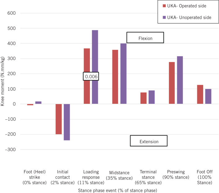
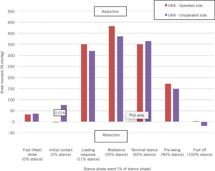
The units of the moments around the joints were in N.mm/kg. The moments were divided by the mass to make the data more comparable between the participants. The direction of the moments is dependent on the numerical value provided by the Vicon system being positive or negative. Moments around the knee joints in the X plane represent: flexion when positive and extension when negative. Moments around the knee in the Y plane represent: adduction when positive and abduction when negative.
Ethics
Ethical approval was granted by the Cambridge South Research Ethics Committee. Caldicott Guardian Approval was obtained. A written consent in accordance to ethics committee guidelines was obtained for all participants.
Results
Demographics
The TKA group consisted of nine males and four females and the UKA group included eight males and five females. There was no significant difference between the groups in terms of age, survival years of prosthesis, knee society score (preoperatively, one year, 2016) and knee society functional score (preoperatively, one year, 2016; Table 1). There was also no significant difference between the spatiotemporal parameters of the groups (Table 2). However, the groups were significantly different in terms of body mass index (BMI). It has been seen that obesity is not associated with increased knee joint torque and power during level ground walking8 and it has been seen that UKA has a higher failure rate only with BMI greater than 35.9
Table 1.
Comparison of demographics and clinical parameters between the unilateral total knee arthroplasty (TKA) and unicondylar knee arthroplasty (UKA) groups.
| Parameter | TKA mean | UKA mean | P-value |
| Age (years) | 65.77 | 65.15 | 0.780 |
| Body mass index (kg/m2) | 30.38 | 26.68 | 0.039a |
| Survival (mean years) | 7.73 | 6.97 | 0.595 |
| Knee score (/100): | |||
| Preoperatively | 37.08 | 42.15 | 0.590 |
| 1 year postoperatively | 88.50 | 91.08 | 0.404 |
| 2016 | 88.46 | 94.62 | 0.183 |
| Knee function score (/100): | |||
| Preoperatively | 55.00 | 64.62 | 0.301 |
| 1 year postoperatively | 92.50 | 94.62 | 0.850 |
| 2016 | 96.15 | 94.62 | 0.711 |
a significant difference (P < 0.05)
Table 2.
Comparison of spatiotemporal data between the unilateral total knee arthroplasty (TKA) and unicondylar knee arthroplasty (UKA) groups.
| Parameter | TKA mean (95% CI) | UKA mean (95% CI) | P-value |
| Cadence (steps/minute) | 102.190 (98.383–105.997) | 105.193 (101.385–109.000) | 0.270 |
| Gait speed (metres/second) | 1.011 (0.953–1.069) | 1.054 (0.996–1.112) | 0.307 |
| Stride: | |||
| Length (metres) | 1.188 (1.142–1.234) | 1.200 (1.154–1.245) | 0.719 |
| Time (seconds) | 1.196 (1.149–1.243) | 1.152 (1.105–1.200) | 0.197 |
| Step length (metres) | 0.600 (0.576–0.623) | 0.599 (0.575–0.622) | 0.953 |
TKA compared with UKA
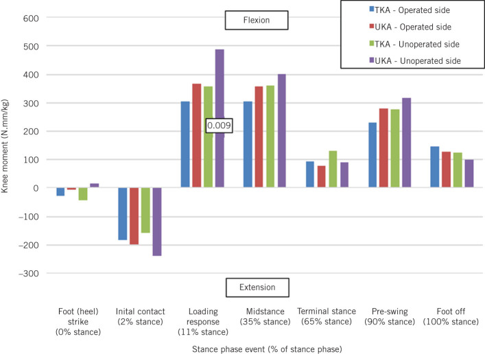
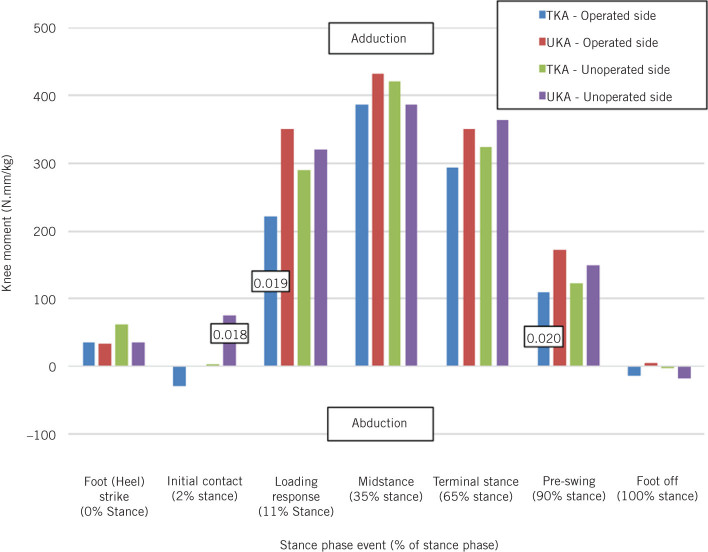
Statistically significant difference was seen in maximum adduction moments between the operated limbs of the TKA and UKA groups. Even in the sagittal plane, the maximum and minimum moments in TKA knees were smaller than that of UKA knees even though it did not reach the level of significance (Table 3). Figure 1 presents the magnitude and the direction of the moments around the knee joint in the sagittal plane. The graph shows a change from extension moments in the earlier stages of the stance phase to flexion moments at the loading response and thereafter. The only significant difference between the TKA and UKA groups is on the unoperated side during loading response when the UKA group had a much higher flexion moment. In the coronal plane, the significant differences between the groups on the operated side occurred during loading response and pre-swing (Fig 2). The UKA groups had significantly greater adduction moments at both of these events. On the unoperated side, there was a significant difference during initial contact. The TKA group adduction moment at this time was close to zero whereas the UKA group was close to 100 N.mm/kg.
Table 3.
Comparison of the mean maximum and minimum moments occurring on the operated side during the stance phase of the gait cycle between the unilateral total knee arthroplasty (TKA) and unicondylar knee arthroplasty (UKA) groups.
| Knee moment | TKA | UKA | P-value |
| X plane (N.mm/kg): | |||
| Max | 555.598 | 636.471 | 0.268 |
| Min | –280.140 | –353.741 | 0.084 |
| Y plane (N.mm/kg): | |||
| Max | 475.947 | 583.518 | 0.029a |
| Min | –96.497 | –97.718 | 0.961 |
a significant difference (P < 0.05)
Figure 1.
Comparison of the moments around the knee in the X plane between the TKA and UKA groups on the operated and unoperated sides (P-values displayed in boxes when TKA and UKA are significantly different for that side)
Figure 2.
Comparison of the moments around the knee in the Y plane between the TKA and UKA groups on the operated and unoperated sides (P-values displayed in boxes when TKA and UKA are significantly different for that side)
TKA: operated side compared with unoperated side
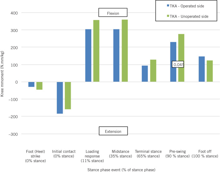
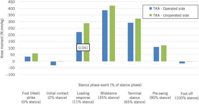
The maximum knee abduction moment was significantly lower on the operated than the unoperated sides (Table 4). In the sagittal plane, the only significant difference between the sides was at pre-swing when the unoperated knee had a significantly higher flexion moment (Fig 3). There is also a significant difference in the coronal plane at loading response. Again, the unoperated side has the greater moment (Fig 4).
Table 4.
Comparison of the mean maximum and minimum moments occurring around the joints on the operated and the unoperated sides in the total knee arthroplasty (TKA) and unicondylar knee arthroplasty (UKA) groups.
| Knee moment | TKA | UKA | |||||
| Operated | Unoperated | P-value | Operated | Unoperated | P-value | ||
| X plane (N.mm/kg): | |||||||
| Max | 555.598 | 599.923 | 0.313 | 636.471 | 765.185 | 0.041a | |
| Min | –280.140 | –268.257 | 0.682 | –353.741 | –375.793 | 0.593 | |
| Y plane (N.mm/kg): | |||||||
| Max | 475.947 | 537.237 | 0.032a | 583.518 | 694.142 | 0.044a | |
| Min | –96.497 | –86.746 | 0.522 | –97.718 | –116.006 | 0.226 | |
a significant difference (P < 0.05)
Figure 3.
Comparison of the moments around the knee in the X plane between the operated and unoperated sides for the TKA group (P-values displayed in boxes when TKA and UKA are significantly different for that side)
Figure 4.
Comparison of the moments around the knee in the Y plane between the operated and unoperated sides for the TKA group (P-values displayed in boxes when TKA and UKA are significantly different for that side)
UKA: Operated side compared with unoperated side
The moments of the opeated side are significantly lower for the knee flexion moments when compared with the unoperated side (Table 4). For the knee, significant differences were identified in the sagittal plane (Fig 5) at loading response and in coronal plane (Fig 6) at initial contact (as there was approximately zero moment on the operated side).
Figure 5.
Comparison of the moments around the knee in the X plane between the operated and unoperated sides for the UKA group (P-values displayed in boxes when TKA and UKA are significantly different for that side)
Figure 6.
Comparison of the moments around the knee in the Y plane between the operated and unoperated sides for the UKA group (P-values displayed in boxes when TKA and UKA are significantly different for that side)
Discussion
The patient groups for each knee replacement were equal in number (13 patients in each) and statistically similar in age, survival years of prosthesis, knee society score and knee society functional score. There was also no significant difference between the spatio-temporal parameters of the groups. This increases the validity of the comparison as differences in these factors could affect results.
TKA compared with UKA
On the operated side, the results displayed a significant difference between the TKA and UKA groups for the knee adduction moment. The UKA group had significantly larger knee adduction moments during the loading phase. The muscles around the joints are required to produce large, equal, opposing moments to stabilise the joints.2 The results suggest less efficient gait in UKA group, as they are required to produce larger opposing moments than the TKA group. Reduction in overall knee adduction moment has been considered to improve loading on medial compartment of the knee.10 Knee adduction moment is related to the distribution of load within the knee. There is a correlation between knee alignment after surgery and peak adduction moments during the stance phase of gait. The peak adduction moments during gait increase in magnitude in direct proportion to the amount of varus of the limb after surgery. The load on the prosthesis significantly increases if residual varus is greater than four degrees.11 Increased knee adduction moments cause increased loads on the medial compartment in relation to the lateral compartment.12
On comparison of the unoperated sides of both groups, the UKA group was found to have significantly larger extension and flexion moments than TKA group. This may be a result of the contralateral side adapting to accommodate the altered gait on the operated side. The increased moments may indicate an overall less efficient gait in the UKA group.
The TKA and UKA groups were compared for the six parameters during the stance phase. This aimed to determine when the differences between the groups were occurring and whether a pattern could be identified. It has been described that cumulative loading of the joint is a more important factor than peak loading in the progression of osteoarthritic changes in the knee.13 The results of this study support these findings. The only significant difference for the knee in the sagittal plane occurred during the loading phase on the unoperated side. The UKA group flexion moment may be higher due to patients favouring their unoperated limb during loading. This would also explain the large flexion moment peak at this point. Overall, it was seen that both the groups of patients in the present study had an increasing extension moment initially in stance phase which by the loading phase changed into flexion moment and towards the end there was increasing extension moment again. Increase in extension moment towards the end of stance phase has been considered to give a better push off which leads to better functioning of the knee.10
The knee moments in the coronal plane were of interest due to the risk of wear in the medial compartment. The differences between the groups on the operated side occurred during the loading response and pre-swing. The UKA groups had significantly greater moments at both of these events. A link has also been noted between increased adduction moments and onset and progression of osteoarthritis, a state of increased wear, particularly in the medial compartment.14,15 Wear in this compartment is a major concern in knee replacement as it can lead to failure and subsequent need for revision. An increased adduction moment may result in increased loads on the medial compartment of the patients’ operated knee leading to increased wear of the patients’ medial compartment prosthesis. Knee kinematics after unicompartmental replacement has been studied in cadavers.16 Similarly, knee kinetics after unicompartmental replacement needs to be studied on cadavers to understand the magnitude of adduction moment beyond which it is detrimental to the survival of the prosthesis.
TKA: operated compared with unoperated sides
The maximum adduction moment was higher on the unoperated side than the operated side. This is due to successful knee replacement operation, as described by Baliunas et al.12 The greater the adduction moment the more the knee becomes offset, exerting greater load on the medial compartment. The lower the moment on the operated side, the more likely the load is to be spread over the whole knee and not just the medial compartment. This is a positive in terms of survival rates of the prosthesis. Reduction in knee adduction moments after TKA on operated side has been reported.17 Benedetti et al. reported that abnormal muscular activity at the trunk on the replaced knee side, together with prolonged tibialis anterior activity during mid stance, led to this reduction in knee adduction moment.17
Moments across the events of the stance phase assesses how the joints on the operated side of a patients’ lower limb in the TKA group are functioning in comparison to the side which has no pathology in the knee, and to discover whether the operated side was following the same moment pattern as the clinically normal side. For the knee in the sagittal plane, the operated and unoperated sides followed a similar pattern, beginning with extension and changing to flexion between initial contact and mid-stance. The only significant difference between the sides was noted at pre-swing, when the un-operated knee had a significantly higher flexion moment. Another finding to note is at initial contact, where the operated side appears to have a substantially larger extension moment exerted on it than the unoperated side (although the difference is not statistically significant). This larger extension moment on the operated side could be due to the loss of proprioception, as hypothesised by Simon et al.18 The larger extension moment seen on the operated side may be due to the leg coming into contact with the ground with greater force. This could be the consequence of a delay in the muscles around the knee producing an opposing flexion moment. This in contrast to what Benedetti et al. found in their study.17 This study found reduced extension moment and prolonged co-contractions of rectus femoris–hamstrings and gastrotibialis anterior, together with abnormal muscular activity at the trunk. As dynamic electromyography was not available, we were not able to monitor the activity of the knee muscles. This delay on the operated side, particularly in the TKA group, may be because more tissue has been removed affecting the intrinsic mechanisms which signal the production of the balancing moment, the end result being a greater extension moment on the operated side. The knee adduction moment was significantly larger on unoperated side than operated side in the coronal plane at loading response. These findings are similar to those of Alnahdi et al.,19 who also studied effect of unilateral TKA on the unoperated side. It can be inferred that after unilateral knee replacement surgery the opposite side is put under greater stress, which could hasten the progress of osteoarthritis.19
UKA: operated vs un-operated side
The comparison between the operated and unoperated sides of patients with UKA displayed some significant differences in the sagittal plane but none in the coronal plane. This may indicate the UKA prosthesis had not significantly offset the joints on the side of operation in the coronal plane as it did not differ from the unoperated knee in terms of coronal moments. This may be attributed to greater postoperative varus angulation in the Oxford unicompartmental knee replacement.20 The larger sagittal plane moment on the unoperated side is possibly due to the patient favouring their unoperated side while walking.
In the sagittal plane at loading response and in the coronal plane at initial contact (as there was approximately zero moment on the operated side), the unoperated knee had significantly greater moments. Apart from these two instances, both sides had no significant difference. The similar moments across gait between the operated and normal sides were reported by Deluzio et al. who noted a return to normal moments postoperatively.21
Limitations of the study
The lack of preoperative data was a major limitation. Measurement of ground reaction forces would help to correlate the other findings. Using a control group of participants with normal knees to obtain two-sided ‘normal’ gait for comparison would support the results of the study. Finally, a larger scale research project in this area can be carried out in future.
Conclusions
Despite the comparison being inconclusive in determining which design provided the closest approximation to ‘normal’ gait, it did provide useful information regarding the differences between the individual knee replacements and ‘normal’ gait. This information can be used to improve the design of the prosthesis and to develop better rehabilitation programmes, which would lead to better functioning of the knee. Although parameters differed slightly between the groups, neither group was consistently more significantly different from normal. If this study was to be repeated with a larger sample size and possibly using a cohort of ‘normal’ participants as a control, more consistent differences might be identified and thus might be able to conclude ‘which is closer to normal’.
References
- 1.National Joint Registry for England, Wales, Northern Ireland and the Isle of Man . Hemel Hempstead: National Joint Registry; 2017. [Google Scholar]
- 2.Bonnin M, Chambat P . Paris: Springer; 2008. [Google Scholar]
- 3.Levine D, Richards J, Whittle MW , 5th ed Edinburgh: Churchill Livingstone; 2012. [Google Scholar]
- 4.Baker R. The history of gait analysis before the advent of modern computers. 2007; (3): 331–342. [DOI] [PubMed] [Google Scholar]
- 5.Komnik I, Weiss S, Pagani CF, Potthast W. Motion analysis of patients after knee arthroplasty during activities of daily living: a systematic review. 2015; (2): 370–377. [DOI] [PubMed] [Google Scholar]
- 6.Zimmer Biomet Oxford® Partial Knee System. http://oxfordknee.com/oxford.cfm (cited December 2017).
- 7.DePuy Synthes LCS® COMPLETE Knee System. https://www.depuysynthes.com/hcp/knee/products/qs/LCS-COMPLETE-Knee-System (cited December 2017).
- 8.DeVita P, Hortobágyi T. Obesity is not associated with increased knee joint torque and power during level walking. 2003; (9): 1,355–1,362. [DOI] [PubMed] [Google Scholar]
- 9.Bonutti PM, Goddard MS, Zywiel MG et al. Outcomes of unicompartmental knee arthroplasty stratified by body mass index. 2011; (8): 1,149–1,153. [DOI] [PubMed] [Google Scholar]
- 10.Hatfield GL, Hubley-Kozey CL, Wilson JL, Dunbar MJ. The effect of total knee arthroplasty on knee joint kinematics and kinetics during gait. 2011; (2): 309–318. [DOI] [PubMed] [Google Scholar]
- 11.Chassin EP, Mikosz RP, Andriacchi TP, Rosenberg AG. Functional analysis of cemented medial unicompartmental knee arthroplasty. 1996; (5): 553–559. [DOI] [PubMed] [Google Scholar]
- 12.Baliunas AJ, Hurwitz DE, Ryals AB et al. Increased knee joint loads during walking are present in subjects with knee osteoarthritis. 2002; (7): 573–579. [DOI] [PubMed] [Google Scholar]
- 13.Maly MR. Abnormal and cumulative loading in knee osteoarthritis. 2008; (5): 547–552. [DOI] [PubMed] [Google Scholar]
- 14.Crenshaw SJ, Pollo FE, Calton EF. Effects of lateral-wedged insoles on kinetics at the knee. 2000; : 185–192. [DOI] [PubMed] [Google Scholar]
- 15.Sharma L, Hurwitz DE, Thonar EJ et al. Knee adduction moment, serum hyaluronan level, and disease severity in medial tibiofemoral osteoarthritis. 1998; (7): 1,233–1,240. [DOI] [PubMed] [Google Scholar]
- 16.Patil S, Colwell CW, Ezzet KA, D’Lima DD. Can normal knee kinematics be restored with unicompartmental knee replacement? 2005; (2): 332–338. [DOI] [PubMed] [Google Scholar]
- 17.Benedetti MG, Catani F, Bilotta TW et al. Muscle activation pattern and gait biomechanics after total knee replacement. 2003; (9): 871–876. [DOI] [PubMed] [Google Scholar]
- 18.Simon SR, Trieshmann HW, Burdett RG et al. Quantitative gait analysis after total knee arthroplasty for monarticular degenerative arthritis. 1983; (5): 605–613. [PubMed] [Google Scholar]
- 19.Alnahdi AH, Zeni JA, Snyder-Mackler L. Gait after unilateral total knee arthroplasty: frontal plane analysis. 2011; (5): 647–652. [DOI] [PMC free article] [PubMed] [Google Scholar]
- 20.Gulati A, Pandit H, Jenkins C et al. The effect of leg alignment on the outcome of unicompartmental knee replacement. 2009; (4): 469–474. [DOI] [PubMed] [Google Scholar]
- 21.Deluzio KJ, Wyss UP, Costigan PA et al. Gait assessment in unicompartmental knee arthroplasty patients: Principal component modelling of gait waveforms and clinical status. 1999; (5): 701–711. [Google Scholar]


