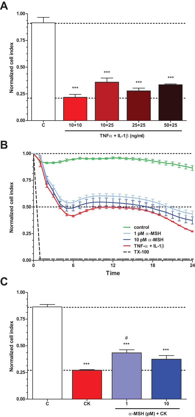
Figure 3. The effect of α-MSH treatment on cell viability of cytokine treated rat brain endothelial cells.
(A) Rat brain endothelial cells were treated with four different combinations of cytokine concentrations (TNF-α + IL-1β: 10 + 10, 10 + 25, 25 + 25, and 50 + 25 ng/ml) for 24 h and the cellular effects were monitored by impedance. Control group received culture medium. (B) Rat brain endothelial cells were treated with cytokines (10 ng/ml TNF-α and 10 ng/ml IL-1β) without or with α-MSH (1 and 10 pM) and the cellular effects were monitored by impedance for 24 h. Control group received culture medium. The cytokines decreased the cell index, which effect could be ameliorated by α-MSH treatment, especially by the lower, 1 pM α-MSH concentration. (C) After 24 h treatment the cell viability significantly decreased due to cytokine treatment, while 1 pM α-MSH significantly blocked the cytokine effect. Mean ± SEM, n = 3–6, ***P < 0.001, #P < 0.05. Asterisks indicate that groups were compared to the control group. Pound signs indicate that groups were compared to the cytokine-treated group. C, control group; CK, cytokine treated group.

