

Abstract
Rationale:
Gallbladder perforation is a serious clinical condition and associated with high morbidity and mortality. A definitive diagnosis is contentious before surgery.
Patient concerns:
We herein report a case of perforation of the gallbladder neck secondary to chemotherapy and radiation for nasopharyngeal carcinoma patient.
Diagnoses:
Gallbladder perforation secondary to chemotherapy and radiation.
Interventions:
To decrease the mortality associated with gallbladder perforation, Laparoscopic cholecystectomy and peritoneal lavage were performed followed for gallbladder perforation patient because of chemotherapy and radiation.
Outcomes:
The patient recovered fully without serious complication and discharged on the 10th postoperative day. A pathological examination of the resected gallbladder revealed cholecystitis in the thinning of the neck.
Lessons:
Early diagnosis and surgical intervention of gallbladder perforation in relation to asopharyngeal carcinoma chemotherapy and radiation are of prime importance. The laparoscopic procedure is safe and feasible in the selected patients.
Keywords: chemotherapy, gallbladder, radiation, spontaneous perforation
1. Introduction
Gallbladder perforation is a rare but serious and potentially fatal disease due to the delayed diagnosis, which usually requires immediate surgical intervention.[1] The main cause of gallbladder perforation is often associated with cholecystitis with or without gallstones.[2] The incidence of gallbladder perforation in acute cholecystitis is about 2% to 18%.[3] However, cases reported as idiopathic or spontaneous gallbladder perforation are rarely seen due to lacking the typical clinical presentation, radiological, and histopathological characteristics.[4] As a result, the diagnosis is often delayed or even missed. This report presents an interesting case of acute acaculous cholecystitis secondary to chemotherapy and radiation with the finding of macroscopic perforation of the gallbladder neck, which has not been reported yet.
2. Case report
This study was approved by the Institutional Review Board of Zhejiang Provincial People's Hospital. Informed consent was obtained from this patient for publication of the unique case report.
A 54-year-old male presented in surgical emergency with upper abdomen pain for 1 day with increase in its intensity over the last 2 hours. The pain was more in the right hypochondrium with occasional vomiting and upper abdominal distention. There was a history of nasopharyngeal carcinoma half 1 year ago, and he has received cisplatin chemotherapy for 5 times and radiation for 30 times (95%PTV boost 8.8 Gy/4 F) until now. He just received cisplatin chemotherapy and radiation 1 day ago. There was nothing particular in the patient's other history. His vital signs were as follows: temperature: 37.7 °C, pulse: 105 beats/minute, respirations: 30 breaths/minute, blood pressure: 138/72 mm Hg, saturation: 94% on room air, and the patient's abdomen was distended, and there was local tenderness in the upper abdomen without muscular defense. On abdominal examination, it was found distended and tender in the upper abdomen with muscular defense. His routine investigations revealed his Hb was 12.2 g, ALT 83 U/L, AST 98 U/L, PT 18.2 seconds, leukocyte count: 2.93 × 10^9 /L, CRP 28 mg/L, sodium 133 mmol/L, T bilirubin, INR, amylase, and lipase were within normal limits. Ultrasonography showed moderate free fluid in peritoneal cavity with thickened gallbladder and no stones within (Fig. 1). A computed tomography (CT) scan was also available (Fig. 2A and B). Diagnostic puncture in right lower abdomen showed 5 mL yellow-brown purulent ascites. Under the diagnosis of the digestive tract perforation, emergency laparoscopic exploration was performed. Contrary to our expectations, the perforation site was not in the alimentary tract, but rather in the gallbladder.
Figure 1.
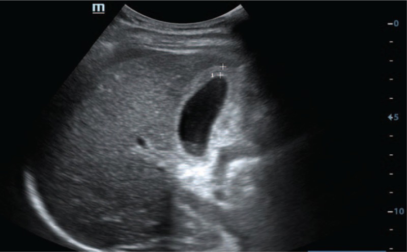
Preoperative imaging findings. Preoperative ultrasonography shows thickened gallbladder with moderate free fluid in peritoneal cavity.
Figure 2.

Preoperative imaging findings. Computed tomography (A, B) shows thickened gallbladder with moderate free fluid in peritoneal cavity.
The following were the operative finding: upper and lower abdomen contained about 0.5 L of bile stained fluid with pus mosses. There was a 0.5 × 0.5 cm ulcerative perforation in the gallbladder neck area (Fig. 3A and B). No gallstone was found in peritoneal cavity. Laparoscopic cholecystectomy was performed followed by thorough peritoneal lavage and a subhepatic drain was placed.
Figure 3.

Intraoperative finding: (A) perforation of gallbladder at neck and (B) moderate free fluid in peritoneal cavity.
In postoperative period, there was about 100 mL bilious drainage daily from subhepatic drain. The patient recovered fully subsequently and discharged on the 10th postoperative day. A pathological examination of the resected gallbladder revealed cholecystitis in the thinning of the neck (Fig. 4).
Figure 4.

Photomicrograph shows cholecystitis of the gallbladder (200×).
3. Discussion
Gallbladder perforation comprises of about 2% cases of acute biliary emergencies and forms a challenge because of high mortality and morbidity.[5,6] Niemeier[7] classified the condition into 3 types: type I, acute free perforation into peritoneal cavity; type II, subacute perforation with pericholecystic abscess formation; and type III, chronic perforation with cholecystoenteric fistula. Type I perforation is in the form of peritonitis, and its treatment is relatively necessary by urgent laparotomy and cholecystectomy, or cholecystostomy. In contrast, type II perforation is far more complex due to chronic nature and the lack of consensus about treatment modality.[8] Our patient had a type I gallbladder perforation. Factors contributing to spontaneous perforation include acute inflammation, infection, lithiasis, congenital obstruction, and anticoagulant therapy. The fundus, the most distant part from the main feeding artery, is the most common site for perforation because of ischemic changes of the gallbladder wall. But interestingly, our case occurs at the neck not the fundus. Systemic vascular disorders, such as atherosclerotic cardiovascular disease, diabetes, and malignancy, are major risk factors for the perforation of gallbladder.[9]
It is relatively rare that gallbladder perforation developed without inflammation, stone, cancer, or body trauma, which makes our research more innovative and unique. Gallbladder perforation may be related to bile duct obstruction or the gallbladder wall destruction, such as acute cholecystitis, gallstones, abdominal surgery, parasites, gallbladder vascular compression, or obstruction. Uncommonly, chemotherapy and radiation can also lead to gallbladder injury. To our knowledge, there is no report of nasopharyngeal carcinoma chemotherapy and radiation-induced gallbladder perforation, especially it occurs at gallbladder neck. It is worth noting that the gallbladder perforation occurred on the second day after completing the cisplatin chemotherapy and radiation, so chemotherapy and radiation-induced gallbladder injury is an important contributing factor. Nasopharyngeal carcinoma is one of the most universal malignant tumors worldwide, and metastasis can cause the majority of cancer-related mortalities. But, our pathological examination of the resected gallbladder revealed chronic cholecystitis and necrosis, without secondary gallbladder cancerous metastasis. In this gallbladder perforation case, gallbladder motility-related hormonal reduce, the decline of enteric nervous system function and immunosuppressed states may also play important roles in gallbladder perforation.
Prior to laparotomy, we often have difficulty in diagnosing the gallbladder perforation. Patients with spontaneous perforation often present with biliary peritonitis[10] as we saw in the patient. The typical presentation includes fever, vomiting, abdominal pain, and sometimes shock.[11] CT and ultrasonography are the sensitive and effective tools to diagnose gallbladder perforation.[12,13] Gallbladder wall thickening, pericholecystic fluid collection, and the streaky omentum or mesentery is common findings of gallbladder perforation.[1,14] Detection on imaging such as CT scans or ultrasonography can be difficult in small perforations and the findings are usually nonspecific. The presence of extra-luminal gallstones would be a suggestive sign, but it was not seen in this case. Confirmatory diagnosis is only after laparotomy and by seeing bile colored peritoneal collection and perforated gallbladder. In the event of the calculus gallbladder perforation, the first choice is urgent surgical treatment. But we also give conservative treatment consisted of puncture drainage, antibiotics, absolute diet, and total parenteral nutrition when there are no symptoms of peritonitis.
4. Conclusions
Our case report is the first presentation of gallbladder perforation associated with nasopharyngeal carcinoma chemotherapy and radiation. In order to prevent gallbladder perforation, we should think seriously about the chemotherapy or radiation adverse event, dose, the weight, gallstones, body surface area, and physical problems. To decrease the morbidity and mortality associated with gallbladder perforation, early diagnosis and surgical intervention are of prime importance. Such cases should always be properly investigated keeping in mind the serious complications.
Acknowledgments
The authors thank the National Science Foundation Committee (NSFC) of China (81502482); Zhejiang Provincial National Science Foundation Committee (NSFC) of China (LQ14H160017); and Zhejiang Provincial research projects of medical and healthy industries (grant number: 2015KYB026, 2017KY193, and 2017KY210) for the support.
Author contributions
Data curation: Guo-liang Shen, Cheng-wu Zhang, Li Jin, hongguo yang.
Formal analysis: Ying Shi.
Funding acquisition: De-fei Hong, weiding wu.
Investigation: jungang zhang, Guo-liang Shen, hanhui cai.
Methodology: wei sun.
Project administration: Li Jin, hongguo yang.
Resources: Cheng-wu Zhang, De-fei Hong, hanhui cai.
Software: Li Jin, wei sun.
Validation: Zhi-ming Hu, weiding wu.
Visualization: weiding wu.
Writing – original draft: jungang zhang.
Writing – review & editing: jungang zhang, Zhi-ming Hu, weiding wu.
Footnotes
Abbreviation: CT = computed tomography.
Funding/support: This study was supported by the National Science Foundation Committee (NSFC) of China (81502482); Zhejiang Provincial National Science Foundation Committee (NSFC) of China (LQ14H160017); and Zhejiang Provincial research projects of medical and healthy industries (grant number: 2015KYB026, 2017KY193, and 2017KY210).
The authors have no conflicts of interest to disclose.
References
- [1].Menakuru SR, Kaman L, Behera A, et al. Current management of gall bladder perforations. ANZ J Surg 2004;74:843–6. [DOI] [PubMed] [Google Scholar]
- [2].Roslyn J, Busuttil RW. Perforation of the gallbladder: a frequently mismanaged condition. Am J Surg 1979;137:307–12. [DOI] [PubMed] [Google Scholar]
- [3].Kalliafas S, Ziegler DW, Flancbaum L, et al. Acute acalculous cholecystitis: incidence, risk factors, diagnosis, and outcome. Am Surg 1998;64:471–5. [PubMed] [Google Scholar]
- [4].Estevao-Costa J, Soares-Oliveira M, Lopes JM, et al. Idiopathic perforation of the gallbladder: a novel differential diagnosis of acute abdomen. J Pediatr Gastroenterol Nutr 2002;35:88–9. [DOI] [PubMed] [Google Scholar]
- [5].MacDonald JA. Perforation of the gallbladder associated with acute cholecystitis: 8-year review of 20 cases. Ann Surg 1966;164:849–52. [DOI] [PMC free article] [PubMed] [Google Scholar]
- [6].Jansen S, Doerner J, Macher-Heidrich S, et al. Outcome of acute perforated cholecystitis: a register study of over 5000 cases from a quality control database in Germany. Surg Endosc 2017;31:1896–900. [DOI] [PubMed] [Google Scholar]
- [7].Niemeier OW. Acute free perforation of the gall-bladder. Ann Surg 1934;99:922–4. [DOI] [PMC free article] [PubMed] [Google Scholar]
- [8].Date RS, Thrumurthy SG, Whiteside S, et al. Gallbladder perforation: case series and systematic review. Int J Surg 2012;10:63–8. [DOI] [PubMed] [Google Scholar]
- [9].Derici H, Kara C, Bozdag AD, et al. Diagnosis and treatment of gallbladder perforation. World J Gastroenterol 2006;12:7832–6. [DOI] [PMC free article] [PubMed] [Google Scholar]
- [10].Badru F, Litton T, Puckett Y, et al. Spontaneous gallbladder perforation in a child secondary to a gallbladder cyst: a rare presentation and review of literature. Pediatr Surg Int 2016;32:629–34. [DOI] [PubMed] [Google Scholar]
- [11].Mirza B, Ijaz L, Saleem M, et al. Management of biliary perforation in children. Afr J Paediatr Surg 2010;7:147–50. [DOI] [PubMed] [Google Scholar]
- [12].Tsai MJ, Chen JD, Tiu CM, et al. Can acute cholecystitis with gallbladder perforation be detected preoperatively by computed tomography in ED? Correlation with clinical data and computed tomography features. Am J Emerg Med 2009;27:574–81. [DOI] [PubMed] [Google Scholar]
- [13].Solva M, Pamilo M, Paivansaio M, et al. Ultasonography in acute gallbladder perforation. Acta Radiol 1988;29:41–4. [PubMed] [Google Scholar]
- [14].Hosaka A, Nagayoshi M, Sugizaki K, et al. Gallbladder perforation associated with carcinoma of the duodenal papilla: a case report. World J Surg Oncol 2010;8:41. [DOI] [PMC free article] [PubMed] [Google Scholar]