Abstract
Interposition of the transverse ligament of the knee between fragments of an intercondylar eminence fracture was diagnosed using magnetic resonance imaging (MRI) in a 11-year-old boy after a sports injury. The interposition was confirmed and corrected during arthroscopy. We report what we believe to be the first published case of isolated interposition of the transverse ligament in a minimally displaced fracture of the tibial eminence.
Keywords: Tibia, Trauma, Transverse ligament of the knee, Interposition
Introduction
Transverse ligament (TL) of the knee, also known as anterior intermeniscal ligament (AIL) or transverse geniculate ligament is an anatomical structure inserting into the anterior horns of the menisci [1, 2]. It is located posterior to Hoffa’s fat pad and may occasionally be visible on lateral plain radiographs of the knee [3]. However, the imaging method of choice of the transverse ligament of the knee is magnetic resonance imaging (MRI). The reported incidence of the TL ranges between 31 and 94% of the population (Table 1) [1–8]. The TL restricts the antero-posterior excursion of the medial meniscus during the early phase of knee flexion [9]. Increased tension of this ligament during knee flexion and rotation may contribute to tears of the anterior horn of the medial meniscus [2].
Table 1.
The incidence of the transverse ligament of the knee
| Reference | Method | Number of specimens / imaged knees | Incidence of the transverse ligament of the knee (%) |
|---|---|---|---|
| [4] | Cadaveric | 92 | 69 |
| [5] | Cadaveric | 34 | 71 |
| [6] | Cadaveric | 50 | 94 |
| [7] | Lateral radiograph | 50 | 12 |
| [7] | MRI | 50 | 68 |
| [1] | MRI | 229 | 53 |
| [1] | MRI + arthroscopy | 36 | MRI 44, arthroscopy 67 |
| [8] | MRI | 100 | 31 |
| [2] | MRI | 49 | 73.5 |
Sports injuries of the tibial eminence are common, and usually result from forced valgus and external rotation of the tibia or hyperflexion and internal rotation of the tibia [10]. The initial diagnosis is usually based on clinical findings and a set of knee radiographs (antero-posterior and lateral projections), or a computed tomography (CT) scan. MRI examination is mandatory, if injury to soft tissues, cartilage, or ligaments is suspected. To the authors’ knowledge this is the first published case of traumatic interposition of the transverse ligament of the knee between the bony fragments in a minimally displaced fracture of the tibial eminence.
Case report
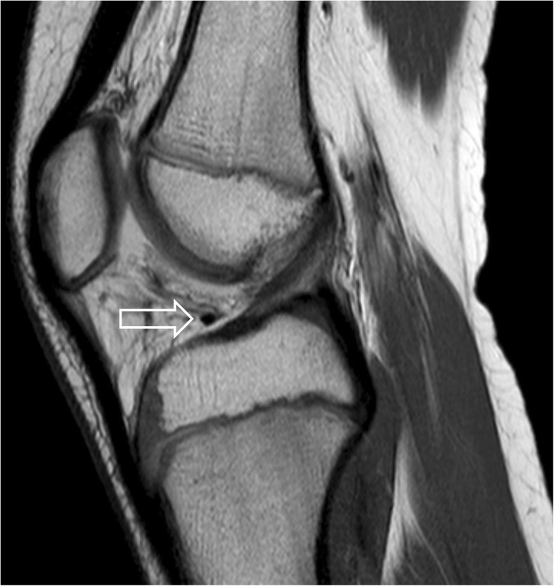
An 11-year-old boy underwent MRI at our clinic to rule out Osgood–Schlatter disease. The images were obtained with an Achieva 1.5-T MRI system (Philips Healthcare, Amsterdam, Netherlands) using a knee coil. No signs of Osgood–Schlatter disease were demonstrated, and the transverse ligament of the knee was present and normal in appearance (Fig. 1).
Fig. 1.
Sagittal proton density (PD)-weighted image of the knee 1 week before the accident. The normal transverse ligament is seen in its anatomical position (arrow). No signs of Osgood–Schlatter disease are visible
One week later the same patient had a sports injury—a direct impaction of the flexed knee against a boulder during a snow-board ride. Radiography performed in the emergency ward at the skiing resort demonstrated a minimally displaced fracture of the tibial eminence, and the patient’s knee was treated with a cast. The diagnosis of a minimally displaced fracture of the tibial eminence was confirmed by CT performed at another institution (not shown).
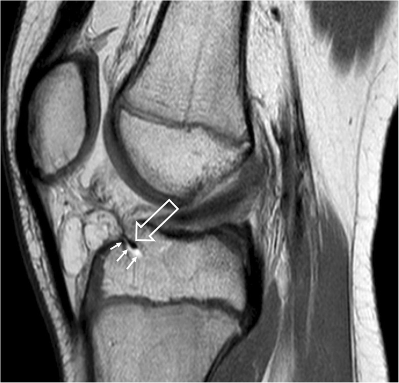
The patient was referred to our institution 7 days after the trauma. MRI performed on the same day, using the same machine and imaging technique as previously, demonstrated a minimally displaced fracture of the intercondylar eminence involving the articular cartilage. Additionally, inferior displacement and interposition of the transverse ligament of the knee between the fragments of the fractured bone were demonstrated (Fig. 2).
Fig. 2.
Sagittal PD image of the knee 1 week after the trauma. A fracture line in the anterior portion of the intercondylar eminence (small arrows) and inferior displacement and interposition of the transverse ligament of the knee between the fragments of the fractured bone is demonstrated (large arrow)
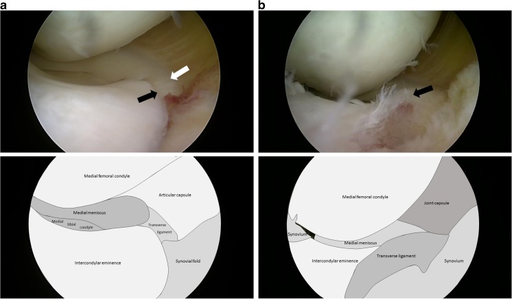
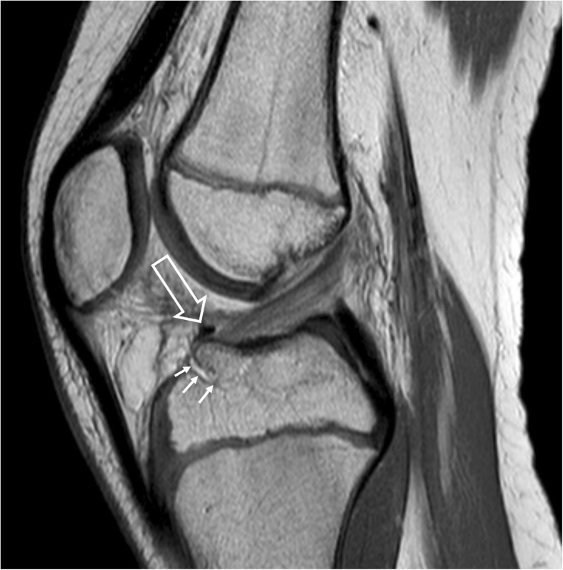
Eleven days after injury the patient underwent arthroscopic surgery. The intraoperative findings confirmed the radiological diagnosis (Fig. 3a). The transverse ligament was successfully repositioned and bony fragment with anterior cruciate ligament insertion was stabilized using an absorbable suture loop (Fig. 3b). A normal position was confirmed in a follow-up MRI performed 6 weeks after the operation (Fig. 4).
Fig. 3.
a Intraoperative image. Transverse ligament of the knee (white arrow) interposed under the tibial eminence (black arrow). b Intraoperative image. Transverse ligament of the knee (arrow) has been restored to its anatomical position after tibial eminence stabilization
Fig. 4.
Control MR examination 6 weeks after the operation, sagittal PD image of the knee. The repositioned transverse ligament (large arrow) is seen in its normal position. The fracture line in the anterior portion of the tibial eminence is still visible (small arrows)
Over a 6-month follow-up, the patient has been pain-free and has had a full range of knee motion.
Discussion
Fractures of the tibial eminence are common in children between 8 and 12 years of age, usually resulting from sports trauma. In most cases, these fractures present as isolated injuries.
Fractures of the tibial eminence are graded according to the modified Meyers and McKeever classification (Table 2). The original classification (three types based on the severity of displacement of the avulsed bone fragment) was proposed by Meyers and McKeever in 1959 [11]. The classification was subsequently modified by the same authors in 1970, creating a new subgroup (III+) encompassing cases with rotated bone fragment [12]. In 1977, Zaricznyj added type IV, including comminuted fractures [13]. The type I and reducible type II lesions can be treated conservatively, the unreducible type II lesions and the type III and IV lesions require surgery [9, 14]. The presented case can be classified as a reducible type II lesion.
Table 2.
Classification of fractures of the intercondylar eminence of the tibia
| Type according to Meyers and McKeever [11] | Type according to Meyers and McKeever [12] | Type according to Zaricznyj [13] | |
|---|---|---|---|
| Minimum displacement of the avulsed fragment and excellent bone apposition | I | I | I |
| Elevation of the anterior third to half of the avulsed fragment producing a beaklike appearance on the lateral roentgenogram | II | II | II |
| Avulsed fragment completely separated from its bone bed, no apposition of the fragment | III | III | IIIA |
| Avulsed fragment completely separated from its bone bed, and rotated so that the cartilaginous surface of the cartilaginous surface of the fragment faces the raw bone of the bone bed making union impossible | – | III+ | IIIB |
| Comminuted fracture | – | – | IV (IIIC) |
The diagnosis is usually based on history, clinical findings, and radiography. If the radiographic image is equivocal, the fracture lines and structure of the bone may be assessed in CT. However, radiography or CT, although performant in assessment of the osseous structures, has only a limited value in the diagnosis of soft-tissue injuries. The reported lesion would have been missed if the imaging were limited only to radiography or CT. In our institution MRI is a gold standard in the assessment of sports injuries of the knee, demonstrating abnormalities of both bone and soft tissues. Ultrasound would not be helpful in this particular case as the transverse ligament was positioned deeply in the fracture. Moreover, the transverse ligament of the knee is not usually assessed in routine ultrasound examinations of the knee and is absent in a large proportion of normal knees.
To the authors’ knowledge no cases of interposition of the TL into a fracture were published. Cadaveric studies show that the TL inhibits posterior translation of the anterior horn of the medial meniscus in the early degrees of the knee flexion (30°) and has no effect on the meniscal motion at extension, 60° flexion, and full flexion [9]. The disruption of the transverse ligament of the knee results in retraction of the anterior horn of the medial meniscus medially and distally over the proximal anterior tibia [6]. The increased frequency of medial meniscal tears was found in patients with a TL attachment to the medial meniscus, compared with patients without this attachment [2]. Therefore, we speculate that limited mobility of the medial meniscus in an untreated case of interposition of the TL into a fracture may result in reduced movement range of the knee and tear of the medial meniscus.
Conclusion
Magnetic resonance imaging should be highly recommended to rule out unexpected pathological findings in patients with fractures of the tibial plateau, even in cases with minimal dislocation of fragments.
Compliance with ethical standards
Conflicts of interest
The authors declare that they have no conflicts of interest.
References
- 1.Aydingöz Ü, Kaya A, Atay ÖA, Öztürk MH, Doral MN. MR imaging of the anterior intermeniscal ligament: classification according to insertion sites. Eur Radiol. 2002;12:824–829. doi: 10.1007/s003300101083. [DOI] [PubMed] [Google Scholar]
- 2.De Abreu MR, Chung CB, Trudell D, Resnick D. Anterior transverse ligament of the knee: MR imaging and anatomic study using clinical and cadaveric material with emphasis on its contribution to meniscal tears. Clin Imaging. 2007;31(3):194–201. 10.1916/j.clinimag.2007.01003. [DOI] [PubMed]
- 3.Sintzoff SA, Gevenois PA, Andrianne Y, Struyven J. Transverse geniculate ligament of the knee: appearance at plain radiography. Radiology. 1991;180(1):259. doi: 10.1148/radiology.180.1.2052706. [DOI] [PubMed] [Google Scholar]
- 4.Kohn D, Moreno B. Meniscus insertion anatomy as a basis for meniscus replacement: a morphological cadaveric study. Arthroscopy. 1995;11(1):96–103. doi: 10.1016/0749-8063(95)90095-0. [DOI] [PubMed] [Google Scholar]
- 5.Berlet GC, Fowler PJ. The anterior horn of the medical meniscus. An anatomic study of its insertion. Am J Sports Med. 1996;26(4):540–543. doi: 10.1177/03635465980260041201. [DOI] [PubMed] [Google Scholar]
- 6.Nelson EW, LaPrade RF. The anterior intermeniscal ligament of the knee—an anatomic study. Am J Sports Med. 2000;28(1):74–76. doi: 10.1177/03635465000280012401. [DOI] [PubMed] [Google Scholar]
- 7.Sintzoff SA, Jr, Stallenberg B, Gillard I, Gevenois PA, Matos C, Struyven J. Transverse geniculate ligament of the knee: appearance and frequency on plain radiographs. Br J Radiol. 1992;65(777):766–768. doi: 10.1259/0007-1285-65-777-766. [DOI] [PubMed] [Google Scholar]
- 8.Erbagci H, Yildirim H, Kizilkan N, Gümüsburun E. An MRI study of the meniscofemoral and transverse ligaments of the knee. Surg Radiol Anat. 2002;24(2):120–124. doi: 10.1007/s00276-002-0023-8. [DOI] [PubMed] [Google Scholar]
- 9.Muhle C, Thompson WO, Sciulli R, et al. Transverse ligament and its effect on meniscal motion: correlation of kinematic MR imaging and anatomic sections. Investig Radiol. 1999;34(9):558–565. doi: 10.1097/00004424-199909000-00002. [DOI] [PubMed] [Google Scholar]
- 10.Fotiadou AN, Karantanas AH. Knee. In: Karantanas AH, editor. Sports Injuries in children and adolescents. Springer, Berlin Heidelberg; 2011. 10.1007/174_2010_16
- 11.Meyers MH, McKeever FM. Fracture of the intercondylar eminence of the tibia. J Bone Joint Surg Am. 1959;41-A(2):209–220. doi: 10.2106/00004623-195941020-00002. [DOI] [PubMed] [Google Scholar]
- 12.Meyers MH, Mc Keever FM. Fracture of the intercondylar eminence of the tibia. J Bone Joint Surg Am. 1970;52(8):1677–1684. doi: 10.2106/00004623-197052080-00024. [DOI] [PubMed] [Google Scholar]
- 13.Zaricznyj B. Avulsion fracture of the tibial eminence: treatment by open reduction and pinning. J Bone Joint Surg Am. 1977;59(8):1111–1114. doi: 10.2106/00004623-197759080-00022. [DOI] [PubMed] [Google Scholar]
- 14.Park HJ, Urabe K, Naruse K. Arthroscopic evaluation after surgical repair of intercondylar eminence fractures. Arch Orthop Trauma Surg. 2007;127(9):753–757. doi: 10.1007/s00402-006-0282-7. [DOI] [PubMed] [Google Scholar]


