



Abstract
Quiescence of myometrium contractile activity allows uterine expansion to accommodate the growing fetus and prevents preterm labor particularly during excessive uterine stretch in multiple pregnancy. However, the mechanisms regulating uterine response to stretch are unclear. We tested the hypothesis that prolonged uterine stretch is associated with decreased myometrium contractile activity via activation of TWIK-related K+ channel (TREK-1). Pregnant women at different gestational age (preterm and term) and uterine stretch (singleton and twin pregnancy) were studied, and uterine strips were isolated for measurement of contractile activity and TREK-1 channel expression/activity. Both oxytocin- and KCl-induced contraction were reduced in term vs preterm pregnancy and in twin vs singleton pregnancy. Oxytocin contraction was reduced in uterine segments exposed to 8 g stretch compared to control tissues under 2 g basal tension. TREK-1 mRNA expression and protein levels were augmented in Singleton-Term vs Singleton-Preterm, and in uterine strips exposed to 8 g stretch. The TREK-1 activator arachidonic acid reduced oxytocin contraction in preterm and term, singleton and twin pregnant uterus. The TREK-1 blocker L-methionine enhanced oxytocin contraction in Singleton-Term and twin pregnant uterus, and reversed the decreases in contraction in uterine strips exposed to prolonged stretch. Carboprost-induced uterine contraction was also reduced by arachidonic acid and enhanced by L-methionine. Thus, myometrium contraction decreases with gestational age and uterine expansion in twin pregnancy. The results suggest that prolonged stretch enhances the expression/activity of TREK-1 channel, leading to decreased myometrium contractile activity and maintained healthy term pregnancy particularly in multiple pregnancy.
Keywords: contraction, potassium channel, pregnancy, uterus, stretch
Graphical abstract

1. Introduction
Rhythmic contraction and relaxation of the myometrium are important for the uterus to perform its function during menstruation, copulation, embryo implantation and pregnancy [1–3]. Successful natural and artificial embryo implantation requires adequate period of myometrium quiescence and reduced contractile activity. Normal pregnancy is also largely associated with uterine quiescence with occasional small rhythmic contractions, thus allowing adequate expansion of the uterus to accommodate the growing fetus and maintain healthy pregnancy to full-term [4]. Disruption of the uterine contraction/relaxation balance during pregnancy could cause preterm labor with serious consequences to mother and fetus particularly during multiple pregnancy [5–8], and thus makes it important to understand the mechanisms controlling myometrium contraction during pregnancy, at different stages of gestation, in singleton vs multiple pregnancy, and in response to prolonged stretch.
Oxytocin is a nonapeptide produced by the hypothalamus-pituitary, the corpus luteum [9, 10], adrenal medulla [11], and placenta [12]. Plasma levels of oxytocin are relatively low in the non-pregnant state, but show progressive increases starting at week 12 of gestation [13]. Oxytocin causes uterine contractions during the second and third stages of labor and promotes cervical dilation before birth. Uterine contraction is also regulated by various contractile proteins, regulatory enzymes, intracellular ions, and ion channels [14]. Uterine contraction is triggered by increases in intracellular Ca2+ [15–17], in part due to Ca2+ entry through voltage-gated channels [18, 19]. On the other hand, smooth muscle relaxation is caused by decreases in intracellular Ca2+ partly due to membrane hyperpolarization and activation of different types of potassium (K+) channels [20–22]. A novel class of K+ channels termed TWIK-related K+ channel (TREK-1) has been identified in the human myometrium [23–28]. TREK-1 channel activity can be modulated by both chemical and mechanical stimuli [29–31]. Polyunsaturated fatty acids such as arachidonic acid and agents that increase membrane tension activate TREK-1, while sulfur amino acids such as L-methionine inhibit TREK-1 channel [32]. Although the role of different K+ channels in relaxation of vascular and gastrointestinal smooth muscle has been well-studied [20–22], little is known regarding the role of TREK-1 in the regulation of myometrium contractile activity at different gestational ages, in term vs preterm pregnancy, in twin vs singleton pregnancy, and in response to uterine stretch.
The objective of the present study was to test the hypothesis that prolonged uterine stretch is associated with altered myometrium contractile activity via changes in uterine TREK-1 channel. We used uterine segments from pregnant women at different gestational ages and with singleton or twin pregnancy to determine whether: 1) Myometrium contractile activity is altered by the gestational age (preterm vs term) and the degree of uterine expansion/stretch (singleton vs twin), 2) Changes in myometrium contractile activity with gestational age and type of pregnancy involve changes in uterine expression of TREK-1 channel, 3) Prolonged stretch of human myometrium is associated with reduced uterine contraction and increased expression of TREK-1, 4) Modulators of TREK-1 influence myometrial contraction at different gestational ages, in twin vs singleton pregnancy, and in response to uterine wall stretch.
2. Materials and Methods
2.1. Ethics statement
The research protocol and procedures were reviewed and approved by the First Affiliated Hospital of Anhui Medical University Ethics Committee for the Protection of Human Subjects in Research and Tissue Collection (No.20140268).
2.2. Tissue collection
Pregnant women 20 to 35 years old, not in labor, and undergoing elective lower segment caesarean section at the First Affiliated Hospital of Anhui Medical University, Hefei, China were recruited and provided informed consent in writing to participate in the study. The gestational age and maternal history were recorded (Table 1). Patients underwent caesarean section because of suspected acute fetal hypoxia, as indicated by elevated umbilical arterial systolic/diastolic velocity ratio under Doppler ultrasound or no reactivity to nonstress tests, or because of patient-specific social reasons. None of the patients had any maternal complications or health factors such as hypertension, diabetes, infection, premature rupture of membrane, polyhydramnios, oligohydramnios or other comorbidities that could affect uterine function or the expression/activity of TREK-1 channels. Exclusion criteria included serious medical illness, anti-hypertensive medication, multiple pregnancy with more than two babies, and polyhydramnios or oligohydramnios. Singleton pregnant women were divided into Singleton-Preterm (28 to <37 gestational weeks) and Singleton-Term (37 to 42 gestational weeks). Twin pregnant women were divided into Twin-Preterm (28 to <37 gestational weeks) and Twin-Term (37 to 42 gestation weeks). Neonatal birth weight for singleton pregnancies and combined neonatal birth weight for twin pregnancies was recorded as an indicator of the extent of uterine expansion and to test if uterine contractile activity changes with the increases in uterine wall stretch [33] (Table 1).
Table 1.
Patient History and Fetal Parameters.
| Parameter | Singleton-Preterm | Singleton-Term | Twin-Preterm | Twin-Term |
|---|---|---|---|---|
| Patient Age (Years) | 27.4±3.3 | 28.8±2.8 | 27.2±1.6 | 27.7±3.8 |
| Gestational Age (Days) | 240.0±2.6 | 275.4±2.1* | 246.7±1.8*# | 263.0±0.8*#† |
| Pregnancy Number | 1.86±0.23 | 1.94±0.42 | 1.33±0.14 | 1.20±0.13* |
| Previous Vaginal Delivery | 0.36±0.13 | 0.19±0.10 | 0.00±0.00* | 0.00±0.00* |
| Previous Caesarean Section | 0.00±0.00 | 0.00±0.00 | 0.00±0.00 | 0.00±0.00 |
| Previous Abortion | 0.50±0.20 | 0.75±0.42 | 0.25±0.13 | 0.20±0.13 |
| Neonatal Weight (g) | 2327.9±166.6 | 3665.6±90.5* | 4588.3±141.3*# (Twins) | 5938.0±207.0*#† (Twins) |
Data are presented as means±SEM, n=10–16 subjects.
Significantly different (P<0.05) from Singleton-Preterm.
Significantly different (P<0.05) from Singleton-Term.
Significantly different (P<0.05) from Twin-Preterm
After safe delivery of the fetus and placenta through elective lower segment caesarean section, specimens were obtained from the edge of the lower uterine segment incision, immediately placed in Krebs solution, and transported to the laboratory in a thermal box. Uterine strips 7 × 3 mm in size were dissected for measurement of myometrium contraction. Because oxytocin is also synthesized within the decidual tissues immediately adjacent to the myometrium [34], the decidua was removed from the myometrial strips prior to myographic assessment. Other uterine strips were cut into 3 mm×3 mm×3 mm segments in preparation for real-time quantitative PCR (qPCR) and Western blots.
2.3. Measurement of uterine contraction
Longitudinal strips of myometrium (7 mm × 3 mm) were dissected. Using silk string, one tie was made 1 mm from each end of the myometrium strip, leaving a functional 5 mm-long strip between two string loops. The myometrial strip was suspended in a water-jacketed tissue bath filled with 5 ml Krebs solution bubbled with 95% O2 5% CO2 at 37°C. One end of the uterine strip was attached via the string loop to a fixed glass hook at the bottom of the tissue bath, and the other end was connected to a force transducer (Beijing SIA Industrial Electronics Co., Beijing, China). Preliminary tension-contraction curves using uterine strips under increasing basal tension 0.5, 1, 2, 3, 4, 5, 6, 7 and 8, maintained under each basal tension for 30 min, then stimulated with 96 mM KCl, demonstrated maximal KCl contraction at 2 g basal tension, no significant change in KCl contraction at 2 to 7 g basal tension, and a decrease in KCl contraction at 8 g stretch. Macroscopical examination did not show any slippage of the string ties, or damage or tear in any of the uterine strips tested, and the strip’s functional length (5 mm) was not different in tissues under 2 g basal tension compared with tissues under 8 g stretch, suggesting structural integrity of the strips. Also, our histological examination of hematoxylin and eosin stained tissue sections showed intact smooth muscle layer in smooth muscle preparations exposed to control 2 g compared with tissues exposed to 8 g stretch [35], supporting structural integrity under these conditions. Furthermore, the decline in contraction in uterine strips under 8 g stretch was completely reversed and even enhanced in tissues treated with the TREK-1 channel blocker L-methionine, supporting functional integrity of the tissues under stretch. Therefore, in most experiments, myometrium strips from different groups were equilibrated under control 2 g basal tension for 1 h. In order to test the effects of prolonged stretch on contractile function and TREK-1 channel expression, in some experiments the uterine strips were equilibrated under 8 g stretch as compared to control 2 g basal tension for 16 h in tissue culture medium, then the bathing solution was changed to Krebs solution. Uterine strips were stimulated twice with 96 mM KCl, and each control KCl contraction was followed by three washes in Krebs, 5 min each. The uterine strips were then stimulated with increasing concentrations of oxytocin (10−12 to 10−7 M) and the contractile response was recorded. At the end of the experiment, each uterine strip was further examined under microscope to ensure lack of damage or tear, the strip was cut at the string ties, and the weight of the tissue between the ties was recorded.
Under resting conditions, the uterine strips showed an oscillatory behavior comprising phasic contractions that spontaneously returned all the way back to baseline. The uterine oscillations increased in frequency and amplitude with increasing oxytocin concentrations, and the phasic contraction did not return all the way back to the baseline leaving a small steady contraction above the baseline (for simplicity will be termed maintained contraction). The distance between the baseline and the maintained response was measured and translated into uterine contraction in grams using a calibration bar [36]. The maintained contraction was normalized to the weight of the myometrium strip and presented as gram per gram tissue weight. Because the phasic contractile response showed significant variability in frequency and amplitude, the total area under the curve (AUC, R1640 data analysis system, Chengdu, China) was measured. AUC was measured at time 0 and was subtracted from AUC measured after 5 min of application of each oxytocin concentration. The AUC representing total contraction was also normalized to the weight of the myometrium strip and presented as AUC per gram tissue weight.
To determine whether the differences in contraction between uterine strips involve TREK-1, uterine strips of all groups were pre-stimulated with oxytocin (10−7 M). Once oxytocin contraction reached steady-state the tissues were treated with arachidonic acid (10−5 M) or the vehicle ethanol, or L-methionine (1 mM) or the vehicle H2O, and their effects on the area under the curve (AUC) were recorded at 0, 5, 10 and 15 min. The AUC of the tissues exposed to tested drugs were compared to the AUC of control tissues nontreated with TREK-modulators, during an equivalent time period. In some experiments, we used carboprost tromethamine (3×10⁻⁴ M) (Pharmacia & Upjohn Company, Kalamazoo, MI, USA) instead of oxytocin to confirm that the effects of TREK-1 modulators on uterine contraction are not due to direct interaction with oxytocin.
2.4. Real-time quantitative PCR (qPCR)
RNA was isolated from the uterine strips using TRIzol (Shanghai Pufei Biotech Co., Shanghai, China). Total RNA (2 μg) was used for RT to synthesize single-strand complimentary DNA (cDNA) in 25 μl reaction mixture following the instructions for the First-Strand cDNA Synthesis Kit (Beyotime Biotechnology, Jiangsu, China). Next, 1 μl of the cDNA dilution (1:20 for TREK-1 and GAPDH) of the RT product was applied to 20 μl of RT-PCR reaction mixture. Quantification of gene expression was performed using a real-time RT-PCR machine (model MX3000p, Agilent, Santa Clara, CA, USA), published oligonucleotide primers for TREK-1 (Shanghai GeneChem Co., Shanghai, China), and SYBR Master Mixture (Bio-Rad, Hercules, CA, USA), which employs the fluorescein compound SYBR-Green for amplicon detection (TAKARA). GAPDH primer was included in the RT-PCR reaction as internal standard to normalize the results. The following primers were used:
TREK-1: forward, 5′-GATTATACCGTTAGGAAACACC-3′; reverse, 5′-TCCCAGTAAGGCATAGATGA-3′.
GAPDH: forward, 5′-TGACTTCAACAGCGACACCCA-3′; reverse, 5′-CACCCTGTTGCTGTAGCCAAA-3′.
PCR was carried out with one cycle for 10 min at 95°C and then 40 cycles of 30 sec of denaturation at 95°C, 45 sec of annealing at 59°C, and 30 sec of extension at 72°C, followed by 1 min of final extension at 95°C. The number of PCR cycles varied according to the expression level of the target gene. An appropriate primer concentration and number of cycles was determined to ensure that the PCR was taking place in the linear range and thereby guarantees a proportional relationship between input RNA and the cycles readout. The relative gene expression was calculated by comparison of cycle thresholds with the housekeeping gene GAPDH.
2.5. Western blot analysis
Uterine strips were homogenized by RIPA lysis buffer (Beyotime Biotechnology). The homogenate was centrifuged at 10,000 g for 5 min. The supernatant was collected, and protein concentration was determined using BCA protein assay kit (Beyotime Biotechnology). Tissue homogenate was subjected to electrophoresis on 10% SDS polyacrylamide gel and then transferred electrophoretically to PVDF membranes (MilliporeSigma, Burlington, MA, USA). The membranes were incubated in 5% dried nonfat milk in PBS-Tween buffer for 1 h to block nonspecific sites, and then in the primary antibody solution containing TREK-1 (1:1000) rabbit polyclonal antibody (Sigma-Aldrich, St. Louis, MO, USA) at 4°C for 24 h. GAPDH was used as an internal control and detected by a monoclonal antibody (1:500,000, Sigma). The PVDF membranes were washed in PBS-Tween three times for 10 min each then incubated in horseradish peroxidase conjugated secondary antibody (1:5000) for 1.5 h. The membrane blots were washed with PBS-Tween and visualized with enhanced chemiluminescence (ECL). The reactive band corresponding to TREK-1 was analyzed by optical densitometry and ImageJ software (NIH). The densitometry value represented the pixel intensity was normalized to GAPDH to correct for loading.
2.6. Solutions and drugs
Normal Krebs solution contained (in mM): 120 NaCl, 5.9 KCl, 25 NaHCO3, 1.2 NaH2PO4, 11.5 dextrose, 2.5 CaCl2, 1.2 MgCl2 (Sigma). The pH of the Krebs solution was 7.3–7.4 when bubbled with 95% O2 5% CO2 at 37.0±0.5 C. High KCl solution (96 mM) was prepared as normal Krebs but with equimolar substitution of NaCl with KCl. Oxytocin (Shanghai Hefeng Pharmaceutical Company, China) and carboprost tromethamine (Pharmacia & Upjohn Company) were dissolved in deionized water. Stock solution of L-methionine (10−1 M) was prepared in deionized water. Stock solution of arachidonic acid (Sigma, 10−3 M) was prepared in ethanol. The final concentration of ethanol was less than 0.1% and had no effect on uterine contraction. The tissue culture medium used for prolonged incubation of the uterine strips for 16 h was composed of Minimum Essential Medium supplemented with penicillin, streptomycin, and amphotericin B (Gibco/Invitrogen, Grand Island, NY). All other chemicals were of reagent grade or better.
2.7. Statistical analysis
Data were analyzed and presented as means±SEM, with “n” representing the number of subjects per group. For uterine contraction experiments, individual concentration-contraction curves were constructed, sigmoidal curves were fitted to the data using the least squares method, and the EC50 values were measured using Prism (v.5.01; GraphPad Software, San Diego, CA). Data were first analyzed using ANOVA with multiple classification criteria [patient group (preterm vs term, singleton vs twin), tissue stretch (control 2 g basal tension vs 8 g stretch), tissue treatment (treated with arachidonic acid or L-methionine vs nontreated control tissues). When a statistical difference was observed, the data were further analyzed using Bonferroni’s post-hoc test for multiple comparisons. Student’s unpaired t-test was used for comparison of two means. Differences were considered statistically significant if P < 0.05.
3. Results
3.1. Neonatal birth weight as an estimate of uterine expansion
We used neonatal birth weight as an estimate of uterine expansion and an indicator of relative uterine stretch at different stages of pregnancy and in singleton vs twin pregnancies. Neonatal birth weight and consequently uterine wall stretch appeared to be increased in term vs preterm pregnancies. Among singleton pregnancies, neonatal birth weight was greater in term vs preterm pregnancies. Similarly among twin pregnancies, neonatal birth weight was significantly increased in term vs preterm pregnancies. Neonatal birth weight also appeared to be increased in twin vs singleton pregnancies. Among preterm-pregnancies, neonatal birth weight was greater in twin vs singleton pregnancies. Similarly, among term pregnancies, neonatal birth weight was greater in twin vs singleton pregnancies. Collectively, neonatal birth weight was in Twin-Term > Twin-Preterm > Singleton-Term > Singleton-Preterm pregnancies (Table 1).
3.2. Oxytocin-induced contraction in preterm vs term and in twin vs singleton pregnancy
In isolated uterine strips, oxytocin caused an increase in the frequency and amplitude of uterine contraction that did not return back to the baseline, leaving a measurable maintained response above baseline (Fig. 1A). Because of the variability of the contractile response in different uterine strips, the maintained contraction was normalized to the uterine strip weight and presented in g/g tissue weight as previously described [36]. In uterine strips from the four groups of women, oxytocin caused concentration-dependent increase in the maintained contraction that reached a maximum at 10−7 M (Fig. 1B). The oxytocin-induced maintained contraction was reduced in term versus preterm pregnancies. Among singleton pregnancies, oxytocin-induced contraction was significantly reduced in uterine strips from term compared to preterm pregnancies. Similarly among twin pregnancies, oxytocin-induced contraction was significantly reduced in uterine strips from term compared to preterm pregnancies. Oxytocin-induced contraction was also reduced in twin vs singleton pregnancies. Among preterm-pregnancies, oxytocin-induced contraction was reduced in twin vs singleton pregnancies. Similarly, among term-pregnancies, oxytocin-induced maintained contraction was reduced in twin compared to singleton pregnancies. Collectively, oxytocin-induced uterine maintained contraction was in Singleton-Preterm > Singleton-Term > Twin-Preterm > Twin-Term pregnancies (Fig. 1B). When the oxytocin-induced maintained contraction was presented as % of maximum, the oxytocin EC50 was not significantly different among the different groups (Fig. 1C, Table 2).
Fig. 1.
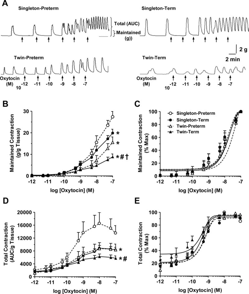
Effect of gestational age and pregnancy type on oxytocin-induced uterine contraction. Uterine strips from Singleton-Preterm, Singleton-Term, Twin-Preterm and Twin-Term pregnant subjects were incubated in normal Krebs solution. The uterine strips were stimulated with oxytocin (10−12 to 10−7 M) and the maintained contraction (in grams) and total contractile response (in AUC) were measured (A). Cumulative oxytocin concentration-contraction curves from uterine strips of different groups were constructed. To correct for differences in the size of uterine segments, maintained contraction was normalized and presented in g/g tissue weight (B), or as % of maximum contraction (C). Because of the variability in the frequency and amplitude of the phasic contractile response, the total contraction area under the curve (AUC) was calculated and presented as AUC/g tissue weight (D) or as % of maximum contractile response (E). Data are presented as means±SEM, n = 7 to 9 subjects.
* Significantly different (P<0.05) from Singleton-Preterm.
# Significantly different (P<0.05) from Singleton-Term.
† Significantly different (P<0.05) from Twin-Preterm
Table 2.
EC50 of Oxytocin-Induced Maintained and Total Contraction in Isolated Uterine Strips of Different Patient Groups.
| Parameter | Singleton Preterm | Singleton Term | Twin Preterm | Twin Term | Singleton Preterm (8 g Stretch) |
|---|---|---|---|---|---|
| Maintained Contraction | 7.72±0.34 | 8.52±0.20 | 7.19±0.5 | 7.96±0.60 | 8.02±0.07 |
| Total Contraction | 9.65±0.18 | 9.53±0.16 | 9.81±0.29 | 9.92±0.30 | 9.87±0.33 |
Oxytocin maintained contraction (in grams) and total contraction (in AUC) was measured as % of maximum contraction and pEC50 (−log EC50) was calculated.
Data are presented as means±SEM, n = 7–9.
Uterine strips were under 2 g basal tension unless indicated otherwise.
To account for the increased amplitude and frequency of the oscillatory uterine contractile response and to assess the changes in total uterine contraction, the oxytocin-induced changes in AUC was measured in uterine strips from the different patient groups. In uterine strips from the four patient groups, oxytocin caused concentration-dependent increase in AUC that reached a maximum at ~10−8 M, then started to decline (Fig. 1D). Among singleton pregnancies, oxytocin-induced AUC was significantly reduced in uterine strips from term compared to preterm pregnancies. Among twin pregnancies, oxytocin-induced AUC was insignificantly reduced in uterine strips from term compared with preterm pregnancies. Among preterm-pregnancies, oxytocin-induced contraction was reduced in twin vs singleton pregnancies. Oxytocin-induced contraction was also reduced in Twin-Term vs Singleton-Term pregnancies. Collectively, oxytocin-induced total uterine contraction was in Singleton-Preterm > Singleton-Term = Twin-Preterm > Twin-Term pregnancies (Fig. 1D). When the oxytocin-induced total contraction was presented as % of maximum, the oxytocin-induced concentration-response curve was slightly shifted to the left and the EC50 was less in Twin-Term compared to the other groups, but collectively the EC50 was not significantly different among the different groups (Fig. 1E, Table 2).
3.3. KCl-induced contraction in preterm vs term and in twin vs singleton pregnancy
High KCl (96 mM) is known to cause membrane depolarization and to stimulate Ca2+ influx through voltage-gated channels [37]. KCl (96 mM) caused an initial followed by steady-state contraction in uterine strips of the different groups (Fig. 2). No oscillatory phasic contraction could be observed during stimulation with KCl. KCl contraction was reduced in term vs preterm pregnancies. Among singleton pregnancies, KCl contraction was significantly reduced in uterine strips from term compared to preterm pregnancies. Similarly among twin pregnancies, KCl contraction was reduced in uterine strips from term compared to preterm pregnancies. Among term pregnancies, KCl-induced contraction was reduced in twin compared to singleton pregnancies. Collectively, KCl-induced uterine contraction was in Singleton-Preterm > Singleton-Term = Twin-Preterm > Twin-Term pregnancies (Fig. 2).
Fig. 2.

Effect of gestational age and pregnancy type on KCl-induced uterine contraction. Uterine strips from Singleton-Preterm, Singleton-Term, Twin-Preterm and Twin-Term pregnant subjects were incubated in normal Krebs solution. The uterine strips were stimulated with 96 mM KCl and the contractile response was recorded and presented in g/g tissue weight. The small contractions preceding the KCl response in uterine strips of Singleton-Term and Twin-Preterm subjects represent spontaneous phasic contractions. Data are presented as means±SEM, n = 7 to 9 subjects.
* Significantly different (P<0.05) from Singleton-Preterm.
# Significantly different (P<0.05) from Singleton-Term.
† Significantly different (P<0.05) from Twin-Preterm.
3.4. Effect of prolonged stretch on uterine contraction
To further test for a possible relationship between uterine stretch and the observed decrease in uterine contraction in term vs preterm, we tested the effects of prolonged stretch on oxytocin-induced contraction in uterine strips. In uterine strips from Singleton-Preterm pregnancies, oxytocin-induced contraction was reduced in uterine strips under prolonged 8 g stretch compared to control tissues under control 2 g basal tension (Fig. 3A). Cumulative data suggest that both the oxytocin-induced maintained contraction in g/g tissue and total contraction in AUC/g tissue were significantly reduced in tissues under 8 g stretch compared to control tissues under 2 g basal tension (Fig. 3B, 3D). When the oxytocin-induced maintained contraction was presented as % of maximum, the concentration-response curve and oxytocin EC50 were not different in tissues under 8 g stretch compared with control tissues under 2 g basal tension (Fig. 3C, Table 2). When the oxytocin-induced total AUC contraction was presented as % of maximum, the concentration-response curve was shifted to the left and the oxytocin EC50 was less in uterine strips from Singleton-Preterm pregnancies under 8 g stretch compared to control tissues under 2 g basal tension; however, the difference did not reach statistical significance (Fig. 3E, Table 2).
Fig. 3.
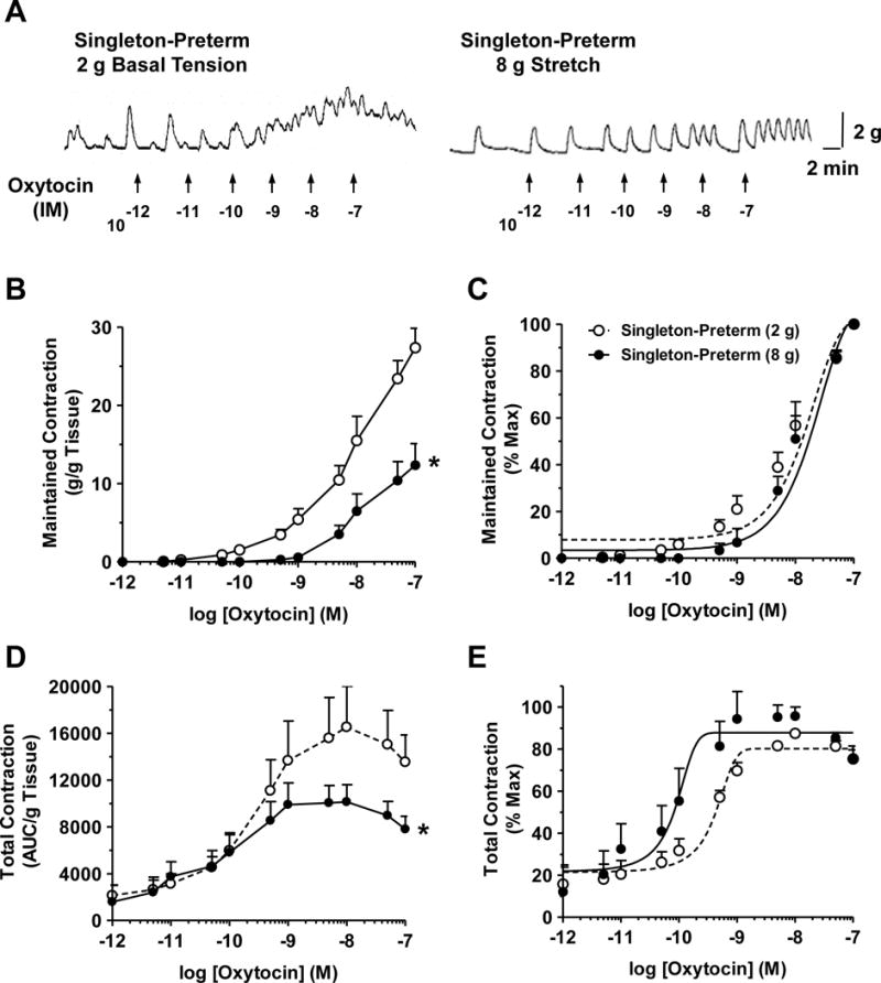
Effect of prolonged stretch on uterine contraction. Uterine strips from Singleton-Preterm pregnant women were incubated under control 2 g basal tension or high 8 g stretch for 16 h, then stimulated with increasing concentrations of oxytocin (10−12 to 10−7 M) (A). Cumulative concentration-contraction curves were constructed and the maintained contraction was normalized and presented in g/g tissue weight (B), or as % of maximum contraction (C). To analyze the changes in the total contractile response, the area under the curve (AUC) was calculated and presented as AUC/g tissue weight (D) or as % of maximum contractile response (E). Data are presented as means±SEM, n = 7.
* Significantly different (P<0.05) from Singleton-Preterm tissues under control 2 g basal tension.
3.5. Uterine TREK-1 expression in term vs preterm and in twin vs singleton pregnancies
Biochemical experiments using qPCR revealed detectable TREK-1 mRNA expression (Fig. 4A), and Western blot analysis revealed a detectable band at 47 kDa corresponding to TREK-1 in uterine strips of the different patient groups (Fig. 4B). PCR analysis revealed that TREK-1 mRNA expression was significantly increased in Singleton-Term compared to Singleton-Preterm pregnant women (Fig. 4A). TREK-1 mRNA expression was significantly decreased in Twin-Preterm vs Singleton-Term pregnancies, and in Twin-Term compared to Singleton-Preterm, Singleton-Term and Twin-Preterm pregnant uterus (Fig. 4A). Western blot analysis revealed that TREK-1 protein levels were significantly increased in Singleton-Term compared to Singleton-Preterm pregnant women (Fig. 4B). TREK-1 protein levels were not significantly different in Twin-Preterm vs Singleton-Preterm, insignificantly decreased in Twin-Preterm vs Singleton-Term, significantly decreased in Twin-Term compared to Singleton-Term, and insignificantly decreased in Twin-Term vs Twin-Preterm pregnant uterus (Fig. 4B).
Fig. 4.

Changes in TREK-1 mRNA expression and protein levels with gestational age and pregnancy type. Uterine tissue homogenate from Singleton-Preterm, Singleton-Term, Twin-Preterm and Twin-Term pregnant women were prepared for q-PCR analysis and TREK-1 mRNA expression was measured relative to the Singleton-Preterm group (A). Tissue homogenate was also prepared for measurement of TREK-1 protein levels using Western blots and antibody to TREK-1 (1:1000). The intensity of the immunoreactive band corresponding to TREK-1 was analyzed using optical densiometry, and normalized to the housekeeping protein GAPDH (B). Data are presented as means±SEM, n=6-8.
* Significantly different (P<0.05) from Singleton-Preterm.
# Significantly different (P<0.05) from Singleton-Term.
† Significantly different (P<0.05) from Twin-Preterm.
3.6. Effect of prolonged uterine stretch on TREK-1 expression
PCR analysis revealed that TREK-1 mRNA expression was increased in uterine strips of Singleton-Preterm pregnant uterus under 8 g stretch compared to control tissues under 2 g basal tension (Fig. 5A). Also, Western blot analysis revealed more prominent immunoreactive band and increases in TREK-1 protein levels in uterine strips of Singleton-Preterm pregnancies under prolonged 8 g stretch compared to control tissues under 2 g basal tension (Fig. 5B).
Fig. 5.

Effect of prolonged uterine stretch on TREK-1 expression. Uterine strips from Singleton-Preterm pregnancies were placed under control 2 g basal tension or prolonged 8 g stretch for 16 h. The tissues were homogenized and prepared for measurement of TREK-1 mRNA using qPCR (A). Other tissues were homogenized and prepared for Western blots using TREK-1 antibody (1:1000). The intensity of the immunoreactive band corresponding to TREK-1 was analyzed using optical densiometry, and normalized to the housekeeping protein GAPDH (B). Data are presented as means±SEM, n=6–8.
* P<0.05, 8 g stretch vs. control 2 g basal tension.
3.7. Effect of TREK-1 modulators on oxytocin-induced uterine contraction
In uterine strips from Singleton-Preterm, Singleton-Term, Twin-Preterm, and Twin-Term pregnancies and precontracted with oxytocin (10−7 M), treatment with the vehicle was associated with a slight time-dependent decline in the AUC representing total uterine contraction, and treatment with modulators of TREK-1 appeared to further modulate oxytocin contraction (Fig. 6A). Cumulative data revealed that the TREK-1 activator arachidonic acid (10−5 M) caused further decline in the AUC representing total contraction of uterine strips of the four patient groups (Fig. 6B, 6C, 6D, 6E). In contrast, treatment with the TREK-1 blocker L-methionine (1 mM) minimized the decline in uterine contraction in the four patient groups, and the uterine contraction appeared to be steadier with time in Singleton-Preterm (Fig. 6B) and Singleton-Term pregnant uterus (Fig. 6C). Treatment with TREK-1 blocker L-methionine significantly enhanced oxytocin contraction in Twin-Preterm (Fig. 6D) and Twin-Term pregnant uterus (Fig. 6E), suggesting functionality of the uterine TREK-1 channel and its responsiveness to both activators and blockers.
Fig. 6.

Effect of modulators of TREK-1 channel on oxytocin-induced uterine contraction. Uterine strips from Singleton-Preterm, Singleton-Term, Twin-Preterm and Twin-term pregnant women were pre-contracted with oxytocin (10−7 M). Once oxytocin contraction reached steady-state, the tissues were treated with TREK-1 activator arachidonic acid (10−5 M), or TREK-1 blocker L-methionine (1 mM), or the vehicle, and the effects on oxytocin contraction were recorded at 0, 5, 10 and 15 min (A). Cumulative data from uterine strips of Singleton-Preterm (B), Singleton-Term (C), Twin-Preterm (D) and Twin-term pregnant women (E) were collected and the magnitude of uterine contraction was presented as % of control AUC (100%) in the absence of TREK-1 modulators. Data represent means±SEM, n=6–7.
* Oxytocin contraction in arachidonic acid treated tissues is significantly reduced (p<0.05) compared to control non-treated tissues.
# Oxytocin contraction in L-methionine treated tissues is significantly enhanced (p<0.05) compared to control non-treated tissues.
3.8. Effect of TREK-1 modulators on carboprost-induced uterine contraction
To rule out possible direct interaction of TREK-1 modulators with oxytocin or the oxytocin receptor, we tested their effects on other stimulants of uterine contraction, namely the prostaglandin analogue carboprost tromethamine. In uterine strips of Singleton-Term pregnancies and precontracted with carboprost tromethamine (3x10⁻⁴ M), the TREK-1 activator arachidonic acid (10−5 M) caused significant decrease of total uterine contraction. In contrast, the TREK-1 blocker L-methionine (1 mM) enhanced the uterine contraction to carboprost tromethamine (Fig. 7).
Fig. 7.

Effect of modulators of TREK-1 on carboprost tromethamine-induced contraction. Uterine strips of Singleton-Term pregnant women were pre-contracted with carboprost tromethamine (3×10⁻⁴ M). Once carboprost tromethamine contraction reached steady-state the tissues were treated with arachidonic acid (10−5 M), L-methionine (1 mM), or the vehicle, and the changes in uterine contraction were measured after 10 min, and presented as % of control AUC (100%) in the absence of TREK-1 modulators. Data are presented as means±SEM, n=6.
* Carboprost contraction in arachidonic acid treated tissues is significantly reduced (p<0.05) compared to control non-treated tissues.
# Carboprost contraction in L-methionine treated tissues is significantly enhanced (p<0.05) compared to control non-treated tissues.
3.9. Effect TREK-1 blockade on uterine strips exposed to prolonged stretch
In uterine strips from Singleton-Preterm pregnancies and exposed to control 2 g basal tension, oxytocin (10−7 M) caused significant contraction that showed some decline over 15 min duration. In uterine strips from Singleton-Preterm pregnancies and exposed to prolonged 8 g stretch, oxytocin-induced total contraction was significantly reduced. In uterine strips from Singleton-Preterm pregnancies exposed to 8 g stretch and treated with the TREK-1 blocker L-methionine, oxytocin-induced contraction was restored and even enhanced above control levels (Fig. 8).
Fig. 8.

Effect of TREK-1 blockade on contraction in uterine strips exposed to prolonged stretch. Uterine strips from Singleton-Preterm pregnant women were incubated under 2 g basal tension or high 8 g stretch for 16 h .The uterine strips were stimulated with oxytocin (10−7 M). Once oxytocin contraction reached steady-state the tissues were treated with TREK-1 blocker L-methionine (1 mM) or the vehicle H2O, and the effects on oxytocin contraction were recorded at 0, 5, 10 and 15 min (A). Cumulative data from uterine strips were collected and the % change of AUC relative to control AUC (100%) in the absence of the TREK-1 blocker L-methionine was measured. Data represent means±weSEM, n=6–7.
* Oxytocin contraction in tissues under 8 g stretch is significantly reduced (p<0.05) compared to control tissues under 2 g basal tension.
# Oxytocin contraction in tissues under 8 g stretch and treated with L-methionine is significantly enhanced (p<0.05) compared to non-treated tissues under 8 g stretch.
† Oxytocin contraction in tissues under 8 g stretch and treated with L-methionine is significantly enhanced (p<0.05) compared to control non-treated tissues under 2 g basal tension.
4. Discussion
The present study in uteri from pregnant women showed that: 1) Uterine contraction is reduced in term compared to preterm and in twin compared to singleton pregnancies. 2) Uterine contraction is reduced during prolonged stretch of the uterine wall. 3) TREK-1 channel is upregulated in Singleton-Term compared to Singleton-Preterm pregnancies, and in response to prolonged stretch. 4) TREK-1 activation reduces uterine contraction, and TREK-1 blockade improves uterine contraction particularly during exposure to prolonged stretch.
Regulation of uterine contraction is critical for normal healthy pregnancy, and safe and uncomplicated labor. During the course of pregnancy, uterine expansion increases progressively and proportionately to fetal growth and the increases in fetal size. In full-term pregnancy, uterine stretch imposed by the fully-developed fetus reaches a threshold level that initiates labor. Studies have suggested that during normal labor, threshold increases in uterine wall stretch could increase myometrial expression of oxytocin receptors, and in turn increases oxytocin-induced uterine contraction [38, 39]. Other studies have suggested that during the final stages of pregnancy, threshold increases in uterine stretch cause upregulation of contraction-associated proteins (CAPs) that could also play a role in the induction of labor [40–44]. While adequate uterine contraction could be beneficial during normal labor, excessive uterine contraction could be detrimental during the course of pregnancy and could lead to abortion or premature labor. Therefore, the uterus is thought to be equipped with adaptive mechanisms that allow it to relax and expand in response to moderate and gradual stretch so that it can accommodate the growing fetus. We hypothesized that the uterine contractile response could be influenced by the size of the pregnant uterus and consequent stretch of the uterine wall pre-birth. Our hypothesis predicted that the size of the uterus and consequently uterine wall stretch would be dependent on gestational age, with uterine wall stretch being greater in term compared to preterm pregnancies. The type of pregnancy (singleton vs twin) could also be a contributing factor, and during twin pregnancies the myometrium is predicted to be exposed to greater stretch due to the excessive distension caused by the second fetus [45]. The present study demonstrated differences in myometrial contractile response in singleton and twin pregnancies at different gestational ages, and these differences appear to be related to uterine wall stretch because: 1) Neonatal birth weight was greater in term vs preterm and in twin vs singleton pregnancies. 2) Oxytocin-induced contraction (analyzed by two different methods in grams and AUC) was reduced in term compared to preterm pregnancy regardless of whether it was singleton or twin pregnancy. 3) Oxytocin-induced contraction was reduced in twin compared to singleton pregnancy regardless of whether it was term or preterm pregnancy. 4) Oxytocin-induced contraction was reduced in uterine strips of Singleton-Preterm pregnancies exposed to prolonged 8 g stretch compared to control tissues under 2 g basal tension. These observations in human uterus are consistent with our previous observations of reduced oxytocin-induced contraction in late pregnant rat uterus, and in virgin rat uterus exposed to prolonged stretch [36]. Collectively these data support a relationship between uterine wall stretch and reduced uterine contraction during the course of pregnancy in order to accommodate the growing fetus.
Previous studies have predicted increases in the uterine contractile response to oxytocin during the final stages of pregnancy and in preparation for natural labor [33, 38, 39]. This is different from the present study which was performed in women with elective cesarean section, and where oxytocin levels may still be at subthreshold levels not sufficient to induce natural labor. Importantly, the observed decrease in uterine contraction was not specific to oxytocin or oxytocin receptor, as the contraction to membrane depolarization by high KCl solution was also reduced in term vs preterm pregnancies and in twin vs singleton pregnancies. Also, contraction to oxytocin was reduced in uterine strips exposed to prolonged 8 g stretch compared to control tissues under 2 g basal tension. Furthermore, the reduced uterine contraction to oxytocin in term vs preterm pregnancies, in twin vs singleton pregnancies, and in response to uterine stretch does not appear to be due to changes in the sensitivity of oxytocin receptors, as the oxytocin EC50 was not different among the different groups and was not significantly altered by stretch. Therefore, the observed reduction in uterine contraction to various agonists in term vs preterm pregnancies, in twin vs singleton pregnancies, and in response to uterine stretch is more likely due to changes in downstream post-receptor mechanisms of uterine smooth muscle contraction and possible changes in the expression/activity of contraction-associated proteins (CAPs).
In search for the molecular mechanisms that could cause the reduced uterine contraction in term and twin pregnancies and in response to prolonged stretch, we have previously shown that the expression/activity of matrix metalloproteinases MMP-2 and MMP-9 are increased during the course of pregnancy in rats, and in late pregnant compared with mid-pregnant and virgin rats [46]. We have also shown that MMP inhibitors improve uterine contraction in uterine strips of late pregnant rats [36]. Also, prolonged stretch of the rat uterus was associated with increased expression/activity of MMP-2 and MMP-9 [36]. Similar increases in MMP expression/activity in response to prolonged stretch have also been observed in small microvessels of the skeletal muscle [47], and smooth muscle of the rat inferior vena cava [35]. MMPs are largely known for their role in tissue remodeling and degradation of extracellular matrix proteins [35, 48]. We and others have recently shown other novel effects of MMPs on cell surface receptors and signaling molecules [48, 49]. Specifically, we have shown that MMP-2 could induce relaxation of rat inferior vena cava via hyperpolarization and activation of large conductance Ca2+-activated K+ channels [50]. These observations made it important to test the potential role of K+ channels in the pregnancy-associated and stretch-related reduction in uterine contraction.
Several types of K+ channels have been identified in different tissues and cells. K+ channels include small conductance Ca2+-activated K+ channels (SKCa), intermediate conductance Ca2+-activated (IKCa), large conductance Ca2+- and voltage-activated (BKCa), voltage-dépendent (KV), ATP-sensitive (KATP) K+ channels, and the inward rectifier (Kir) [20–22, 51, 52]. The TREK-1 channel belongs to two-pore K+ channels (K2P), which are largely expressed in visceral smooth muscle [27]. Studies have suggested that TREK-1 is a class of CAPs that is expressed in human myometrium particularly during pregnancy [23, 28]. Based on the topology of the K+ channel subunits, these K2P channels are thought to maintain background outward K+ current and resting membrane potential and thereby counterbalance membrane depolarization and muscle contraction [29–31]. We tested whether the pregnancy-associated and stretch-related reduction in uterine contraction reflect changes in TREK-1 expression/activity. We found that the mRNA expression and protein levels of TREK-1 were enhanced in Singleton-Term compared with Singleton-Preterm pregnancies. This is consistent with previous reports that TREK-1 is up-regulated in the human myometrium during pregnancy [23–27]. The increases in TREK-1 expression appear to be related to stretch, as the mRNA expression and protein levels were increased in Singleton-Preterm uterus under prolonged 8 g stretch compared to control tissues under 2 g basal tension. However, the mRNA expression and protein levels of TREK-1 appeared to decrease in Twin-Term compared to Singleton-Term and Twin-Preterm pregnancies. The causes of these decreases are unclear, but it is possible that during Twin-Term pregnancies the uterus may have reached a critical size, and a decrease in the relaxing TREK-1 channels may be needed in preparation for parturition. This is supported by reports that TREK-1 may play a role in the differences in uterine contraction in multiple vs singleton pregnancy [33]. Thus while the expression of TREK-1 may increase during most of the gestational period, it may abruptly decrease during the intrapartum period. In other words, the increased TREK-1 expression may help to stabilize the membrane potential and reduce uterine excitability during the course of pregnancy, while a decrease in TREK-1 expression may increase uterine excitability during imminent labor. Another possible explanation is that modulation of TREK-1 with stretch involves not only changes in its expression but also its activity, and an exaggerated TREK-1 activity during Twin-Term pregnancies may prompt a feed-back mechanism to reduce its mRNA expression and protein levels. Future experiments should further assess TREK-1 activity at different stages of pregnancy and in response to different degrees of stretch.
In addition to changes in TREK-1 expression, the present data suggest that TREK-1 function is influenced by gestational age and type of pregnancy as well as in response to stretch because: 1) The TREK-1 activator arachidonic acid further decreased oxytocin-induced myometrial contraction in both preterm and term pregnancies and in singleton and twin pregnancies. 2) The effects of arachidonic acid are not due direct interaction with oxytocin or oxytocin receptor, as arachidonic acid also reduced uterine contraction to the synthetic prostaglandin F2α analogue carboprost tromethamine. 3) The TREK-1 inhibitor L-methionine enhanced oxytocin-induced contraction. The reversal of inhibition of myometrial contraction by L-methionine was more pronounced in Twin-Preterm and Twin-Term pregnancies compared to Singleton-Preterm and Singleton-Term pregnancies, suggesting greater TREK-1 activity that causes greater reduction in uterine contraction in twin compared to singleton pregnancies. 4) The effects of L-methionine are not due direct interaction with oxytocin or oxytocin receptor, as L-methionine also enhanced uterine contraction to carboprost. 5) The decreased contraction in uterine strips from Singleton-Preterm pregnancy and exposed to prolonged stretch was reversed and the contraction was even enhanced in tissues treated with the TREK-1 inhibitor L-methionine, supporting up-regulation of TREK-1 expression/activity in response to stretch. We should note that stretch of smooth muscle tissues such as the myometrium could affect the actin cytoskeleton and focal adhesion sites, potentially altering the sensitivity of TREK-1 channels to stretch and the myometrial contractile activity. While reciprocal regulation of the uterine cytoskeleton and TREK-1 channels by stretch can not be ruled out, the L-methionine-induced improved contraction in uterine strips exposed to prolonged stretch supports functional integrity of the stretched tissues, and argues against potential damage to the uterine strips under stretch.
Consistent with previous reports in the rat uterus [19, 39], the present study demonstrates that oxytocin increased the contractile response in the human uterus. Studies in human myometrial cells have shown that oxytocin causes an initial [Ca2+]i transient followed by uniform relatively low frequency [Ca2+]i oscillations [15–17]. The oxytocin-induced [Ca2+]i oscillations in myometrial cells are attenuated by caffeine and the voltage-dependent Ca2+ channel antagonist verapamil and blocked by the inorganic Ca2+ antagonist La3+ and the Ca2+-ATPase inhibitor 2.5-di-tert-butylhydroquinone [18]. Similarly, in isolated rat uterine segments, oxytocin induced simultaneous [Ca2+]i oscillations and phasic contractions that were inhibited by the Ca2+ channel blocker nifedipine [19]. Collectively, these studies support oxytocin-induced [Ca2+]i oscillations that are mediated by intracellular Ca2+ release from inositol 1,4,5-trisphosphate (IP3)-sensitive Ca2+ stores combined with voltage-dependent and capacitative Ca2+ influx. On the other hand, activation of K+ channels and outward K+ efflux would lead to membrane repolarization, and inhibition of Ca2+ influx through voltage-gated channels. In support, we have previously shown that MMP-2 causes hyperpolarization and reduction in Ca2+ influx in rat inferior vena cava [50, 53]. Whether the observed pregnancy-associated and stretch-related changes in uterine contraction and TREK-1 channels involve parallel changes in uterine [Ca2+]i and Ca2+ regulatory mechanisms should be examined in future experiments. Also, whether the stretch-related changes in TREK-1 channels are reversible with the decrease in uterine stretch during the postpartum period needs to be examined.
In conclusion, the present data support the concept that contraction of the human myometrium is influenced by gestation age and type of pregnancy, and that the changes of uterine contraction involve changes in expression/activity of TREK-1 channels in response to uterine wall stretch. The present data could have important clinical implications as the differences in the uterine contractile response and TREK-1 expression/activity in preterm vs term and in singleton vs twin pregnancies may explain the inability of current tocolytic regimens to delay labor in certain pregnancies. Preterm delivery complicates 10% to 15% of all pregnancies, and is a leading cause of perinatal morbidity and death [5]. The incidence of premature labor increases during multiple pregnancy, and approximately 50% of multiple births occur prematurely, with twins being delivered on average three weeks earlier than singletons, and the fetus in twin pregnancy weighing approximately 1 kg less than that in singleton pregnancy [6–8]. Modulators of TREK-1 channels could be useful in prolonging gestation and reducing uterine contraction in subjects prone to premature labor and during multiple pregnancy.
Acknowledgments
This work was funded by National Natural Science Foundation of China (81300514), China Postdoctoral Science Foundation (2016M592039) and Postdoctoral Science Foundation of Anhui Province (2015B080) to ZY, and partly supported by a grant from The National Heart, Lung, and Blood Institute (HL-65998) to RAK. We thank Dr. Ziquan Lv, Shenzhen Center for Disease Control and Prevention, Shenzhen, China, for critical reading of the manuscript and providing feedback.
List of Non-standard Abbreviations
- AUC
Area under the curve
- CAPs
contraction-associated proteins
- TREK-1
TWIK-related K+ channel
Footnotes
Publisher's Disclaimer: This is a PDF file of an unedited manuscript that has been accepted for publication. As a service to our customers we are providing this early version of the manuscript. The manuscript will undergo copyediting, typesetting, and review of the resulting proof before it is published in its final citable form. Please note that during the production process errors may be discovered which could affect the content, and all legal disclaimers that apply to the journal pertain.
Conflict of Interest
None
Author Contribution
Participated in research design: Yin, Cao, Khalil. Conducted experiments: Yin, He, Li Y, Li D, Li H, Yang, Wei, Shen. Performed data analysis: Yin, He, Li Y, Li, D, Li H, Yang, Wei, Shen, Khalil. Wrote or contributed to the writing of the manuscript: Yin, Wang, Cao, Khalil. Drs. Yin and He equally contributed to this research.
References
- 1.Arrowsmith S, Kendrick A, Hanley JA, Noble K, Wray S. Myometrial physiology–time to translate? Exp Physiol. 2014;99:495–502. doi: 10.1113/expphysiol.2013.076216. [DOI] [PubMed] [Google Scholar]
- 2.de Ziegler D, Bulletti C, Fanchin R, Epiney M, Brioschi PA. Contractility of the nonpregnant uterus: the follicular phase. Annals of the New York Academy of Sciences. 2001;943:172–84. doi: 10.1111/j.1749-6632.2001.tb03801.x. [DOI] [PubMed] [Google Scholar]
- 3.Kunz G, Beil D, Deiniger H, Einspanier A, Mall G, Leyendecker G. The uterine peristaltic pump. Normal and impeded sperm transport within the female genital tract. Adv Exp Med Biol. 1997;424:267–77. [PubMed] [Google Scholar]
- 4.Parkington HC, Coleman HA. Ionic mechanisms underlying action potentials in myometrium. Clin Exp Pharmacol Physiol. 1988;15:657–65. doi: 10.1111/j.1440-1681.1988.tb01125.x. [DOI] [PubMed] [Google Scholar]
- 5.Fortunato SJ, Menon R. Distinct molecular events suggest different pathways for preterm labor and premature rupture of membranes. Am J Obstet Gynecol. 2001;184:1399–405. doi: 10.1067/mob.2001.115122. discussion 405-6. [DOI] [PubMed] [Google Scholar]
- 6.Santana DS, Cecatti JG, Surita FG, Silveira C, Costa ML, Souza JP, et al. Twin Pregnancy and Severe Maternal Outcomes: The World Health Organization Multicountry Survey on Maternal and Newborn Health. Obstet Gynecol. 2016;127:631–41. doi: 10.1097/AOG.0000000000001338. [DOI] [PubMed] [Google Scholar]
- 7.Fuchs F, Senat MV. Multiple gestations and preterm birth. Semin Fetal Neonatal Med. 2016;21:113–20. doi: 10.1016/j.siny.2015.12.010. [DOI] [PubMed] [Google Scholar]
- 8.Hayes EJ, Paul D, Ness A, Mackley A, Berghella V. Very-low-birthweight neonates: do outcomes differ in multiple compared with singleton gestations? Am J Perinatol. 2007;24:373–6. doi: 10.1055/s-2007-981852. [DOI] [PubMed] [Google Scholar]
- 9.Wathes DC, Swann RW, Pickering BT, Porter DG, Hull MG, Drife JO. Neurohypophysial hormones in the human ovary. Lancet. 1982;2:410–2. doi: 10.1016/s0140-6736(82)90441-x. [DOI] [PubMed] [Google Scholar]
- 10.Wathes DC, Swann RW. Is oxytocin an ovarian hormone? Nature. 1982;297:225–7. doi: 10.1038/297225a0. [DOI] [PubMed] [Google Scholar]
- 11.Ang VT, Jenkins JS. Neurohypophysial hormones in the adrenal medulla. J Clin Endocrinol Metab. 1984;58:688–91. doi: 10.1210/jcem-58-4-688. [DOI] [PubMed] [Google Scholar]
- 12.Fields PA, Eldridge RK, Fuchs AR, Roberts RF, Fields MJ. Human placental and bovine corpora luteal oxytocin. Endocrinology. 1983;112:1544–6. doi: 10.1210/endo-112-4-1544. [DOI] [PubMed] [Google Scholar]
- 13.Stock S, Bremme K, Uvnas-Moberg K. Plasma levels of oxytocin during the menstrual cycle, pregnancy and following treatment with HMG. Hum Reprod. 1991;6:1056–62. doi: 10.1093/oxfordjournals.humrep.a137484. [DOI] [PubMed] [Google Scholar]
- 14.Shynlova O, Tsui P, Jaffer S, Lye SJ. Integration of endocrine and mechanical signals in the regulation of myometrial functions during pregnancy and labour. European journal of obstetrics, gynecology, and reproductive biology. 2009;144(Suppl 1):S2–10. doi: 10.1016/j.ejogrb.2009.02.044. [DOI] [PubMed] [Google Scholar]
- 15.Burghardt RC, Barhoumi R, Sanborn BM, Andersen J. Oxytocin-induced Ca2+ responses in human myometrial cells. Biol Reprod. 1999;60:777–82. doi: 10.1095/biolreprod60.4.777. [DOI] [PubMed] [Google Scholar]
- 16.Yue C, Ku CY, Liu M, Simon MI, Sanborn BM. Molecular mechanism of the inhibition of phospholipase C beta 3 by protein kinase C. J Biol Chem. 2000;275:30220–5. doi: 10.1074/jbc.M004276200. [DOI] [PubMed] [Google Scholar]
- 17.Barhoumi R, Awooda I, Mouneimne Y, Safe S, Burghardt RC. Effects of benzo-a-pyrene on oxytocin-induced Ca2+ oscillations in myometrial cells. Toxicol Lett. 2006;165:133–41. doi: 10.1016/j.toxlet.2006.02.005. [DOI] [PubMed] [Google Scholar]
- 18.Fu X, Liu YJ, Ciray N, Olovsson M, Ulmsten U, Gylfe E. Oxytocin-induced oscillations of cytoplasmic Ca2+ in human myometrial cells. Acta Obstet Gynecol Scand. 2000;79:174–9. [PubMed] [Google Scholar]
- 19.Phillippe M, Basa A. Effects of sodium and calcium channel blockade on cytosolic calcium oscillations and phasic contractions of myometrial tissue. J Soc Gynecol Investig. 1997;4:72–7. [PubMed] [Google Scholar]
- 20.Mazzuca MQ, Mata KM, Li W, Rangan SS, Khalil RA. Estrogen receptor subtypes mediate distinct microvascular dilation and reduction in [Ca2+]I in mesenteric microvessels of female rat. The Journal of pharmacology and experimental therapeutics. 2015;352:291–304. doi: 10.1124/jpet.114.219865. [DOI] [PMC free article] [PubMed] [Google Scholar]
- 21.Jackson WF. Potassium Channels in Regulation of Vascular Smooth Muscle Contraction and Growth. Adv Pharmacol. 2017;78:89–144. doi: 10.1016/bs.apha.2016.07.001. [DOI] [PMC free article] [PubMed] [Google Scholar]
- 22.Nelson MT, Quayle JM. Physiological roles and properties of potassium channels in arterial smooth muscle. The American journal of physiology. 1995;268:C799–822. doi: 10.1152/ajpcell.1995.268.4.C799. [DOI] [PubMed] [Google Scholar]
- 23.Bai X, Bugg GJ, Greenwood SL, Glazier JD, Sibley CP, Baker PN, et al. Expression of TASK and TREK, two-pore domain K+ channels, in human myometrium. Reproduction. 2005;129:525–30. doi: 10.1530/rep.1.00442. [DOI] [PubMed] [Google Scholar]
- 24.Tichenor JN, Hansen ET, Buxton IL. Expression of stretch-activated potassium channels in human myometrium. Proc West Pharmacol Soc. 2005;48:44–8. [PubMed] [Google Scholar]
- 25.Buxton IL, Singer CA, Tichenor JN. Expression of stretch-activated two-pore potassium channels in human myometrium in pregnancy and labor. PloS one. 2010;5:e12372. doi: 10.1371/journal.pone.0012372. [DOI] [PMC free article] [PubMed] [Google Scholar]
- 26.Wu YY, Singer CA, Buxton IL. Variants of stretch-activated two-pore potassium channel TREK-1 associated with preterm labor in humans. Biology of reproduction. 2012;87:96. doi: 10.1095/biolreprod.112.099499. [DOI] [PMC free article] [PubMed] [Google Scholar]
- 27.Monaghan K, Baker SA, Dwyer L, Hatton WC, Sik Park K, Sanders KM, et al. The stretch-dependent potassium channel TREK-1 and its function in murine myometrium. The Journal of physiology. 2011;589:1221–33. doi: 10.1113/jphysiol.2010.203869. [DOI] [PMC free article] [PubMed] [Google Scholar]
- 28.Heyman NS, Cowles CL, Barnett SD, Wu YY, Cullison C, Singer CA, et al. TREK-1 currents in smooth muscle cells from pregnant human myometrium. American journal of physiology Cell physiology. 2013;305:C632–42. doi: 10.1152/ajpcell.00324.2012. [DOI] [PMC free article] [PubMed] [Google Scholar]
- 29.Chemin J, Patel AJ, Duprat F, Sachs F, Lazdunski M, Honore E. Up- and downregulation of the mechano-gated K(2P) channel TREK-1 by PIP (2) and other membrane phospholipids. Pflugers Archiv: European journal of physiology. 2007;455:97–103. doi: 10.1007/s00424-007-0250-2. [DOI] [PubMed] [Google Scholar]
- 30.Chemin J, Patel AJ, Delmas P, Sachs F, Lazdunski M, Honore E. Regulation of the Mechano-Gated K2P Channel TREK-1 by Membrane Phospholipids. Curr Top Membr. 2007;59:155–70. doi: 10.1016/S1063-5823(06)59007-6. [DOI] [PubMed] [Google Scholar]
- 31.Maingret F, Patel AJ, Lesage F, Lazdunski M, Honore E. Mechano- or acid stimulation, two interactive modes of activation of the TREK-1 potassium channel. The Journal of biological chemistry. 1999;274:26691–6. doi: 10.1074/jbc.274.38.26691. [DOI] [PubMed] [Google Scholar]
- 32.Park KJ, Baker SA, Cho SY, Sanders KM, Koh SD. Sulfur-containing amino acids block stretch-dependent K+ channels and nitrergic responses in the murine colon. British journal of pharmacology. 2005;144:1126–37. doi: 10.1038/sj.bjp.0706154. [DOI] [PMC free article] [PubMed] [Google Scholar]
- 33.Turton P, Arrowsmith S, Prescott J, Ballard C, Bricker L, Neilson J, et al. A comparison of the contractile properties of myometrium from singleton and twin pregnancies. PloS one. 2013;8:e63800. doi: 10.1371/journal.pone.0063800. [DOI] [PMC free article] [PubMed] [Google Scholar]
- 34.Chibbar R, Miller FD, Mitchell BF. Synthesis of oxytocin in amnion, chorion, and decidua may influence the timing of human parturition. The Journal of clinical investigation. 1993;91:185–92. doi: 10.1172/JCI116169. [DOI] [PMC free article] [PubMed] [Google Scholar]
- 35.Raffetto JD, Qiao X, Koledova VV, Khalil RA. Prolonged increases in vein wall tension increase matrix metalloproteinases and decrease constriction in rat vena cava: Potential implications in varicose veins. Journal of vascular surgery. 2008;48:447–56. doi: 10.1016/j.jvs.2008.03.004. [DOI] [PMC free article] [PubMed] [Google Scholar]
- 36.Yin Z, Sada AA, Reslan OM, Narula N, Khalil RA. Increased MMPs expression and decreased contraction in the rat myometrium during pregnancy and in response to prolonged stretch and sex hormones. American journal of physiology Endocrinology and metabolism. 2012;303:E55–70. doi: 10.1152/ajpendo.00553.2011. [DOI] [PMC free article] [PubMed] [Google Scholar]
- 37.Murphy JG, Herrington JN, Granger JP, Khalil RA. Enhanced [Ca2+]i in renal arterial smooth muscle cells of pregnant rats with reduced uterine perfusion pressure. American journal of physiology Heart and circulatory physiology. 2003;284:H393–403. doi: 10.1152/ajpheart.00247.2002. [DOI] [PubMed] [Google Scholar]
- 38.Liedman R, Hansson SR, Igidbashian S, Akerlund M. Myometrial oxytocin receptor mRNA concentrations at preterm and term delivery - the influence of external oxytocin. Gynecol Endocrinol. 2009;25:188–93. doi: 10.1080/09513590802573213. [DOI] [PubMed] [Google Scholar]
- 39.Arthur P, Taggart MJ, Zielnik B, Wong S, Mitchell BF. Relationship between gene expression and function of uterotonic systems in the rat during gestation, uterine activation and both term and preterm labour. The Journal of physiology. 2008;586:6063–76. doi: 10.1113/jphysiol.2008.164004. [DOI] [PMC free article] [PubMed] [Google Scholar]
- 40.Korita D, Sagawa N, Itoh H, Yura S, Yoshida M, Kakui K, et al. Cyclic mechanical stretch augments prostacyclin production in cultured human uterine myometrial cells from pregnant women: possible involvement of up-regulation of prostacyclin synthase expression. The Journal of clinical endocrinology and metabolism. 2002;87:5209–19. doi: 10.1210/jc.2002-020531. [DOI] [PubMed] [Google Scholar]
- 41.Terakawa K, Itoh H, Sagawa N, Yura S, Yoshida M, Korita D, et al. Site-specific augmentation of amnion cyclooxygenase-2 and decidua vera phospholipase-A2 expression in labor: possible contribution of mechanical stretch and interleukin-1 to amnion prostaglandin synthesis. J Soc Gynecol Investig. 2002;9:68–74. doi: 10.1016/s1071-5576(01)00157-5. [DOI] [PubMed] [Google Scholar]
- 42.Terzidou V, Sooranna SR, Kim LU, Thornton S, Bennett PR, Johnson MR. Mechanical stretch up-regulates the human oxytocin receptor in primary human uterine myocytes. The Journal of clinical endocrinology and metabolism. 2005;90:237–46. doi: 10.1210/jc.2004-0277. [DOI] [PubMed] [Google Scholar]
- 43.Li Y, Reznichenko M, Tribe RM, Hess PE, Taggart M, Kim H, et al. Stretch activates human myometrium via ERK, caldesmon and focal adhesion signaling. PloS one. 2009;4:e7489. doi: 10.1371/journal.pone.0007489. [DOI] [PMC free article] [PubMed] [Google Scholar]
- 44.Mohan AR, Sooranna SR, Lindstrom TM, Johnson MR, Bennett PR. The effect of mechanical stretch on cyclooxygenase type 2 expression and activator protein-1 and nuclear factor-kappaB activity in human amnion cells. Endocrinology. 2007;148:1850–7. doi: 10.1210/en.2006-1289. [DOI] [PubMed] [Google Scholar]
- 45.Corsello G, Piro E. The world of twins: an update. The journal of maternal-fetal & neonatal medicine: the official journal of the European Association of Perinatal Medicine, the Federation of Asia and Oceania Perinatal Societies, the International Society of Perinatal Obstet. 2010;23(Suppl 3):59–62. doi: 10.3109/14767058.2010.508218. [DOI] [PubMed] [Google Scholar]
- 46.Dang Y, Li W, Tran V, Khalil RA. EMMPRIN-mediated induction of uterine and vascular matrix metalloproteinases during pregnancy and in response to estrogen and progesterone. Biochemical pharmacology. 2013;86:734–47. doi: 10.1016/j.bcp.2013.06.030. [DOI] [PMC free article] [PubMed] [Google Scholar]
- 47.Milkiewicz M, Haas TL. Effect of mechanical stretch on HIF-1{alpha} and MMP-2 expression in capillaries isolated from overloaded skeletal muscles: laser capture microdissection study. American journal of physiology Heart and circulatory physiology. 2005;289:H1315–20. doi: 10.1152/ajpheart.00284.2005. [DOI] [PubMed] [Google Scholar]
- 48.Cui N, Hu M, Khalil RA. Biochemical and Biological Attributes of Matrix Metalloproteinases. Prog Mol Biol Transl Sci. 2017;147:1–73. doi: 10.1016/bs.pmbts.2017.02.005. [DOI] [PMC free article] [PubMed] [Google Scholar]
- 49.Chen Y, Peng W, Raffetto JD, Khalil RA. Matrix Metalloproteinases in Remodeling of Lower Extremity Veins and Chronic Venous Disease. Prog Mol Biol Transl Sci. 2017;147:267–99. doi: 10.1016/bs.pmbts.2017.02.003. [DOI] [PMC free article] [PubMed] [Google Scholar]
- 50.Raffetto JD, Ross RL, Khalil RA. Matrix metalloproteinase 2-induced venous dilation via hyperpolarization and activation of K+ channels: relevance to varicose vein formation. Journal of vascular surgery. 2007;45:373–80. doi: 10.1016/j.jvs.2006.10.041. [DOI] [PMC free article] [PubMed] [Google Scholar]
- 51.Crane GJ, Gallagher N, Dora KA, Garland CJ. Small- and intermediate-conductance calcium-activated K+ channels provide different facets of endothelium-dependent hyperpolarization in rat mesenteric artery. J Physiol. 2003;553:183–9. doi: 10.1113/jphysiol.2003.051896. [DOI] [PMC free article] [PubMed] [Google Scholar]
- 52.Quayle JM, Nelson MT, Standen NB. ATP-sensitive and inwardly rectifying potassium channels in smooth muscle. Physiological reviews. 1997;77:1165–232. doi: 10.1152/physrev.1997.77.4.1165. [DOI] [PubMed] [Google Scholar]
- 53.Raffetto JD, Barros YV, Wells AK, Khalil RA. MMP-2 induced vein relaxation via inhibition of [Ca2+]e-dependent mechanisms of venous smooth muscle contraction. Role of RGD peptides. J Surg Res. 2010;159:755–64. doi: 10.1016/j.jss.2008.09.022. [DOI] [PMC free article] [PubMed] [Google Scholar]