

Abstract
This study aims to present the surgical technique for reconstructing both the acetabular labrum and the ligamentum teres and to describe the early outcomes of this procedure in a 15-year-old male with recurrent hip instability. A 15-year-old patient with recurrent left hip dislocation, hip joint instability and failed non-operative intervention presented following two left hip dislocations. A labral reconstruction was performed utilizing an iliotibial band allograft tissue with a concomitant ligamentum teres reconstruction using a tibialis anterior allograft. The patient was assessed pre- and postoperatively using modified Harris Hip Score, Lower Extremity Functional Scale and Visual Analogue Scale for pain and satisfaction. The patient reported improvement on all measures, including hip stability 14 months following surgery. The patient has not reported any episodes or subjective feelings of instability, has not required further surgical procedures in the hip and has returned to full sports participation. This case report demonstrates a technique for and early outcomes of simultaneous arthroscopic ligamentum teres and acetabular labrum reconstruction in a patient with recurrent hip instability. Short-term outcomes suggest improved hip stability, reduced pain, high patient satisfaction and return to pre-injury activities at 14 months postoperative in this single case report.
INTRODUCTION
The acetabular labrum has been demonstrated to play a key role in the hip [1–4]. The labrum’s main function is to create a tight seal with the femoral head to prevent fluid flow outside of the joint space, enhance acetabular volume and stability of the joint [2–5]. Degeneration or sudden tearing of the labrum can cause microinstability and pain [2, 3], and treatment options that restore the fluid seal surrounding the hip joint have shown promise for improving joint stability and alleviating pain among patients with labral pathology [6–8]. In particular, complete arthroscopic allograft labral reconstruction is an emerging technique that allows the surgeon to directly influence the size, quality and length of a labral graft, which consistently restores functional labral tissue [8, 9].
While the biomechanical role and importance of the labrum have been recently established in the literature, other intra-articular structures of the hip have been studied less frequently, including the ligamentum teres. The ligamentum teres arises from the acetabular ligament along the inferior margin of the acetabulum and inserts into the fovea capitis of the femur [10, 11]. The ligamentum teres tightens in positions of flexion and external rotation, as well as extension and internal rotation [12]. This finding suggests that the ligamentum teres may be particularly important for stabilization in the setting of osseous hip instability. While individuals with normal hip anatomy have osseous constraints protecting against instability, the ligamentum teres tension in positions where the hip is most vulnerable to instability may be critical for those with acetabular insufficiency [12].
Historically, the ligamentum teres in the immature hip has been shown to provide a source of vascularity, while in the adult hip it has long been regarded as a vestigial remnant without any biomechanical or biological function [10, 13]. However, recent studies have determined the histological presence of both nociceptors and mechanoreceptors in the ligamentum teres, indicating that the ligamentum teres is part of the reflex pathway of the hip that is responsible for proprioception, pain sensation and overall stability of the joint [10, 14–16].
The increased understanding of the role of the ligamentum teres has led to the belief that degeneration or tearing of this structure may contribute to significant pain and general hip dysfunction in the pre-arthritic hip. In certain instances, reconstruction of this ligament may be recommended to mitigate pain and enhance stability of the hip [16, 17]. The importance of this case report stems from the novelty of performing a complete allograft ligamentum teres reconstruction in combination with a complete allograft labral reconstruction to treat severe hip instability. The purpose of this article is to present the surgical technique and early outcomes for a case in a 15-year-old male with recurrent hip instability who underwent reconstruction of both the acetabular labrum and ligamentum teres.
MATERIALS AND METHODS
Patient presentation
This case involves a 15-year-old male patient with no pertinent orthopaedic past medical or surgical history. He sustained an initial injury to his left hip six weeks prior to presentation following a direct lateral impact during a lacrosse match. The patient felt pain but was able to return to play. Two weeks later, he was running in a lacrosse match and sustained a non-contact left hip injury. He was unable to ambulate and was taken by ambulance to the emergency room, where he was diagnosed with a posterior hip dislocation. He underwent uncomplicated closed reduction within two hours of the injury and was referred for follow-up to the senior author’s practice. Upon examination, his left hip had good range of motion, demonstrated no joint laxity and had a negative dial test. There was no laxity in his elbows, wrist, hands or knees. His hip range of motion was symmetric to his contralateral side with internal rotation to 10 degrees and external rotation to 65 degrees. His radiographs showed a concentric reduction with no fracture (Fig. 1). He was instructed to limit weight bearing to 30% for 4 weeks, and to start a physical therapy program focusing primarily on hip stabilization and gluteus medius activation. He was also given standard instructions for posterior hip precautions to limit flexion to 90 degrees, adduction to neutral and to limit internal rotation. He was not braced, and the risks of avascular necrosis were discussed. He was instructed to follow-up with our clinic in 6–8 weeks.
Fig. 1.
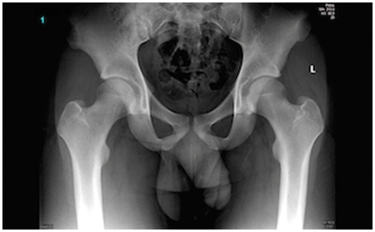
Preoperative AP pelvis radiograph showing a left hip that is concentrically reduced with a lateral center edge angle of 32 degrees.
Approximately 4 weeks after his clinical evaluation, he dislocated his hip in his sleep. He was evaluated in the Emergency Room where a magnetic resonance imaging (MRI) scan was ordered due to the lack of clinical suspicion for hip dislocation. It showed the left hip to be dislocated posteriorly with the ligamentum teres absent (Fig. 2). The hip was reduced with closed reduction techniques, and he was instructed to follow-up with our clinic. Radiographic evaluation in our clinic showed a non-arthritic joint with reasonable lateral coverage and a lateral centre edge angle of 32 degrees with no crossover sign. His overall acetabular volume was low with small anterior and posterior walls and neutral acetabular version. A CT scan was ordered to assess the volume of the cup and, specifically, integrity of the posterior acetabular wall (Fig. 3a). Distal cuts were not made through the knee so femoral version was not analysed. Both the anterior and posterior walls were small and there was a mild flattening to the posterior wall, on a cephalad cut on the CT scan there was small ossification (Fig. 3b). It was not felt to be a fragment of the acetabular wall, but rather possibly some early heterotopic ossification. He was sent to an Orthopaedic Traumatologist for confirmation, and he was sent back to our clinic for definitive treatment.
Fig. 2.
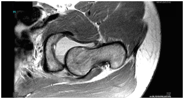
Preoperative MRI of the left hip indicating posterior hip dislocation.
Fig. 3.
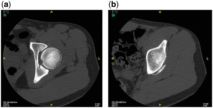
Preoperative CT scan of the left hip evaluating integrity of the posterior acetabular wall (a) and indicating mild flattening and small ossification of the posterior wall (b), which were determined to be negative for posterior wall fracture by an Orthopaedic Traumatologist.
Both he and his family were very concerned about the stability of his hip and requested a surgical procedure to correct the problem and prevent it from happening again. A periacetabular osteotomy to increase acetabular coverage and bony stability was ruled out as it was felt that his acetabulum was in an optimal position for a low volume cup. His lateral X-ray showed mild CAM-type femoroacetabular impingement with an alpha angle exceeding 60 degrees (Fig. 4). His MRI showed a labral tear and no evidence of avascular necrosis. He and his family were offered hip arthroscopy to address both the labral tear and the ligamentum teres deficiency, understanding the possibility of reconstruction of both.
Fig. 4.
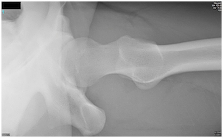
Preoperative cross table lateral radiograph showing mild CAM-type FAI.
Data collection
The patient completed a pre- and postoperative subjective questionnaire that included a Visual Analogue Scale (VAS) for average pain at rest, average pain with activities of daily living (ADL) and average pain with athletic activities in addition to the Lower Extremity Functional Scale (LEFS) [18] and the modified Harris Hip Score (MHHS) [19]. At follow-up the patient also completed a satisfaction scale, where 1 was very dissatisfied and 10 was very satisfied. Failure was defined as subsequent intra-articular hip surgery or recurrent hip instability.
Arthroscopic findings
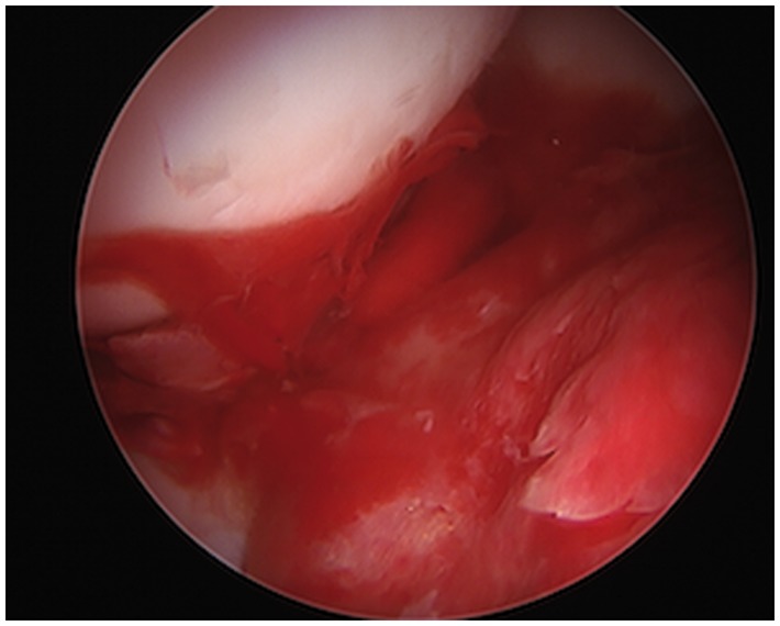
Hip arthroscopy was performed with a standard supine approach. His hip capsule was grossly intact and was opened roughly 3 cm with a linear capsulotomy between the 10:30 and 2:30 position on the acetabulum for exposure and a 1 cm medial sleeve of capsular tissue was preserved for later repair. His labrum was extensively torn, shredded, degenerative and severely bruised from the 7:0–4:30 position of the acetabulum. It also appeared very small, measuring 2 mm in diameter throughout (Fig. 5). Additionally, it did not form a seal with a femoral head, so it was non-functional and did not contribute to the stability or volume of the acetabulum (Fig. 6). The labrum was deemed not suitable for repair as it was grossly insufficient in size, inflamed and chronically torn. In the posterior compartment the labrum and capsule complex was torn off of the posterior aspect of the acetabular wall (Fig. 7). The posterior wall of the acetabulum was shallow, but there was no evidence of a fracture from the previous instability episodes. In the peripheral compartment there was reactive wear over the neck from the underlying cam type impingement. There was also an indentation medial to the head/neck junction on the anterior femoral head in a crescent shape that likely resulted from the previous posterior dislocations (Fig. 8). A grade 2 area of chondromalacia measuring 5 × 15 mm was noted on the posterior inferior aspect of the acetabulum, and diffuse grade 1 chondromalacia and softening of the cartilage on the posterior femoral head was also noted. Otherwise the cartilage was normal. As expected, the ligamentum teres was completely absent and a large amount of inflammatory tissue was present in the fovea (Fig. 9). Additionally, in the peripheral compartment any adduction in a flexed position caused the femur to violently dislocate posteriorly. This indicated substantial joint instability and reinforced the need for combined labral and ligamentum teres reconstruction.
Fig. 5.
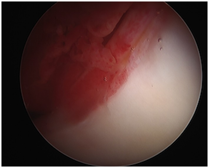
Arthroscopic view from the anterolateral portal showing an extensively torn, shredded and bruised labrum measuring 2 mm in diameter.
Fig. 6.
Arthroscopic view from the anteromedial portal of the anterior aspect of the reduced hip showing a severely torn and bruised labrum with no seal between the femoral head and acetabulum.
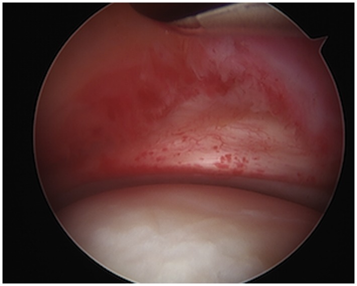
Fig. 7.
Arthroscopic view from the anteromedial portal of the posterior aspect of the hip joint showing a severely torn and inflamed labrum and capsule.
Fig. 8.
Arthroscopic view from the anteromedial portal with the hip flexed showing an indentation in the femoral head, suggestive of previous hip dislocation.
Fig. 9.
Arthroscopic view from the anterolateral portal showing a severely inflamed fovea and deficient ligamentum teres.
Surgical technique
Arthroscopically, femoral osteoplasty was performed, removing 4–7 mm of excess bone over a 3.5 mm width to create a more anatomic concavity, reducing the alpha angle to approximately 45 degrees and removing the cam deformity. The labrum was resected and the acetabular rim was excoriated, which, in addition to the femoral osteoplasty, provided a nice bleeding environment for incorporation of the labral graft. The goal was to prepare the acetabular edge without reducing the acetabular volume, so less than 1 mm was removed to achieve this.
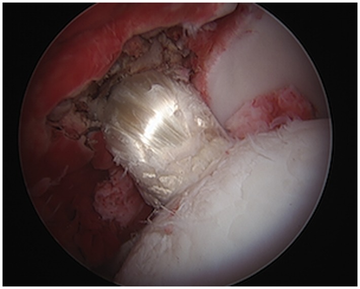
The torn, damaged labrum was removed from the origin of the transverse acetabulum (7:30) to approximately 2/3 down the back of the acetabulum (4:00). The labral reconstruction was performed using a front-to-back fixation technique described previously [20]. The extent of the labral deficiency was first measured to estimate graft length and anchors were placed in the acetabular rim in preparation of securing the labral graft. Frozen iliotibial band allograft was used for this technique. The graft was rolled and sutured using an accordion-type suturing technique to create a final tubularized graft measuring approximately 5.5 mm in diameter and 12.5 cm in length. After the graft was secured to the acetabular rim utilizing the nine previously placed suture anchors, the hip was reduced and a perfect seal was noted between the reconstructed labrum and the femoral head (Figs 10 and 11). The hip was also more stable in the peripheral compartment.
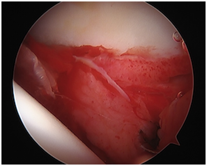
Fig. 10.
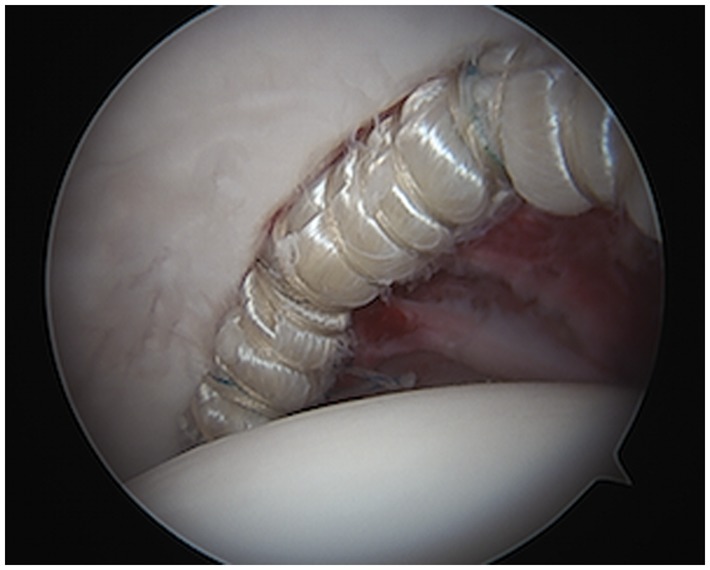
Arthroscopic view from the anteromedial portal of the posterior aspect of the joint showing the 12.5 cm labral reconstruction with allograft from approximately the 1: 00–4: 00 position.
Fig. 11.
Arthroscopic view from the anteromedial portal showing a perfect seal between the allograft labral reconstruction and a concentrically reduced femoral head.
Following the labral reconstruction, the ligamentum teres reconstruction began using a similar technique as described previously [21, 22]. The joint remained in a reduced position while a guide wire was placed over the lateral aspect of the femur to establish an acceptable guide to the inferocentral aspect of the femoral head over the fovea (2:00 position). Once the guide wire was in an acceptable position, the hip was distracted to view entry of the guide wire through the femoral head. Parallel guides were used to make small adjustments and a 10 mm drill was used to create a tunnel from the lateral aspect of the femur to the femoral head. Once this was completed, the synovium and the remnant of the ligamentum teres over the posterior aspect of the fovea down to the bone were then debrided working either down the tunnel in the femoral neck or through the arthroscopic portals. A 4.5 mm drill was inserted down the femoral neck tunnel and angled posteriorly into the fovea. This was used to drill through the fovea and fluoroscopic assistance was used to avoid plunging through the medial wall of the pelvis.
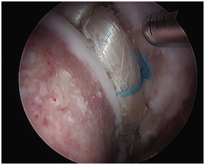
Once the tunnel was created, a Zimmer Biomet ToggleLoc (Zimmer Biomet, Warsaw, Indiana) was passed to the medial aspect of the pelvis and flipped under fluoroscopic guidance. The ligamentum teres graft was created from a tibialis anterior allograft measuring approximately 5 mm in diameter and 18 cm in length (Allosource, Centennial, Colorado). The graft was then looped through the suture construct on the end of the ToggleLoc and doubled over to create a final graft diameter of 10 mm. The suture was then used to pull the graft down the femoral tunnel to the base of the fovea. This zip loop construct locked the medial aspect of the graft to the fovea of the acetabulum. With the graft fixed, the hip joint was reduced and a perfect seal was noted between the labral reconstruction and the femoral head. The anterior portion of the capsulotomy was then closed with #1 vicryl to preserve the stability gained with an intact hip capsule. With the hip extended and externally rotated slightly, the ligamentum teres graft was tightened and a 9 × 30 mm PEEK interference screw (Arthrex, Naples, Florida) was inserted in the femoral tunnel to secure the graft (Fig. 12).
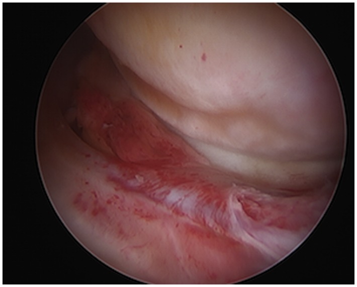
Fig. 12.
Arthroscopic view from the anteromedial portal showing the ligamentum teres reconstruction with allograft.
Postoperative management
The rehabilitation program following the labral and ligamentum teres reconstruction was similar to that following labral reconstruction [8]. The patient was cautioned regarding the aneural properties of the grafts and began a supervised physical therapy program within 1 week of surgery. Limited weight bearing at 30% for 6 weeks and posterior hip precautions were maintained, including no flexion more than 90 degrees, no adduction across midline or internal rotation and an abduction pillow for 4 weeks. The focus of physical therapy was to regain motion, strengthen the gluteus medius muscle and establish a normal gait pattern. The long-term goal was to return to the full pre-injury level of physical activity approximately after 6 months.
RESULTS
Subjective follow-up questionnaires were obtained from the patient at 14 months postoperative. At the time of follow-up his MHHS improved from 58 preoperatively to 100 postoperatively and his LEFS improved from 56 to 79. Similarly, his VAS pain scores decreased; at rest he improved from 3 to 1, with activity he improved from 3 to 1, and with athletics he improved from 5 to 1. Patient satisfaction was 10 out of 10. The only subjective complaint was ‘a little bit of difficulty’ with ‘making sharp turns while running fast’ on the LEFS. The patient has not reported any episodes or subjective feelings of instability and has not required further surgical procedures in the hip.
Imaging, including anteroposterior and lateral radiographs, was obtained at 3 months posteroperatively. The radiographs showed a non-arthritic joint that is well reduced and hardware for the ligmentum teres reconstruction in the appropriate position (Fig. 13).
Fig. 13.

Final AP pelvis radiograph showing anatomic tunnel placement and medial fixation of the ligamentum teres reconstruction and a concentric, stable reduction of the left hip.
DISCUSSION
At 14 months postoperative, simultaneous arthroscopic labral reconstruction and ligamentum teres reconstruction in a young patient with recurrent, severe hip instability resulted in excellent patient-reported outcomes. While arthroscopic labral reconstruction and ligamentum teres reconstruction techniques have been reported separately in the literature [(20–22], to our knowledge, simultaneous reconstructions have been rarely reported. Consistent with the results identified in a previous systematic review on ligamentum teres injuries [16, 17] and published outcomes following arthroscopic labral reconstruction of the hip [8, 23, 24], the postoperative results in this case suggest benefits of surgical reconstruction in the presence of recurrent hip instability.
While the acetabular labrum’s role in hip stability is increasingly recognized [2, 3], the ligamentum teres has recently emerged as a key stabilizer of the hip that may also contribute to hip symptomology [10, 16, 17, 25–27]. Nociceptive and proprioceptive nerve endings have been identified in the ligamentum teres, indicating that damage to this structure may produce pain [10, 16, 27, 28]. Additionally, although the published data regarding mechanical and biological properties of the ligamentum teres are still limited, increasing evidence suggests that it may be critical for hip stability [16, 17]. Data from animal models demonstrated a significant increase in the number of hip dislocations when the ligamentum teres was resected compared with the control group with intact ligamentum teres [16, 25]. The ligamentum teres is believed to contribute to the stability of the hip most substantially when externally rotated and flexed or internally rotated and extended, and when other stabilizers of the hip such as the acetabular labrum are deficient [12, 16].
As our understanding of hip anatomy evolves and improves, arthroscopic treatment options for both labral and ligamentum teres pathology have also advanced. Historically, surgical options for both labral and ligamentum teres pathology included predominantly debridement and resection [17, 29, 30]. In recent years, arthroscopic techniques for labral pathology have progressed to primarily labral preserving procedures. Complete labral reconstruction, in particular, has recently emerged as a first-line treatment option in the setting of insufficient or poor-quality labral tissue [8]. Several advantages for labral reconstruction over labral repair include the recreation of a normal labral size and quality and recreate a tight seal between the femoral head and the labrum, which results in improved stabilization of the hip and reduced pain [8, 13, 21, 31]. Similarly, while short-term successes in reducing pain following ligamentum teres debridement have been appreciated, complete reconstruction may provide further benefits for pain reduction and improved hip stability, especially in positions of extension or external rotation while squatting [13, 16].
Due to the important stabilization role of both the acetabular labrum and the ligamentum teres, arthroscopic ligamentum teres reconstruction coupled with labrum reconstruction represents a novel way of approaching severe hip instability. In this case, the patient had severe recurrent hip instability with appropriate acetabular coverage following a lacrosse injury. Subsequently, the preferred treatment choice was soft tissue reconstruction as opposed to osteotomy. The potential for continued pain and instability with debridement, coupled with the evidence for stability provided by both the ligamentum teres and labrum, provided the basis for performing both a labrum and a ligamentum teres reconstruction on the patient. In reconstructing the patient’s acetabular labrum and ligamentum teres using allografts the patient was able to retain physiological integrity in the hip joint, and short-term outcomes have been excellent.
LIMITATIONS
Early outcomes recorded for this patient appear promising, but further long-term assessment is necessary to continue to monitor hip stability, ADL and future joint health. In addition, this procedure was performed on a young, otherwise healthy adolescent. This population can represent a particularly challenging group and may not be representative of other age groups. As a case report, this study also does not include a comparative group; therefore, it is not possible to determine if labral reconstruction or ligamentum teres reconstruction alone would have resulted in the same outcome. Given the limited reports of ligamentum teres reconstructions in the literature, we believe this case report is an important addition that highlights success of a novel procedure for a complex presentation. Finally, it is important to note that this procedure is technically demanding, requires advanced hip arthroscopy training, and has a steep learning curve. As such, these results may not be generalizable to all patients or surgeons.
CONCLUSIONS
This case report demonstrates a technique for and early outcomes of simultaneous arthroscopic ligamentum teres and acetabular labrum reconstruction in a patient with recurrent hip instability. Short-term outcomes suggest improved hip stability, reduced pain, high patient satisfaction and return to pre-injury activities at 14 months postoperative in this single case report.
FUNDING
None.
CONFLICT OF INTEREST STATEMENT
Brian J. White is a consultant for Smith & Nephew, Biomet, and Conmed Linvatec, where he holds a role in surgeon education and product development for hip arthroscopy.
REFERENCES
- 1. Stubbs AJ, Howse EA, Mannava S.. Tissue engineering and the future of hip cartilage, labrum and ligamentum teres. J Hip Preserv Surg 2016; 3: 23–9. [DOI] [PMC free article] [PubMed] [Google Scholar]
- 2. Philippon MJ, Nepple JJ, Campbell KJ. et al. The hip fluid seal–Part I: the effect of an acetabular labral tear, repair, resection, and reconstruction on hip fluid pressurization. Knee Surg Sports Traumatol Arthrosc 2014; 22: 722–9. [DOI] [PubMed] [Google Scholar]
- 3. Nepple JJ, Philippon MJ, Campbell KJ. et al. The hip fluid seal–Part II: the effect of an acetabular labral tear, repair, resection, and reconstruction on hip stability to distraction. Knee Surg Sports Traumatol Arthrosc 2014; 22: 730–6. [DOI] [PubMed] [Google Scholar]
- 4. Ferguson SJ, Bryant JT, Ganz R, Ito K.. An in vitro investigation of the acetabular labral seal in hip joint mechanics. J Biomech 2003; 36: 171–8. [DOI] [PubMed] [Google Scholar]
- 5. Ferguson SJ, Bryant JT, Ganz R, Ito K.. The acetabular labrum seal: a poroelastic finite element model. Clin Biomech (Bristol, Avon) 2000; 15: 463–8. [DOI] [PubMed] [Google Scholar]
- 6. Ayeni OR, Adamich J, Farrokhyar F. et al. Surgical management of labral tears during femoroacetabular impingement surgery: a systematic review. Knee Surg Sports Traumatol Arthrosc 2014; 22: 756–62. [DOI] [PubMed] [Google Scholar]
- 7. Philippon MJ, Briggs KK, Yen YM, Kuppersmith DA.. Outcomes following hip arthroscopy for femoroacetabular impingement with associated chondrolabral dysfunction: minimum two-year follow-up. J Bone Joint Surg Br 2009; 91-B: 16–23. [DOI] [PubMed] [Google Scholar]
- 8. White BJ, Stapleford AB, Hawkes TK. et al. Allograft use in arthroscopic labral reconstruction of the hip with front-to-back fixation technique: minimum 2-year follow-up. Arthroscopy 2016; 32: 26–32. [DOI] [PubMed] [Google Scholar]
- 9. White BJ, Patterson J, Herzog MM.. Revision arthroscopic acetabular labral treatment: repair or reconstruct? Arthroscopy 2016; 32: 2513–20. [DOI] [PubMed] [Google Scholar]
- 10. Dehao BW, Bing TK, Young JL.. Understanding the ligamentum teres of the hip: a histological study. Acta Ortop Bras 2015; 23: 29–33. [DOI] [PMC free article] [PubMed] [Google Scholar]
- 11. Mikula JD, Slette EL, Chahla J. et al. Quantitative anatomic analysis of the native ligamentum teres. Orthop J Sports Med 2017; 5: 2325967117691480.. [DOI] [PMC free article] [PubMed] [Google Scholar]
- 12. Martin RL, Palmer I, Martin HD.. Ligamentum teres: a functional description and potential clinical relevance. Knee Surg Sports Traumatol Arthrosc 2012; 20: 1209–14. [DOI] [PubMed] [Google Scholar]
- 13. Leunig M, Beck M, Stauffer E. et al. Free nerve endings in the ligamentum capitis femoris. Acta Orthop Scand 2000; 71: 452–4. [DOI] [PubMed] [Google Scholar]
- 14. Hosalkar HS, Varley ES, Glaser D. et al. Isocentric reattachment of ligamentum teres: a porcine study. J Pediatr Orthop 2011; 31: 847–52. [DOI] [PubMed] [Google Scholar]
- 15. Lindner D, Sharp KG, Trenga AP. et al. Arthroscopic ligamentum teres reconstruction. Arthrosc Tech 2013; 2:e21–5. [DOI] [PMC free article] [PubMed] [Google Scholar]
- 16. O'Donnell JM, Pritchard M, Salas AP, Singh PJ.. The ligamentum teres – its increasing importance. J Hip Preserv Surg 2014; 1: 3–11. [DOI] [PMC free article] [PubMed] [Google Scholar]
- 17. de SD, Phillips M, Philippon MJ. et al. Ligamentum teres injuries of the hip: a systematic review examining surgical indications, treatment options, and outcomes. Arthroscopy 2014; 30: 1634–41. [DOI] [PubMed] [Google Scholar]
- 18. Binkley JM, Stratford PW, Lott SA, Riddle DL.. The Lower Extremity Functional Scale (LEFS): scale development, measurement properties, and clinical application. North American Orthopaedic Rehabilitation Research Network. Phys Ther 1999; 79: 371–83. [PubMed] [Google Scholar]
- 19. Byrd JW, Jones KS.. Prospective analysis of hip arthroscopy with 2-year follow-up. Arthroscopy 2000; 16: 578–87. [DOI] [PubMed] [Google Scholar]
- 20. White BJ, Herzog MM.. Arthroscopic labral reconstruction of the hip using iliotibial band allograft and front-to-back fixation technique. Arthrosc Tech 2016; 5:e89–97. [DOI] [PMC free article] [PubMed] [Google Scholar]
- 21. Simpson JM, Field RE, Villar RN.. Arthroscopic reconstruction of the ligamentum teres. Arthroscopy 2011; 27: 436–41. [DOI] [PubMed] [Google Scholar]
- 22. Menge TJ, Mitchell JJ, Briggs KK, Philippon MJ.. Anatomic arthroscopic ligamentum teres reconstruction for hip instability. Arthrosc. Tech 2016; 5:e737–42. [DOI] [PMC free article] [PubMed] [Google Scholar]
- 23. Philippon MJ, Briggs KK, Hay CJ. et al. Arthroscopic labral reconstruction in the hip using iliotibial band autograft: technique and early outcomes. Arthroscopy 2010; 26: 750.. [DOI] [PubMed] [Google Scholar]
- 24. Geyer MR, Philippon MJ, Fagrelius TS, Briggs KK.. Acetabular labral reconstruction with an iliotibial band autograft: outcome and survivorship analysis at minimum 3-year follow-up. Am J Sports Med 2013; 41: 1750–6. [DOI] [PubMed] [Google Scholar]
- 25. Wenger D, Miyanji F, Mahar A, Oka R.. The mechanical properties of the ligamentum teres. A pilot study to assess its potential for improving stability in children's hip surgery. J Pediatr Orthop 2007; 27: 408–10. [DOI] [PubMed] [Google Scholar]
- 26. Cerezal L, Kassarjian A, Canga A. et al. Anatomy, biomechanics, imaging, and management of ligamentum teres injuries. Radiographics 2010; 30: 1637–51. [DOI] [PubMed] [Google Scholar]
- 27. Dierckman BD, Guanche CA.. Anterior hip capsuloligamentous reconstruction for recurrent instability after hip arthroscopy. Am J Orthop (Belle Mead NJ) 2014; 43:E319–23. [PubMed] [Google Scholar]
- 28. Gray AJ, Villar RN.. The ligamentum teres of the hip: an arthroscopic classification of its pathology. Arthroscopy 1997; 13: 575–8. [DOI] [PubMed] [Google Scholar]
- 29. Maradit Kremers H, Schilz SR, Van Houten HK. et al. Trends in utilization and outcomes of hip arthroscopy in the United States between 2005 and 2013. J Arthroplasty 2017; 32: 750–55. [DOI] [PubMed] [Google Scholar]
- 30. Ng VY, Arora N, Best TM. et al. Efficacy of surgery for femoroacetabular impingement. A systematic review. Am J Sports Med 2010; 38: 2337–45. [DOI] [PubMed] [Google Scholar]
- 31. Lee S, Wuerz TH, Shewman E. et al. Labral reconstruction with iliotibial band autografts and semitendinosus allografts improves hip joint contact area and contact pressure. An in vitro analysis. Am J Sports Med 2015; 43: 98–104. [DOI] [PubMed] [Google Scholar]