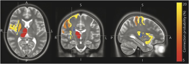
Figure 2. Postmortem brain R2 and total daily physical activity proximate to death.
Axial, coronal, and sagittal postmortem brain MRI sections depicting the regions (insular cortex, precentral and postcentral gyrus, and putamen) in which white matter tissue integrity (R2) is most strongly associated with total daily physical activity proximate to death (blue) and the most common gray matter terminals of white matter fiber passing through those regions (red/orange/yellow). The red-to-yellow coloring of each gray matter region corresponds to the probability that a white matter fiber passing through the blue R2 region has one of its terminals at that region. A = anterior; I = inferior; P= posterior; S = superior.

